CureRays Innovation & Education Institute

Research · Innovation · Education

Doctoring is discovering

The CureRays Innovation & Education Institute exists to make radiation medicine safer, gentler, and available to everyone — by funding the research that improves it, publishing what we learn without a paywall, teaching the clinicians who deliver it, and placing working equipment in the hands of communities that have none.

A California nonprofit corporation with 501(c)(3) status. Contributions are tax-deductible to the extent allowed by law.

Our mission

Why the Institute exists

The CureRays Innovation & Education Institute exists to make radiation medicine safer, gentler, and available to everyone — by funding the research that improves it, publishing what we learn without a paywall, teaching the clinicians who deliver it, and placing working equipment in the hands of communities that have none.

The Institute is the non-profit partner of CureRays Radiation Medicine. The clinic treats patients. The Institute studies whether that treatment can be made better, teaches what is learned, and works to put it within reach of people who currently have no access to it at all. The two are separate organisations with separate books.

Open knowledge

Everything we teach, free to read

The patient education library

Plain-language guides to cancers and benign conditions treated with radiation — what it is, how it is staged, what the standard of care is, how radiation works on it, and which studies are changing that. No account, no paywall, no advertising.

Browse the library

IJRM-SSS, the Institute’s journal

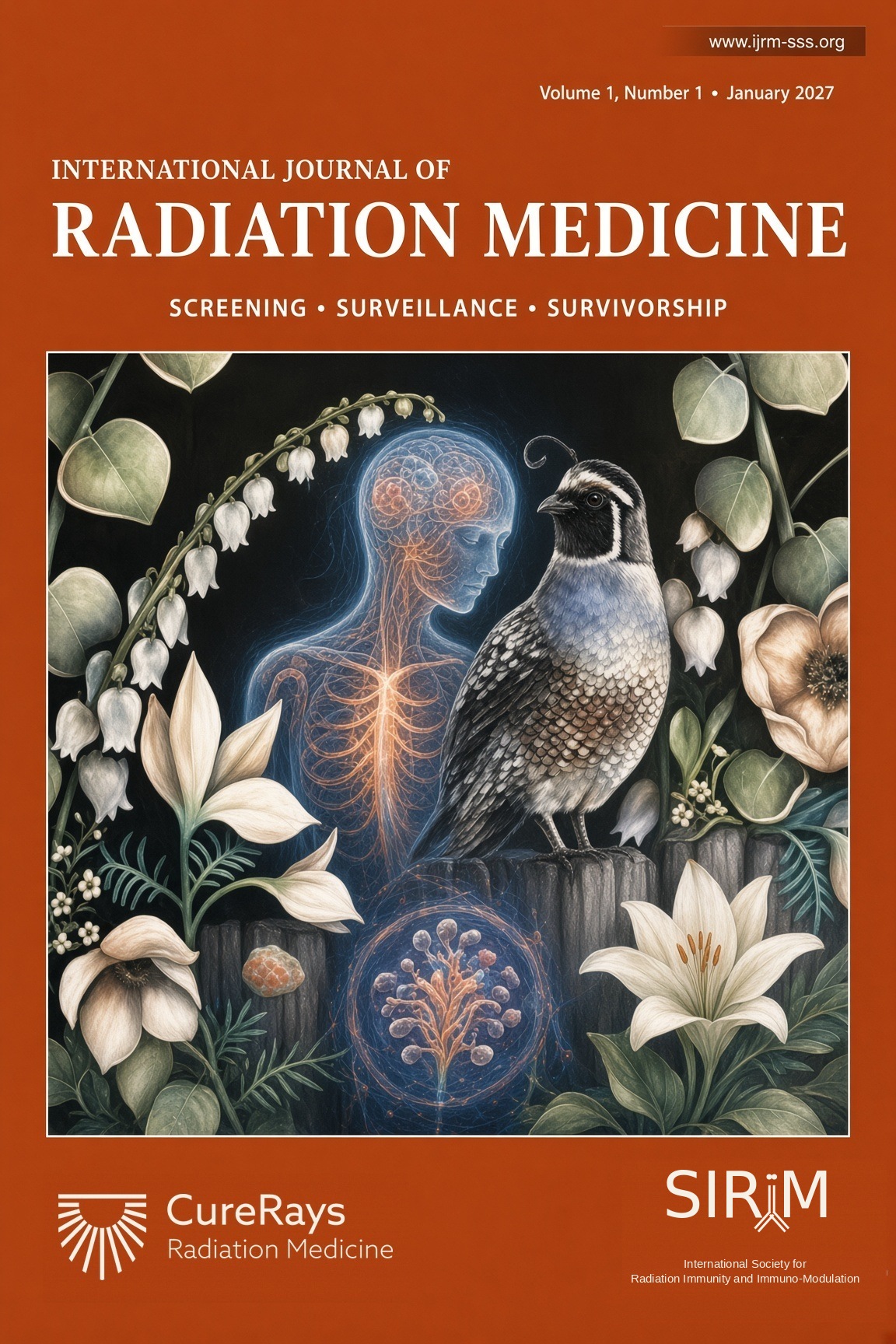
The International Journal of Radiation Medicine — Screening, Surveillance, Survivorship, published with the International Society for Radiation Immunity and Immuno-Modulation (SIRiiM). Authors retain ownership of their work, and proceeds fund the researchers and peer reviewers directly.

Read the journal

Access

Getting this care to where it isn’t

A hub-and-spoke model

Radiation oncologists at central facilities supervise and collaborate with primary-care and dermatology partners in community clinics — extending specialist expertise closer to where patients live.

Equipment where there is none

Hospitals, clinics and private practices donate superficial radiation units they no longer use. The Institute coordinates with partners in the Philippines and Pakistan to place that equipment where it will train therapists and treat patients — and helps donors establish fair market value for the deduction.

How equipment donation works

Accountability

How the Institute is organised

A 501(c)(3) nonprofit

The CureRays Innovation & Education Institute is a California nonprofit corporation with 501(c)(3) status. Donations qualify under Section 170 of the Internal Revenue Code.

Separate from the clinic

CureRays Radiation Medicine is a for-profit medical practice and a separate entity. The Institute does not bill for patient care, and charitable funds do not subsidise clinical operations.

What we publish

Research findings are published through IJRM-SSS whether or not they favour the treatments we use. Patient education is free and carries no advertising.

Conflict of interest

The Institute’s founder also runs the affiliated for-profit practice. That is a real conflict, and we would rather state it than have you find it.

Read the disclosure

Support the mission

Ways to give

Donate equipment

A superficial unit you have retired can become a regional cancer centre’s only machine.

Start a donation

Fund research

Support the trials and translational work that test whether this care can be made gentler.

See the research

Partner with us

Institutions, societies and industry partners collaborate on education, publishing and equipment placement abroad.

About the Institute

Looking for treatment?

Patient care is delivered by CureRays Radiation Medicine, a separate clinical practice, at www.curerays.com.

Patient Education

Understand your cancer — in plain language

Facing a cancer diagnosis can be overwhelming. We're building a comprehensive library for every cancer: staging explained simply, the standard of care, how each treatment works, and the latest research — paired with short, AI-narrated explainer videos.

▶  Watch on our YouTube channel

Start here

Two short videos to begin with

The basics of cancer — what it is, in three minutes.
Organ of origin — why where a cancer starts matters more than where it spreads.

The library

Guides by cancer type

Each guide follows the same trusted structure: what it is · staging in plain terms · the standard of care · how each treatment works · latest studies · your questions answered.

Breast Cancer

Breast cancer begins when cells in the breast's ducts or lobules grow out of control; caught early, it is highly treatable.

Full guide live

Prostate Cancer

Prostate cancer grows in the walnut-sized gland below the bladder; most cases grow slowly and are highly curable.

Full guide live

Lung Cancer

Lung cancer begins when cells in the lungs grow out of control; found early, many cases can be cured — and low-dose CT screening is finding more of them in time.

Full guide live

Colorectal Cancer

Colorectal cancer starts in the colon or rectum, usually from a small growth called a polyp; screening can catch and remove polyps before they ever become cancer.

Full guide live

Brain Cancer

Brain tumors form when cells in or around the brain grow abnormally; treatment combines surgery, radiation, and medicine, tailored to the tumor type.

Full guide live

Skin Cancer: Non-Melanoma vs. Melanoma

Skin cancer is the most common cancer of all; the everyday non-melanoma types are highly curable, often without surgery.

Full guide live

Head & Neck Cancer

Head and neck cancers arise in the mouth, throat, voice box, and related areas; many — especially HPV-related throat cancers — are very curable.

Full guide live

Bladder Cancer

Bladder cancer starts in the lining of the bladder and often shows up as blood in the urine; when it is caught before it grows into the muscle, it is very treatable.

Full guide live

Lymphoma

Lymphoma is a cancer of the immune system's white blood cells; many forms are among the most curable of all cancers.

Full guide live

Melanoma

Melanoma is a serious skin cancer that begins in pigment-making cells; found early it is almost always curable, and new medicines have transformed advanced disease.

Full guide live

Kidney Cancer

Kidney cancer forms in the filtering organs of the urinary system; many are found early by chance on scans and cured with surgery — and precise radiation is a newer option.

Full guide live

Uterine (Endometrial) Cancer

Uterine cancer usually starts in the lining of the womb and most often announces itself early with abnormal bleeding — which is why it is frequently caught and cured.

Full guide live

Cervical Cancer

Cervical cancer starts in the lower part of the uterus and is caused by HPV — making it one of the few cancers we can largely prevent through vaccination and screening.

Full guide live

Pancreatic Cancer

Pancreatic cancer forms in the digestive gland behind the stomach; it is challenging because it often hides until it grows, but treatments and precise radiation keep improving.

Full guide live

Anal Cancer

Anal cancer is usually cured without surgery, using a proven combination of radiation and chemotherapy that preserves normal function.

Full guide live

Metastatic Cancer

Metastatic cancer has spread beyond where it started; when only a few spots exist (oligometastatic), focused radiation can sometimes treat it for cure.

Full guide live

Liver Cancer

Liver cancer starts in the cells of the liver; when found early it can often be cured, and precise radiation now gives many patients a non-surgical path to control.

Full guide live

Stomach (Gastric) Cancer

Stomach cancer begins in the lining of the stomach; caught early it is highly curable, and combined treatment — including radiation — improves cure rates for more advanced disease.

Full guide live

Esophageal Cancer

Esophageal cancer forms in the tube that carries food to the stomach; radiation combined with chemotherapy is central to treatment, and can even cure some patients without surgery.

Full guide live

Ovarian Cancer

Ovarian cancer starts in the ovaries or nearby tissue; surgery and chemotherapy lead treatment, with newer targeted drugs and selective radiation helping control disease and relieve symptoms.

Full guide live

Thyroid Cancer

Thyroid cancer forms in the butterfly-shaped gland in the neck; most types are highly curable, often with surgery and radioactive iodine rather than external radiation.

Full guide live

Sarcoma (Soft-Tissue & Bone)

Sarcomas are rare cancers of the body's connective tissues — muscle, fat, nerve, and bone; combining surgery with precisely targeted radiation offers the best chance to cure them while preserving the limb.

Full guide live

Leukemia

Leukemia is cancer of the blood and bone marrow; treated mainly with medicines, with radiation playing key supporting roles — especially in preparing for a stem-cell transplant.

Full guide live

Multiple Myeloma

Multiple myeloma is a cancer of plasma cells in the bone marrow; modern medicines control it for many years, and targeted radiation is highly effective for relieving bone pain.

Full guide live

Gastrointestinal Stromal Tumor (GIST)

GIST is an uncommon tumor of the digestive tract's own pacemaker cells; surgery and targeted pill medicines control it remarkably well, and radiation has a focused, supportive role.

Full guide live

Gallbladder & Bile Duct Cancer

These uncommon cancers of the biliary system are best treated by a coordinated team; surgery offers the best chance of cure, and radiation paired with chemotherapy helps control disease and relieve blockage.

Full guide live

Neuroendocrine Tumors (NETs)

Neuroendocrine tumors grow from the body's hormone-producing cells and often grow slowly; many are highly treatable, and a special form of targeted radiation can find and treat them throughout the body.

Full guide live

Testicular Cancer

Testicular cancer is among the most curable of all cancers, even when it has spread; for one common type, low-dose radiation is a proven, well-tolerated option after surgery.

Full guide live

Vaginal & Vulvar Cancer

These uncommon gynecologic cancers are often linked to HPV and are very treatable when found early; radiation, sometimes with chemotherapy, can cure many cases while preserving function.

Full guide live

Penile Cancer

Penile cancer is rare and often curable, especially when caught early; modern, organ-preserving treatments — including radiation — aim to cure while keeping as much normal form and function as possible.

Full guide live

Thymoma & Thymic Cancer

These rare tumors of the thymus gland behind the breastbone are often slow-growing and very treatable; surgery is the cornerstone, and radiation helps secure control after surgery or when removal isn't complete.

Full guide live

Mesothelioma

Mesothelioma is an uncommon cancer of the lining around the lungs or abdomen, usually linked to past asbestos exposure; a coordinated team using surgery, chemotherapy, immunotherapy, and radiation can meaningfully control it.

Full guide live

Adrenal Cancer (Adrenocortical Carcinoma)

Adrenal cancer is a rare tumor of the small hormone-making glands above the kidneys; surgery is the main cure, and radiation and medicine help control it and ease symptoms.

Full guide live

Small Intestine Cancer

Small intestine cancer is an uncommon tumor of the long middle section of the gut; surgery is the main treatment, with chemotherapy and focused radiation in supporting roles.

Full guide live

Appendix Cancer (Appendiceal Cancer)

Appendix cancer is a rare tumor of the small pouch off the colon, often found by surprise during appendix surgery; treatment centers on surgery, sometimes with heated chemotherapy in the abdomen.

Full guide live

Salivary Gland Cancer

Salivary gland cancer is an uncommon tumor of the glands that make saliva; surgery is the main treatment, and radiation — sometimes with specialized particle beams — plays a major supporting role.

Full guide live

Nasopharyngeal Cancer

Nasopharyngeal cancer forms high in the throat behind the nose; because surgery there is difficult, radiation — usually with chemotherapy — is the main, often curative treatment.

Full guide live

Merkel Cell Carcinoma

Merkel cell carcinoma is a rare, fast-growing skin cancer; it is very sensitive to radiation, and surgery, radiation, and immunotherapy together give the best control.

Full guide live

Ocular Melanoma (Eye Melanoma)

Ocular melanoma is a rare cancer inside the eye; precise radiation, often a small implant placed on the eye, controls most tumors while preserving the eye itself.

Full guide live

Osteosarcoma (Bone Cancer)

Osteosarcoma is the most common bone cancer, often in teenagers and young adults; chemotherapy and surgery are the cure, with radiation used in specific, hard-to-reach situations.

Full guide live

Pheochromocytoma & Paraganglioma

Pheochromocytoma and paraganglioma are rare tumors that can release adrenaline-like hormones; surgery is the main treatment, and special targeted radiation can control tumors that have spread.

Full guide live

Ureteral & Upper Tract Cancer

Ureteral cancer is a rare cancer of the thin tubes that carry urine from the kidneys to the bladder; surgery is the main treatment, with radiation and medicine added in selected cases.

Full guide live

Urethral Cancer

Urethral cancer is a rare cancer of the tube that carries urine out of the body; treatment combines surgery, radiation, and medicine, often aiming to preserve function.

Full guide live

Ewing Sarcoma

Ewing sarcoma is a rare bone and soft-tissue cancer mostly in children and young adults; it is very sensitive to chemotherapy and radiation, which work together with surgery to cure many patients.

Full guide live

Chordoma

Chordoma is a rare, slow-growing bone cancer of the spine and skull base; surgery and high-dose particle radiation are the cornerstones of treatment.

Full guide live

Eyelid & Conjunctival Cancer

Eyelid and conjunctival cancers are skin and surface cancers of the eye area; most are highly curable, and radiation helps treat the eye while preserving sight.

Full guide live

Parathyroid Cancer

Parathyroid cancer is a very rare cancer of the tiny calcium-controlling glands in the neck; surgery is the main cure, and radiation helps lower the chance of return.

Full guide live

Wilms Tumor (Childhood Kidney Cancer)

Wilms tumor is the most common kidney cancer in children; it responds very well to surgery, chemotherapy, and radiation, and most children are cured.

Full guide live

Gestational Trophoblastic Disease

Gestational trophoblastic disease is a rare group of tumors that grow from the tissue that would normally become the placenta; it is highly treatable and one of the most curable of all cancers.

Full guide live

Retinoblastoma (Childhood Eye Cancer)

Retinoblastoma is a rare eye cancer of young children that is highly curable, with modern treatment focused on saving the child's life, the eye, and vision whenever possible.

Full guide live

Hypopharyngeal Cancer

Hypopharyngeal cancer forms in the lower part of the throat near the voice box; modern chemoradiation can often cure it while preserving the ability to speak and swallow.

Full guide live

Laryngeal (Voice Box) Cancer

Laryngeal cancer forms in the voice box; when caught early it is highly curable with radiation that preserves the voice, and even advanced cases can often be cured while keeping the larynx.

Full guide live

Nasal Cavity & Paranasal Sinus Cancer

Nasal and paranasal sinus cancers form in the space behind the nose and the air pockets around it; they are treated with surgery and precisely targeted radiation that protects the nearby eyes and brain.

Full guide live

Pituitary Tumors

Pituitary tumors grow in the small hormone-control gland at the base of the brain; most are benign and very treatable, with focused radiation reserved for tumors that persist after surgery or medication.

Full guide live

Plasmacytoma (Solitary Plasma Cell Tumor)

A plasmacytoma is a single tumor made of plasma cells; it is highly sensitive to radiation, which is the main, often curative treatment for this localized disease.

Full guide live

Neuroblastoma (Childhood Nerve Cancer)

Neuroblastoma is a cancer of early nerve cells that affects young children; treatment is matched to risk, ranging from observation to intensive therapy, and radiation plays an important role in high-risk disease.

Full guide live

Oropharyngeal (Throat) Cancer

Oropharyngeal cancer affects the middle of the throat — the tonsils and base of the tongue — and the most common form today is caused by HPV, which responds especially well to radiation and is highly curable.

Full guide live

Oral Cavity (Mouth) Cancer

Oral cavity cancer forms in the mouth — most often the tongue, gums, or floor of the mouth — and is highly curable when found early, with surgery and radiation as the main treatments.

Full guide live

Rhabdomyosarcoma

Rhabdomyosarcoma is a soft-tissue cancer that most often affects children and is treated with a combination of chemotherapy, radiation, and sometimes surgery — an approach that cures the majority of patients.

Full guide live

Anaplastic Thyroid Cancer

Anaplastic thyroid cancer is a rare, fast-growing thyroid cancer that requires urgent, coordinated treatment — and new targeted drugs matched to its genetics are improving outcomes for some patients.

Full guide live

Primary CNS Lymphoma

Primary CNS lymphoma is a lymphoma that begins in the brain, spinal cord, or eyes; it is treated mainly with specialized chemotherapy that reaches the brain, with radiation used in selected situations.

Full guide live

Bile Duct Cancer (Cholangiocarcinoma)

Bile duct cancer (cholangiocarcinoma) arises in the tubes that carry bile from the liver; treatment depends on where it forms, and radiation plays an important role in controlling tumors that cannot be removed.

Full guide live

Cutaneous (Skin) Lymphoma

Cutaneous lymphoma is a lymphoma that begins in the skin rather than the lymph nodes; it is usually slow-growing, and radiation is one of the most effective treatments for clearing skin lesions.

Full guide live

Spinal Cord & Spinal Tumors

Spinal tumors grow in or around the spinal cord and spine; treatment protects nerve function, and precise radiation — including spine SBRT — can control tumors while sparing the delicate spinal cord.

Full guide live

Medullary Thyroid Cancer

Medullary thyroid cancer starts in the thyroid's hormone-making C cells, is tracked with the blood markers calcitonin and CEA, is often linked to the RET gene, and is treated mainly with surgery — with radiation and targeted pills for harder-to-control disease.

Full guide live

Medulloblastoma

Medulloblastoma is a fast-growing brain tumor of the cerebellum, most common in children; it is highly curable with surgery, craniospinal radiation, and chemotherapy, and modern radiation is carefully shaped to protect the developing brain.

Full guide live

Ependymoma

Ependymoma is a tumor that grows from the lining of the brain's fluid spaces and the spinal cord; surgery to remove it as completely as possible plus focused radiation gives the best chance of long-term control.

Full guide live

Craniopharyngioma

Craniopharyngioma is a noncancerous but troublesome brain tumor near the pituitary gland and optic nerves; precise radiation, often after limited surgery, controls it while protecting vision and hormones.

Full guide live

Intracranial Germ Cell Tumors (Germinoma)

Intracranial germ cell tumors arise deep in the brain in children and young adults; germinomas are exquisitely sensitive to radiation and among the most curable brain tumors, with modern treatment carefully reducing radiation to protect the developing brain.

Full guide live

Small Cell Lung Cancer

Small cell lung cancer is a fast-growing lung cancer strongly tied to smoking; it responds quickly to chemotherapy and radiation, and timely combined treatment — sometimes with radiation to prevent spread to the brain — offers the best chance of control.

Full guide live

Ampullary Cancer

Ampullary cancer forms at the small junction where the bile and pancreatic ducts drain into the intestine; because it blocks bile early, it is often found sooner than nearby cancers, and surgery offers the best chance of cure, sometimes with radiation and chemotherapy.

Full guide live

Primary Peritoneal Cancer

Primary peritoneal cancer arises from the lining of the abdomen and behaves much like ovarian cancer; it is treated with surgery and chemotherapy, with radiation reserved for relieving symptoms or treating specific spots.

Full guide live

Choroid Plexus Tumors

Choroid plexus tumors grow from the tissue that makes the brain's fluid; most are curable with surgery alone, while the aggressive carcinoma form also needs chemotherapy and carefully shaped radiation to protect a child's developing brain.

Full guide live

Atypical Teratoid/Rhabdoid Tumor (ATRT)

ATRT is a rare, fast-growing brain tumor of very young children; cure is possible with intensive, coordinated treatment — surgery, multi-drug chemotherapy, and carefully shaped radiation — delivered urgently and tailored to the child's age.

Full guide live

Pineal Region Tumors & Pineoblastoma

Tumors of the pineal region sit deep in the center of the brain near the pineal gland; treatment ranges from surgery alone for benign types to surgery, chemotherapy, and craniospinal radiation for the aggressive pineoblastoma, with modern beams shaped to protect the developing brain.

Full guide live

Peritoneal Mesothelioma

Peritoneal mesothelioma is a rare cancer of the lining of the abdomen; the main treatment is specialized surgery to remove visible tumor combined with heated chemotherapy bathed directly in the abdomen, with systemic therapy and, occasionally, targeted radiation for symptom relief.

Full guide live

Cancer of Unknown Primary (CUP)

Cancer of unknown primary is cancer found in the body when doctors cannot pinpoint where it started; modern tissue and molecular testing increasingly reveal the likely origin, allowing treatment — including targeted radiation — to be tailored to the most probable cancer type.

Full guide live

Desmoid Tumors (Aggressive Fibromatosis)

Desmoid tumors are non-cancerous but locally invasive growths of connective tissue; many are simply watched, and when treatment is needed, options now include newer targeted medicine, surgery, and radiation for tumors that cannot be removed.

Full guide live

Kaposi Sarcoma

Kaposi sarcoma is a cancer of blood-vessel-lining cells caused by a herpes virus, appearing as skin lesions and sometimes affecting internal organs; the most important treatment is restoring the immune system, and the tumor itself is very sensitive to radiation.

Full guide live

Angiosarcoma

Angiosarcoma is a rare, aggressive cancer of the cells that line blood and lymph vessels; treatment combines surgery, radiation, and chemotherapy, and it can arise in skin previously treated with radiation or in long-standing lymphedema.

Full guide live

Basal Cell Carcinoma (Skin Cancer)

Basal cell carcinoma is the most common cancer in people — a slow-growing skin cancer that almost never spreads, is highly curable, and can often be treated without surgery using precise radiation that preserves the look and function of the skin.

Full guide live

Squamous Cell Skin Cancer (Cutaneous SCC)

Cutaneous squamous cell carcinoma is the second most common skin cancer — usually very curable, but with a small chance of spreading, which is why high-risk cases get extra attention, and radiation offers a precise, non-surgical cure for delicate areas.

Full guide live

Meningioma (Brain & Spine Membrane Tumor)

Meningioma is the most common tumor of the brain and spine and is usually benign — many are simply watched, and when treatment is needed, focused radiation, including a single-session radiosurgery, can control them precisely while protecting the brain.

Full guide live

Acoustic Neuroma (Vestibular Schwannoma)

An acoustic neuroma is a benign tumor on the hearing-and-balance nerve — many are simply monitored, and when treatment is needed, single-session radiosurgery can control it precisely while protecting hearing and facial movement.

Full guide live

Carcinoid Tumors (Well-Differentiated Neuroendocrine Tumors)

Carcinoid tumors are slow-growing neuroendocrine tumors, most often in the lung or digestive tract — many are cured by surgery, and when they spread, hormone-blocking medicine and a special targeted radiation called PRRT can control them for years.

Full guide live

Ovarian Germ Cell Tumors

Ovarian germ cell tumors mostly affect girls and young women and are among the most curable cancers — usually treated with fertility-sparing surgery and, when needed, highly effective chemotherapy, with radiation reserved for select situations.

Full guide live

Extragonadal Germ Cell Tumors

Extragonadal germ cell tumors are uncommon cancers that arise from germ cells outside the ovary or testicle — usually in the chest or back of the abdomen — and, like their testicular cousins, are highly treatable, mainly with chemotherapy and surgery.

Full guide live

Dermatofibrosarcoma Protuberans (DFSP)

DFSP is a rare, slow-growing skin sarcoma that almost never spreads but can come back locally — usually cured by specialized surgery, with radiation and a targeted pill available for difficult cases.

Full guide live

Neurofibroma (including Plexiform)

Neurofibromas are usually benign tumors that grow on nerves; most need no treatment, larger or plexiform ones are watched or removed, and a newer targeted pill can shrink them — radiation is used sparingly because it can raise long-term risk in nerve-tumor syndromes.

Full guide live

Hemangioblastoma

Hemangioblastomas are benign, blood-vessel-rich tumors of the brain, brainstem, or spinal cord; many are cured by surgery, focused radiosurgery controls those that are hard to reach, and finding several can point to the inherited VHL syndrome.

Full guide live

Glomus Tumor (Head & Neck Paraganglioma)

Glomus tumors are usually benign, slow-growing tumors of the head and neck that wrap around blood vessels and nerves; focused radiation (radiosurgery) often controls them with less risk than surgery, and several occurring together can point to an inherited cause.

Full guide live

Sebaceous Carcinoma

Sebaceous carcinoma is an uncommon, aggressive skin cancer that most often arises on the eyelid; surgery is the main cure, radiation treats tumors that can't be fully removed or have higher risk, and several cases can signal an inherited syndrome.

Full guide live

Microcystic Adnexal Carcinoma

Microcystic adnexal carcinoma is a rare, slow-growing skin cancer — usually on the face — that almost never spreads but burrows deeply and along nerves, so it is treated with margin-controlled surgery and radiation for difficult cases.

Full guide live

Atypical Fibroxanthoma

Atypical fibroxanthoma is a low-grade skin cancer of sun-damaged skin, usually on the head and neck of older adults; it rarely spreads and is almost always cured by margin-controlled surgery, with radiation reserved for difficult cases.

Full guide live

Hepatoblastoma (Childhood Liver Cancer)

Hepatoblastoma is the most common liver cancer in young children; most are cured with chemotherapy to shrink the tumor followed by surgery or, for the most extensive cases, a liver transplant — radiation is used selectively, with proton therapy favored to protect a growing child.

Full guide live

Undifferentiated Pleomorphic Sarcoma (UPS)

Undifferentiated pleomorphic sarcoma is one of the most common soft-tissue sarcomas of adults, usually a deep mass in an arm or leg; it is treated with limb-sparing surgery combined with radiation, which works microscopically beyond the tumor's edge to make removal safer and lower the chance of recurrence.

Full guide live

Liposarcoma

Liposarcoma is a cancer that arises from the body's fat cells, most often as a deep, painless mass in a limb or deep in the back of the abdomen; treatment centers on careful surgery, with radiation used to control the microscopic disease that extends beyond the visible tumor and to lower the chance of it coming back.

Full guide live

Leiomyosarcoma

Leiomyosarcoma is a cancer of smooth muscle — the involuntary muscle found in blood vessel walls, the uterus, and the digestive tract — that can appear in a limb, deep in the abdomen, or in the uterus; treatment centers on surgery, with radiation used to control microscopic disease and lower the chance of local recurrence.

Full guide live

Synovial Sarcoma

Synovial sarcoma is a soft-tissue cancer that most often appears as a deep mass near a joint in a younger adult; it is defined by a specific gene fusion, and it is treated with limb-sparing surgery and radiation, with chemotherapy playing a larger role than in many other sarcomas.

Full guide live

Myxofibrosarcoma

Myxofibrosarcoma is a soft-tissue sarcoma of older adults that usually appears as a slow-growing mass in or just under the skin of an arm or leg; it is notorious for spreading microscopic 'tails' far beyond the visible tumor, which is exactly why radiation is paired with surgery to lower the high chance of local recurrence.

Full guide live

Epithelioid Sarcoma

Epithelioid sarcoma is a rare, slow-growing soft-tissue cancer of younger adults that usually starts as a firm nodule on a hand, forearm, or lower leg; it is treated with surgery and radiation, with a newer targeted pill now available for advanced disease.

Full guide live

Clear Cell Sarcoma

Clear cell sarcoma is a rare soft-tissue cancer of younger adults that usually arises near tendons of the foot or ankle; though it makes the same pigment as melanoma, it is a sarcoma defined by its own gene fusion, and it is treated with surgery and radiation, with attention to the lymph nodes.

Full guide live

Malignant Rhabdoid Tumor

Malignant rhabdoid tumor is a rare, aggressive cancer of infancy and early childhood that arises in the kidney or other soft tissues; it is defined by loss of the SMARCB1 gene and is treated urgently with surgery, intensive chemotherapy, and radiation, with proton therapy favored to protect a young child's growing body.

Full guide live

Infantile Fibrosarcoma

Infantile fibrosarcoma is a soft-tissue tumor of babies and toddlers that, despite looking aggressive, rarely spreads and is highly curable; it is driven by a specific gene fusion that a targeted pill can shut down, so treatment now often spares infants from intensive chemotherapy and radiation.

Full guide live

Alveolar Soft Part Sarcoma

Alveolar soft part sarcoma is a rare, slow-growing soft-tissue cancer of young people that often forms a painless deep mass but has a strong tendency to spread quietly to the lungs and brain; because it resists ordinary chemotherapy, treatment relies on complete surgery, radiation to control the tumor where it started, and newer targeted and immune therapies for disease that has spread.

Full guide live

Malignant Peripheral Nerve Sheath Tumor (MPNST)

A malignant peripheral nerve sheath tumor is an aggressive soft-tissue cancer that grows from the protective lining of a nerve, often arising in people with the inherited condition neurofibromatosis type 1; treatment centers on complete surgery, with radiation used to control the microscopic disease that spreads along the nerve and to lower the chance of it coming back.

Full guide live

Solitary Fibrous Tumor

A solitary fibrous tumor is an uncommon growth of connective tissue — once called hemangiopericytoma — that most often arises around the lining of the lung or in the brain's coverings; most behave gently and are cured by surgery, but some can return or spread years later, so radiation is used to control disease that can't be fully removed and to lower the chance of it coming back.

Full guide live

Epithelioid Hemangioendothelioma

Epithelioid hemangioendothelioma is a rare cancer of the cells that line blood vessels, often appearing in the liver, lungs, or bone as several spots at once; its pace varies widely — from years of stability that may only need watching to disease that needs treatment — so care is individualized, with radiation used to control specific painful or problem areas.

Full guide live

Desmoplastic Small Round Cell Tumor (DSRCT)

Desmoplastic small round cell tumor is a rare, aggressive cancer that mostly strikes teenagers and young men, scattering tumors across the lining of the abdomen; because it spreads within the belly so widely, treatment is intensive and combines chemotherapy, surgery to remove as much tumor as possible, and radiation to the whole abdomen to control the microscopic disease left behind.

Full guide live

Adenoid Cystic Carcinoma

Adenoid cystic carcinoma is a slow-growing but persistent cancer of the salivary and other secretory glands that has a striking tendency to creep along nerves; treatment combines surgery with radiation — often using precise photon, neutron, or proton beams — to control the microscopic disease that travels beyond the visible tumor.

Full guide live

Porocarcinoma

Porocarcinoma is a rare skin cancer that arises from the cells of a sweat-gland duct, usually as a slowly changing bump on the head, leg, or trunk of an older adult; most are cured by complete surgical removal, with radiation used to lower the chance of return for high-risk tumors or to treat disease that can't be fully removed.

Full guide live

Eccrine Carcinoma (Sweat Gland Carcinoma)

Eccrine carcinoma is a rare group of skin cancers that arise from sweat glands, usually appearing as a slowly growing lump on the head, neck, or limbs; treatment centers on complete surgical removal, with radiation used to control microscopic disease — especially for tumors, like microcystic adnexal carcinoma, that creep along nerves.

Full guide live

Chondrosarcoma

Chondrosarcoma is a cancer that forms in cartilage, the smooth tissue that cushions bones; most grow slowly and are cured by surgery, but they resist ordinary chemo and radiation.

Full guide live

Giant Cell Tumor of Bone

Giant cell tumor of bone is usually benign but locally aggressive, eating into bone near a joint; surgery cures most, and a targeted drug (denosumab) can shrink tumors that can't be removed.

Full guide live

Pancreatic Neuroendocrine Tumor

Pancreatic neuroendocrine tumors grow from the pancreas's hormone-making cells; many grow slowly, and a targeted radioactive medicine (PRRT) can treat tumors that have spread.

Full guide live

Phyllodes Tumor of the Breast

Phyllodes tumors are rare breast tumors that grow from the breast's supporting tissue; most are benign, and surgery with a clear margin is the main treatment.

Full guide live

Extramammary Paget Disease

Extramammary Paget disease is a slow-growing skin cancer of sweat-gland-rich areas like the groin; it spreads in the top layer of skin, and radiation can cure it when surgery can't.

Full guide live

Primary Cutaneous Mucinous Carcinoma

Primary cutaneous mucinous carcinoma is a rare, slow-growing sweat-gland skin cancer that often appears on the eyelid or face; surgery is the main cure, and it can recur locally.

Full guide live

Apocrine Carcinoma of the Skin

Primary cutaneous apocrine carcinoma is a rare sweat-gland skin cancer of the underarm and groin; surgery is the main treatment, with radiation for high-risk or node-positive disease.

Full guide live

Hidradenocarcinoma

Hidradenocarcinoma is a rare, aggressive sweat-gland skin cancer; complete surgery is the main treatment, and radiation is used for high-risk, recurrent, or unresectable disease.

Full guide live

Parosteal & Periosteal Osteosarcoma (Surface Osteosarcoma)

Surface osteosarcomas grow on the outside of a bone rather than deep inside it; most are low-grade, slow-growing, and cured by surgery alone, which makes them very different from ordinary (conventional) osteosarcoma.

Full guide live

Adamantinoma

Adamantinoma is a rare, slow-growing bone cancer that almost always starts in the shin bone (tibia); surgery to remove it completely is the only effective treatment, because it does not respond to radiation or chemotherapy.

Full guide live

Benign Notochordal Cell Tumor

A benign notochordal cell tumor is a harmless growth inside a spinal or skull-base bone, made of leftover cells from the notochord; most are found by chance, cause no symptoms, and need only watchful imaging rather than surgery or radiation.

Full guide live

Bronchial Carcinoid (Lung Neuroendocrine Tumor)

Bronchial carcinoids are slow-growing neuroendocrine tumors of the lung airways; most are cured by surgery, and when they spread there is a targeted 'radiation from within' treatment (PRRT) that homes in on the tumor cells.

Full guide live

Large Cell Neuroendocrine Carcinoma of the Lung

Large cell neuroendocrine carcinoma (LCNEC) is an aggressive, fast-growing lung cancer that behaves much like small cell lung cancer; treatment combines surgery or radiation for localized tumors with chemotherapy, and increasingly immunotherapy, for advanced disease.

Full guide live

Spiradenocarcinoma

Spiradenocarcinoma is a rare sweat-gland skin cancer that usually develops inside a long-standing benign skin lump; surgery to remove it completely is the cornerstone, with radiation added for high-risk or spreading tumors.

Full guide live

Malignant Chondroid Syringoma (Malignant Mixed Tumor of Skin)

Malignant chondroid syringoma is a very rare, aggressive sweat-gland skin cancer made of both gland-like and cartilage-like tissue; wide surgical removal is the main treatment, with radiation added when surgery can't fully control it or the cancer reaches lymph nodes.

Full guide live

Uterine Carcinosarcoma (Malignant Mixed Müllerian Tumor)

Uterine carcinosarcoma is an aggressive cancer of the uterus that contains two different cancer types blended together; treatment combines surgery, chemotherapy with carboplatin and paclitaxel, and often pelvic radiation.

Full guide live

Pleuropulmonary Blastoma

Pleuropulmonary blastoma (PPB) is a rare cancer of the lung and the lining around it that occurs in young children, and most cases are part of an inherited condition called DICER1 syndrome.

Full guide live

Pancreatoblastoma

Pancreatoblastoma is a rare cancer of the pancreas that occurs mostly in young children, and it is often curable when the tumor can be removed by surgery and supported with chemotherapy.

Full guide live

Fibrolamellar Carcinoma

Fibrolamellar carcinoma is a rare liver cancer that strikes teens and young adults who usually have healthy livers, and surgery to remove the tumor is the main path to cure.

Full guide live

Low-Grade Fibromyxoid Sarcoma

Low-grade fibromyxoid sarcoma is a slow-growing soft-tissue cancer that looks harmless under the microscope but can quietly return or spread to the lungs many years later, so complete surgery and long-term follow-up are essential.

Full guide live

Intimal Sarcoma

Intimal sarcoma is a rare, aggressive cancer that grows inside the wall of large blood vessels or the heart — most often the pulmonary artery — and is frequently mistaken at first for a blood clot.

Full guide live

Uterine Adenosarcoma

Uterine adenosarcoma is a rare cancer of the uterus made of a benign glandular part and a cancerous connective-tissue part; most behave gently, but a feature called sarcomatous overgrowth makes them far more dangerous.

Full guide live

Vulvovaginal Melanoma

Vulvovaginal melanoma is a rare, aggressive melanoma that arises on the vulva or in the vagina rather than on sun-exposed skin; treatment combines surgery, radiation, and modern immunotherapy.

Full guide live

Renal Medullary Carcinoma

Renal medullary carcinoma is a rare, aggressive kidney cancer that almost always occurs in young people with sickle cell trait, and it is treated mainly with platinum-based chemotherapy because surgery alone is rarely enough.

Full guide live

Collecting Duct Carcinoma

Collecting duct carcinoma is a rare, aggressive kidney cancer that begins deep in the kidney's drainage tubes and behaves more like an aggressive urinary-tract cancer, so it is treated mainly with chemotherapy alongside surgery rather than with the targeted pills used for common kidney cancers.

Full guide live

Pseudomyxoma Peritonei

Pseudomyxoma peritonei is a rare condition in which a usually slow-growing tumor — most often from the appendix — fills the belly with jelly-like mucus, and it is best treated with extensive surgery to remove the deposits combined with heated chemotherapy washed through the abdomen.

Full guide live

Clear Cell Adenocarcinoma of the Vagina and Cervix

Clear cell adenocarcinoma of the vagina or cervix is a rare gland-cell cancer historically linked to a medication (DES) given to some pregnant women decades ago, and it is treated with surgery and radiation, often combined, with very good results when caught early.

Full guide live

Esthesioneuroblastoma (Olfactory Neuroblastoma)

Esthesioneuroblastoma is a rare cancer that starts in the smell nerves high in the nose and is treated with surgery and radiation — often together — with radiation playing a central, curative role because of the tumor's delicate location near the brain and eyes.

Full guide live

Sinonasal Undifferentiated Carcinoma (SNUC)

Sinonasal undifferentiated carcinoma is a rare, fast-growing cancer of the nasal cavity and sinuses that is treated aggressively by combining chemotherapy, radiation, and surgery, with radiation a central part of cure because of its delicate location near the eyes and brain.

Full guide live

Sclerosing Epithelioid Fibrosarcoma

Sclerosing epithelioid fibrosarcoma is a rare, slow-but-stubborn soft-tissue sarcoma defined by a specific gene fusion, treated mainly with wide surgery and radiation because it resists ordinary chemotherapy and tends to come back or spread late.

Full guide live

Myoepithelial Carcinoma of Soft Tissue

Myoepithelial carcinoma is a rare soft-tissue cancer arising from specialized 'myoepithelial' cells; it is treated mainly with wide surgery and radiation, with the tumor's appearance under the microscope guiding how aggressively it is managed.

Full guide live

Secretory Carcinoma (Salivary Gland)

Secretory carcinoma is a usually low-grade salivary-gland cancer driven by a specific gene fusion (ETV6-NTRK3), treated mainly with surgery and radiation — and, importantly, with a targeted pill that can shrink advanced disease.

Full guide live

NUT Carcinoma

NUT carcinoma is a rare, fast-growing cancer defined by a single gene change (a NUTM1 fusion) that most often starts in the chest or the head and neck, and is treated with a combination of surgery, radiation, and chemotherapy alongside newer targeted drugs being tested in trials.

Full guide live

Acinic Cell Carcinoma (Salivary Gland)

Acinic cell carcinoma is a usually slow-growing, low-grade salivary-gland cancer most often found in the parotid gland, treated mainly with surgery and, when needed, radiation — with an excellent outlook for most patients.

Full guide live

Epithelial-Myoepithelial Carcinoma (Salivary Gland)

Epithelial-myoepithelial carcinoma is a rare, usually low-grade salivary-gland cancer made of two cell types, treated mainly with surgery and, for higher-risk features, radiation — with a generally favorable outlook but a tendency to recur locally.

Full guide live

Sinonasal Neuroendocrine Carcinoma

Sinonasal neuroendocrine carcinoma is a rare, aggressive cancer of the nasal cavity and sinuses arising from hormone-sensing (neuroendocrine) cells, usually treated with chemotherapy and radiation — often with proton or intensity-modulated radiation to protect the eyes and brain.

Full guide live

Sclerosing Rhabdomyosarcoma

Sclerosing rhabdomyosarcoma is a rare subtype of muscle-forming soft-tissue sarcoma with a distinctive dense, scar-like background, treated with the rhabdomyosarcoma approach of chemotherapy plus local control by surgery and radiation.

Full guide live

Malignant Glomus Tumor (Glomangiosarcoma)

Malignant glomus tumor is a very rare cancerous form of the usually-benign glomus tumor — a growth of the tiny temperature-regulating structures in the skin — treated primarily with wide surgical removal, with radiation added for high-risk or unresectable cases.

Full guide live

PEComa (Perivascular Epithelioid Cell Tumor)

PEComa is a rare family of soft-tissue tumors driven by overactive mTOR signaling; most are benign or borderline and cured by surgery, while the rare malignant ones are now treated with an FDA-approved mTOR-inhibitor drug, nab-sirolimus, alongside surgery and radiation when useful.

Full guide live

Mucinous Tubular and Spindle Cell Carcinoma (Kidney)

Mucinous tubular and spindle cell carcinoma is a rare, usually low-grade kidney cancer with an excellent outlook, treated primarily with surgery — with radiation reserved for the uncommon cases that spread.

Full guide live

Polymorphous Adenocarcinoma

Polymorphous adenocarcinoma is a slow-growing salivary-gland cancer that almost always starts in the minor glands of the mouth — especially the roof of the mouth — and is usually cured by surgery alone.

Full guide live

Salivary Duct Carcinoma

Salivary duct carcinoma is an aggressive salivary-gland cancer that closely resembles a high-grade breast cancer — and, remarkably, can often be treated with the same kinds of hormone-blocking and HER2-targeted drugs.

Full guide live

Basal Cell Adenocarcinoma

Basal cell adenocarcinoma is a rare, low-grade salivary-gland cancer — usually in the parotid — that grows slowly and is cured in most people by complete surgery.

Full guide live

Alveolar Rhabdomyosarcoma

Alveolar rhabdomyosarcoma is an aggressive soft-tissue cancer of muscle-forming cells, most often in older children and teens, treated with intensive chemotherapy plus surgery and radiation to cure the disease.

Full guide live

Extraskeletal Myxoid Chondrosarcoma

Extraskeletal myxoid chondrosarcoma is a rare, slow-growing soft-tissue sarcoma — not a true bone or cartilage cancer despite its name — that is controlled with surgery and radiation but tends to recur and spread very late.

Full guide live

Malignant Solitary Fibrous Tumor

Malignant solitary fibrous tumor is the aggressive form of a usually-benign fibrous tumor that can grow almost anywhere in the body; it is treated with surgery and radiation and watched closely because it can recur or spread many years later.

Full guide live

Translocation Renal Cell Carcinoma

Translocation renal cell carcinoma is a distinct kidney cancer driven by a TFE3 or TFEB gene fusion that tends to affect younger people, can spread to lymph nodes early, and is treated mainly with surgery plus modern targeted and immune therapies.

Full guide live

Small Cell Carcinoma of the Ovary

Small cell carcinoma of the ovary, hypercalcemic type (SCCOHT) is a very rare, aggressive ovarian cancer of young women driven by loss of the SMARCA4 gene, treated with intensive surgery, chemotherapy, and radiation — and increasingly with targeted and immune approaches.

Full guide live

Primary Urethral Adenocarcinoma

Primary urethral adenocarcinoma is a rare gland-forming cancer that begins in the urethra itself, treated with surgery and often organ-preserving chemotherapy and radiation depending on where it sits and how far it has spread.

Full guide live

Sertoli-Leydig Cell Tumor of the Ovary

Sertoli-Leydig cell tumors are rare ovarian tumors that often make male hormones, usually affect young women, are mostly cured by fertility-sparing surgery, and only occasionally need chemotherapy or radiation.

Full guide live

Steroid Cell Tumor of the Ovary

Ovarian steroid cell tumors are rare hormone-producing ovarian tumors, most of which are benign and cured by surgery, with chemotherapy or radiation reserved for the minority that behave aggressively.

Full guide live

Extraskeletal Osteosarcoma

Extraskeletal osteosarcoma is a rare bone-forming cancer that grows in the soft tissues rather than in bone, treated as a high-grade soft-tissue sarcoma with surgery and radiation, often with chemotherapy.

Full guide live

Inflammatory Myofibroblastic Tumor

Inflammatory myofibroblastic tumor is a rare borderline tumor of children and young adults, often driven by an ALK gene change, that is usually cured by surgery and increasingly treated with targeted pills when it cannot be fully removed.

Full guide live

Lymphoepithelial Carcinoma

Lymphoepithelial carcinoma is a rare cancer that looks and behaves like nasopharyngeal cancer, is often linked to the Epstein-Barr virus, and is notably sensitive to radiation, which (with chemotherapy) is the mainstay of treatment.

Full guide live

Trichilemmal Carcinoma

Trichilemmal carcinoma is a rare skin cancer that arises from the outer root sheath of hair follicles, usually on sun-exposed skin of older adults; it is generally low-grade and cured by surgery, with radiation reserved for difficult cases.

Full guide live

Malignant Mixed Salivary Tumor (Carcinosarcoma & Carcinoma ex Pleomorphic Adenoma)

Malignant mixed salivary tumors are aggressive cancers that arise within or alongside a benign salivary gland tumor; they are treated with surgery plus radiation, with chemotherapy or targeted therapy added for high-risk and advanced disease.

Full guide live

Granulosa Cell Tumor of the Ovary

Granulosa cell tumors are uncommon, usually slow-growing ovarian tumors that often make estrogen, are mostly cured by surgery when found early, and are known for sometimes coming back many years later — so long-term follow-up matters.

Full guide live

Gonadoblastoma

Gonadoblastoma is a rare, mostly benign gonadal tumor that develops in people with certain differences of sex development who carry Y-chromosome material; it is important mainly because it can give rise to a true germ-cell cancer, so the standard treatment is preventive removal of the gonads.

Full guide live

Immature Teratoma of the Ovary

Immature teratoma is a rare ovarian germ-cell cancer of children and young women that is highly curable — usually with fertility-sparing surgery, sometimes followed by chemotherapy — while radiation is essentially not used.

Full guide live

Adrenocortical Carcinoma

Adrenocortical carcinoma is a rare, aggressive cancer of the adrenal gland's outer layer that often overproduces hormones; complete surgery offers the best chance of cure, usually followed by the adrenal-specific drug mitotane, with radiation used to reduce local recurrence.

Full guide live

Ossifying Fibromyxoid Tumor

Ossifying fibromyxoid tumor is a rare soft-tissue tumor that is usually benign and cured by complete surgical removal; a minority are malignant and can recur or spread, so the plan depends on the tumor's grade and how completely it is removed, with radiation reserved for higher-risk or difficult cases.

Full guide live

Phosphaturic Mesenchymal Tumor

Phosphaturic mesenchymal tumor is a rare, usually benign tumor that secretes a hormone (FGF23) and causes bone-softening 'tumor-induced osteomalacia'; complete surgical removal cures most cases and reverses the bone disease, with the FGF23-blocking drug burosumab and radiation reserved for tumors that can't be removed.

Full guide live

Gestational Choriocarcinoma

Gestational choriocarcinoma is a rare, fast-growing cancer that arises from placental tissue after a pregnancy; despite spreading quickly, it is one of the most curable cancers because it responds dramatically to chemotherapy, with surgery and radiation in supporting roles.

Full guide live

Tenosynovial Giant Cell Tumor

Tenosynovial giant cell tumor is a benign but locally aggressive growth of the joint lining (also called PVNS) that can damage a joint; surgery is the mainstay, while new targeted pills and, in selected cases, radiation help when surgery alone isn't enough.

Full guide live

Dysgerminoma

Dysgerminoma is the most common cancerous ovarian germ-cell tumor, usually in teens and young women — and although it can grow quickly, it is one of the most curable cancers there is, with surgery and chemotherapy that almost always preserve the chance to have children.

Full guide live

Yolk Sac Tumor

Yolk sac tumor is a fast-growing germ-cell cancer of the ovary or testicle (and sometimes other sites) that mostly affects children and young adults — it reliably raises the blood marker AFP, and modern surgery plus chemotherapy cure the large majority of patients.

Full guide live

Placental-Site Trophoblastic Tumor

Placental-site trophoblastic tumor is a rare cancer that grows from leftover placenta cells after a pregnancy — unlike most pregnancy-related tumors it resists chemotherapy, so surgery (usually hysterectomy) is the main cure.

Full guide live

Paraganglioma

Paraganglioma is a rare tumor of nerve-related hormone tissue found outside the adrenal gland — often slow-growing and frequently inherited — treated mainly with surgery, with a special 'radiation from within' therapy (Lutetium-177 DOTATATE) for tumors that have spread.

Full guide live

Parathyroid Carcinoma

Parathyroid carcinoma is a very rare cancer of one of the tiny neck glands that control calcium — most of its harm comes from sky-high calcium levels, and the best chance of cure is complete removal in one careful operation.

Full guide live

Periosteal Osteosarcoma

Periosteal osteosarcoma is an uncommon bone cancer that grows on the surface of a bone rather than deep inside it — it is intermediate-grade, sits between the gentler parosteal type and classic osteosarcoma, and is treated with surgery, usually with chemotherapy.

Full guide live

Desmoplastic Melanoma

Desmoplastic melanoma is an uncommon, often colorless form of melanoma that grows as a firm scar-like patch on sun-damaged skin and tends to creep along nerves — surgery is the main treatment, and unlike most melanomas it is notably responsive to radiation.

Full guide live

Mucosal Melanoma

Mucosal melanoma is a rare melanoma that starts on the moist inner linings of the body — such as the nose, mouth, or anorectal area — rather than the skin; it is not caused by sun, is often found late, and is treated with surgery plus radiation, with immunotherapy for advanced disease.

Full guide live

Non-Gestational Choriocarcinoma

Non-gestational choriocarcinoma is a rare, aggressive germ-cell cancer that makes the pregnancy hormone beta-hCG but does not arise from a pregnancy — it starts in the ovary, testicle, or chest — and is treated mainly with cisplatin-based chemotherapy, with surgery and occasionally radiation in support.

Full guide live

Polyembryoma

Polyembryoma is an extremely rare germ-cell tumor — made of structures that resemble very early embryos — that almost always appears as part of a mixed germ-cell tumor of the ovary or testicle and is treated, like its relatives, with surgery plus cisplatin-based chemotherapy.

Full guide live

Low-Grade Central Osteosarcoma

Low-grade central osteosarcoma is a rare, slow-growing bone cancer that forms inside the marrow cavity and behaves far more gently than ordinary osteosarcoma; complete surgical removal alone usually cures it, with chemotherapy added only if it has changed into a high-grade tumor.

Full guide live

Telangiectatic Osteosarcoma

Telangiectatic osteosarcoma is a rare, high-grade bone cancer made largely of blood-filled spaces that can be mistaken for a benign cyst; treated like conventional osteosarcoma with chemotherapy before and after surgery, its outlook is now similar to that of standard osteosarcoma.

Full guide live

Dedifferentiated Chondrosarcoma

Dedifferentiated chondrosarcoma is a rare, aggressive bone cancer in which a slow-growing cartilage tumor develops a separate high-grade sarcoma; treatment centers on wide surgical removal, with osteosarcoma-type chemotherapy and high-dose particle radiation used because ordinary cartilage tumors resist both.

Full guide live

Angiomatoid Fibrous Histiocytoma

Angiomatoid fibrous histiocytoma is a rare, slow-growing soft-tissue tumor of children and young adults that usually behaves gently; complete surgical removal cures most people, and only a small minority ever recur or spread.

Full guide live

Lacrimal Gland Carcinoma

Lacrimal gland carcinoma is a rare cancer of the tear-producing gland above the eye — most often the adenoid cystic type — that tends to invade along nerves; treatment combines eye-preserving surgery with high-dose proton or neutron radiation to control disease while protecting vision.

Full guide live

Ceruminous Adenocarcinoma

Ceruminous adenocarcinoma is a rare cancer of the wax-producing glands lining the outer ear canal; treatment is complete surgical removal, often followed by radiation, with long-term follow-up because it can recur locally.

Full guide live

Embryonal Carcinoma

Embryonal carcinoma is an aggressive type of germ-cell cancer, most often found in the testicle, that is highly curable with surgery and cisplatin-based chemotherapy; radiation has only a limited, selective role.

Full guide live

Spermatocytic Tumor

Spermatocytic tumor is a rare, slow-growing testicular tumor of older men that almost never spreads and is cured by surgery alone; radiation and chemotherapy are not needed in the usual case.

Full guide live

Mixed Germ-Cell Tumor

A mixed germ-cell tumor contains two or more germ-cell types in one mass and is the most common form of testicular cancer; it is highly curable with surgery and cisplatin-based chemotherapy, with radiation used only in select situations.

Full guide live

Mesenchymal Chondrosarcoma

Mesenchymal chondrosarcoma is a rare, aggressive cartilage-forming cancer of younger people that — unlike ordinary chondrosarcoma — responds to chemotherapy and radiation, so it is treated with surgery plus chemotherapy and often radiation.

Full guide live

Clear Cell Chondrosarcoma

Clear cell chondrosarcoma is a rare, slow-growing, low-grade cartilage cancer usually at the ends of long bones; it is cured by complete surgical removal, and because it resists radiation and chemotherapy, those are reserved for rare unresectable cases.

Full guide live

Malignant Cylindroma

Malignant cylindroma is a rare skin-appendage cancer that usually arises from a long-standing benign scalp tumor; treatment is complete surgical removal, often with radiation, and people with the inherited CYLD syndrome need genetic counseling and monitoring.

Full guide live

Conjunctival Squamous Cell Carcinoma

Conjunctival squamous cell carcinoma is a surface cancer of the clear membrane covering the white of the eye; it is treated with surgery plus topical chemotherapy eye drops, and radiation (often plaque brachytherapy) for higher-risk tumors, with excellent eye-preserving outcomes.

Full guide live

Clear Cell Sarcoma of the Kidney

Clear cell sarcoma of the kidney is a rare childhood kidney cancer known for spreading to bone; it is treated with surgery, intensive chemotherapy, and radiation, and modern combined treatment has greatly improved survival.

Full guide live

What's here

A guide for every human cancer

Whatever the diagnosis, a patient should be able to find a clear, trustworthy explanation here — written the same way every time, so the second guide you read is as easy as the first.

200 guides, one structure

Every guide covers what it is, staging in plain terms, the standard of care, how each treatment works, the latest studies, and your questions answered.

The common cancers in depth

Breast, prostate, lung, colorectal, skin, brain, bladder, lymphoma, melanoma, kidney, uterine, pancreatic, cervical, head & neck, and metastatic disease.

And the rare ones too

Sarcomas, neuroendocrine tumors, germ-cell and trophoblastic disease, childhood cancers, and the uncommon histologies most sites leave out.

Have a question about your diagnosis?

Our team is here to explain your options — no pressure, just clarity.

Talk to our team
Breast Cancer

Breast Cancer, explained simply

Everything a patient or caregiver wants to understand: what breast cancer is, how doctors describe its stage, the standard treatment plan, how radiation works, and the research shaping care today.

On this page

Watch: the CureRays® explainer series

Watch Dr. Hess explain radiation therapyPlain-language videos on how radiation works, what treatment feels like, and what to expect — on the CureRays® YouTube channel.

Prefer to read? The full guide below is complete on its own, with tables you can revisit any time.

What is breast cancer?

The breast is made of ducts that carry milk and lobules that make it. Breast cancer starts when cells lining these structures grow uncontrollably, forming a lump that can invade nearby tissue and, over time, spread elsewhere. Found early, the 5-year survival rate is about 99%.

In one line: Breast cancer begins when cells in the breast's ducts or lobules grow out of control; caught early, it is highly treatable.

The main types

Doctors group breast cancer by where it starts and how it behaves:

TypeWhat it means, simply
DCIS (ductal carcinoma in situ)The earliest, stage 0 form — abnormal cells still inside a milk duct.
Invasive ductal carcinomaMost common type; started in a duct and grew into surrounding tissue.
Invasive lobular carcinomaStarted in a milk-making lobule; can be harder to feel as a lump.
Triple-negative & HER2-positiveSubtypes defined by lab tests that guide which medicines work best.

Staging, in plain terms

Staging describes how much cancer there is and where it has gone, using T (tumor size), N (lymph nodes), and M (metastasis/distant spread).

TNMWhat it generally means
Stage 0Pre-invasive (DCIS). Abnormal cells confined inside a duct. Very high cure rate.
Stage IA small invasive cancer (up to ~2 cm) with little or no lymph-node involvement.
Stage IIA larger tumor and/or a few nearby lymph nodes, still confined to the breast area.
Stage IIILocally advanced — larger and/or more nodes involved, not yet in distant organs.
Stage IVMetastatic — spread to distant organs. Treatable and manageable, often for years.
Plain-language takeaway: Staging tells your team how much disease there is and where — but your tumor's biology matters too. Two people described the same way can still have different plans, and that's a good thing.

The standard of care

Breast Cancer is almost always treated by a team that may include a surgeon, a medical oncologist, and a radiation oncologist, combining therapies for the best result. The usual building blocks are:

Surgery

Lumpectomy (remove the tumor, keep the breast) or mastectomy (remove the whole breast); lymph nodes are often sampled.

Radiation therapy

After lumpectomy, radiation lowers the chance the cancer returns — the standard partner to breast-conserving surgery.

Systemic medicine

Hormone-blocking pills, chemotherapy, and/or targeted drugs (like HER2 therapy) based on tumor biology.

How radiation treatment works

Radiation uses focused high-energy x-rays to damage cancer-cell DNA so the cells can no longer divide. Healthy cells repair this damage better than cancer cells, so fractionated treatment clears cancer while sparing normal tissue. Sessions are painless and brief; the main side effects are a sunburn-like skin reaction and some fatigue that settle after treatment.

The main ways radiation is delivered for breast cancer:

Whole-breast irradiation (WBI)

Treats the whole breast after lumpectomy. Standard today is hypofractionation — slightly larger daily doses over ~3 weeks (e.g., 40 Gy in 15 fractions).

Accelerated partial-breast irradiation (APBI)

Targets only the area around the tumor bed for select early cancers, over 1-2 weeks, with fewer side effects.

Advanced external-beam (IMRT/IGRT/breath-hold)

Shapes and aims the dose precisely; breath-hold moves the heart away for left-sided cancers.

Latest studies shaping care

Care keeps improving — often toward getting the same excellent results with less burden on patients. A few developments:

LUMINA (2022, 10-yr data 2025): Women 55+ with small low-risk luminal-A cancers on hormone therapy could safely skip radiation — only 2.3% local recurrence at 5 years.[1]

The Oncologist (2025) · PMID 37585627 (opens in a new tab)

PRIME II (10-year results): Omitting radiation in women 65+ with small hormone-receptor-positive cancers did not affect survival, with about a 10% in-breast recurrence at 10 years.[2]

PRIME II randomized trial · PMID 36791159 (opens in a new tab)

Hypofractionation as standard: NCCN and ASTRO now prefer shorter hypofractionated whole-breast radiation — fewer visits, equal control, comparable cosmetics.[3]

NCCN / ASTRO guidelines

Common questions

Will radiation make me radioactive? No. External-beam radiation passes through and leaves no radioactivity. You are safe around family, including children.

How many treatments will I need? Often 15-20 short daily sessions over about 3 weeks, and sometimes fewer with partial-breast approaches.

Can I keep working? Most people continue normal activities, fitting brief daily visits around their day.

References

Numbered sources for the studies cited above. Links open the primary publication on PubMed or the publisher’s site.

  1. Whelan TJ, Smith S, Parpia S, Fyles AW, Bane A, Liu FF, et al. Omitting radiotherapy after breast-conserving surgery in luminal A breast cancer. N Engl J Med. 2023;389(7):612-619. (opens in a new tab) PMID 37585627
  2. Kunkler IH, Williams LJ, Jack WJL, Cameron DA, Dixon JM. Breast-conserving surgery with or without irradiation in early breast cancer. N Engl J Med. 2023;388(7):585-594. (opens in a new tab) PMID 36791159
  3. NCCN / ASTRO guidelines (no indexed identifier — see your care team)
Medical disclaimer: This guide is general patient education, not medical advice, and reflects widely accepted standards as of 2026. Your situation is unique — always discuss your diagnosis and options with your own care team. CureRays clinicians are here to help you understand your choices.

Talk to a CureRays radiation oncologist Back to all guides

Prostate Cancer

Prostate Cancer, explained simply

Everything a patient or caregiver wants to understand: what prostate cancer is, how doctors describe its stage, the standard treatment plan, how radiation works, and the research shaping care today.

On this page

Watch: the CureRays® explainer series

Watch Dr. Hess explain radiation therapyPlain-language videos on how radiation works, what treatment feels like, and what to expect — on the CureRays® YouTube channel.

Prefer to read? The full guide below is complete on its own, with tables you can revisit any time.

What is prostate cancer?

The prostate is a walnut-sized gland that sits below the bladder and makes part of the fluid in semen. Prostate cancer begins when cells in the gland start to grow out of control. Many prostate cancers grow so slowly that they never cause harm, while others are more aggressive. Because of this, treatment is matched carefully to how risky your specific cancer looks. When caught early, prostate cancer is one of the most curable cancers there is.

In one line: Prostate cancer grows in the walnut-sized gland below the bladder; most cases grow slowly and are highly curable.

The main types

Doctors group prostate cancer by where it starts and how it behaves:

TypeWhat it means, simply
AdenocarcinomaOver 95% of cases — cancer that starts in the gland cells that make prostate fluid.
Low-risk / favorableSlow-growing cancer that may only need monitoring (active surveillance) rather than immediate treatment.
Intermediate-riskA middle group where treatment is usually recommended, guided by the Gleason score and PSA.
High-risk / aggressiveFaster-growing cancer that often needs combined treatment such as radiation plus hormone therapy.

Staging, in plain terms

Doctors describe prostate cancer with TNM (tumor, nodes, metastasis) plus your PSA blood level and Gleason score (how abnormal the cells look). Together these sort you into low-, intermediate-, or high-risk — which matters more for choosing treatment than the stage number alone.

TNM + risk groupsWhat it generally means
Stage IA small cancer found only inside the prostate, often by chance. Frequently low-risk and watched closely.
Stage IIStill confined to the prostate but more substantial; sorted into favorable or unfavorable by PSA and Gleason score.
Stage IIILocally advanced — has grown just outside the prostate or into the seminal vesicles, but not to distant organs.
Stage IVSpread to lymph nodes or distant sites like bone. Very treatable and often controlled for many years.
Plain-language takeaway: Staging tells your team how much disease there is and where — but your tumor's biology matters too. Two people described the same way can still have different plans, and that's a good thing.

The standard of care

Prostate Cancer is almost always treated by a team that may include a surgeon, a medical oncologist, and a radiation oncologist, combining therapies for the best result. The usual building blocks are:

Active surveillance

For many low-risk cancers, the safest choice is careful monitoring with PSA tests, exams, and occasional biopsies — treating only if it changes.

Radiation therapy

Highly effective and non-surgical. Given as external beam (including SBRT) or as seeds/implants placed in the gland (brachytherapy).

Surgery (prostatectomy)

Removing the prostate, often robotically — another standard cure for localized disease.

Hormone therapy

Lowers testosterone, which prostate cancer feeds on. Added to radiation for intermediate- and high-risk disease.

How radiation treatment works

Radiation uses focused, high-energy x-rays to damage the DNA of cancer cells so they can no longer divide. Healthy cells repair this damage better than cancer cells, so carefully aimed, fractionated treatment clears the cancer while protecting the bladder and rectum nearby. Modern image guidance and techniques like a gel spacer between the prostate and rectum make treatment more precise and reduce side effects. Sessions are painless and take only minutes.

The main ways radiation is delivered for prostate cancer:

Stereotactic body radiation (SBRT)

Delivers a precise, high dose in just 5 sessions over about two weeks. NCCN now recognizes SBRT as a standard option across all risk groups, with 5-year cancer-control rates above 90%.

Moderate hypofractionation

External-beam radiation over about 4-5 weeks (fewer, slightly larger doses than the old 8-9 week course) — now a standard schedule with equal control.

Brachytherapy

Radioactive seeds or temporary implants placed directly in the prostate, delivering dose from the inside while sparing nearby tissue. Can be used alone or as a boost.

Latest studies shaping care

Care keeps improving — often toward getting the same excellent results with less burden on patients. A few developments:

SBRT as a standard option (NCCN): Five-session stereotactic radiation is now endorsed across all risk groups, with reported 5-year biochemical control of roughly 91-97% depending on risk — equal to longer courses but far more convenient.[1]

NCCN Guidelines; PACE-B and HYPO-RT-PC trials

HYPO-RT-PC (ultrahypofractionation): A phase III trial showed seven larger radiation sessions worked as well as a conventional 8-week course for intermediate-risk disease, supporting much shorter treatment.[2]

HYPO-RT-PC randomized trial · PMID 31227373 (opens in a new tab)

Moderate hypofractionation guidelines: AUA/ASTRO/ASCO guidelines confirm 4-5 week schedules as standard, sparing patients weeks of daily visits with no loss of effectiveness.[3]

AUA/ASTRO/ASCO hypofractionation guideline

Common questions

Do I have to treat it right away? Not always. Many low-risk prostate cancers are safely watched with active surveillance, and treated only if they show signs of growing. Your team will help you decide.

Will radiation make me incontinent or impotent? Modern, image-guided radiation is designed to protect the bladder, rectum, and nerves. Side effects vary, and many men keep normal urinary and sexual function — discuss your specific risks with your team.

How long is treatment? It ranges from just 5 sessions with SBRT to about 4-5 weeks with moderate hypofractionation, or a single brachytherapy procedure.

References

Numbered sources for the studies cited above. Links open the primary publication on PubMed or the publisher’s site.

  1. NCCN Guidelines; PACE-B and HYPO-RT-PC trials (no indexed identifier — see your care team)
  2. Widmark A, Gunnlaugsson A, Beckman L, Thellenberg-Karlsson C, Hoyer M, Lagerlund M, et al. Ultra-hypofractionated versus conventionally fractionated radiotherapy for prostate cancer: 5-year outcomes of the HYPO-RT-PC randomised, non-inferiority, phase 3 trial. Lancet. 2019;394(10196):385-395. (opens in a new tab) PMID 31227373
  3. AUA/ASTRO/ASCO hypofractionation guideline (no indexed identifier — see your care team)
Medical disclaimer: This guide is general patient education, not medical advice, and reflects widely accepted standards as of 2026. Your situation is unique — always discuss your diagnosis and options with your own care team. CureRays clinicians are here to help you understand your choices.

Talk to a CureRays radiation oncologist Back to all guides

Lung Cancer

Lung Cancer, explained simply

Everything a patient or caregiver wants to understand: what lung cancer is, how doctors describe its stage, the standard treatment plan, how radiation works, and the research shaping care today.

On this page

Watch: the CureRays® explainer series

Watch Dr. Hess explain radiation therapyPlain-language videos on how radiation works, what treatment feels like, and what to expect — on the CureRays® YouTube channel.

Prefer to read? The full guide below is complete on its own, with tables you can revisit any time.

What is lung cancer?

The lungs are two sponge-like organs that bring oxygen into the body and remove carbon dioxide. Lung cancer starts when cells lining the airways or air sacs grow uncontrollably, forming a tumor that can block breathing, invade nearby tissue, and spread elsewhere. Smoking is the biggest cause, but lung cancer also occurs in people who never smoked. Caught early — often through low-dose CT screening — small lung cancers can frequently be cured without surgery.

In one line: Lung cancer begins when cells in the lungs grow out of control; found early, many cases can be cured — and low-dose CT screening is finding more of them in time.

The main types

Doctors group lung cancer by where it starts and how it behaves:

TypeWhat it means, simply
Non-small cell lung cancer (NSCLC)About 85% of cases. Includes adenocarcinoma (most common, often in non-smokers and outer lung), squamous cell, and large-cell types.
Small cell lung cancer (SCLC)About 15% of cases; grows and spreads fast and is strongly linked to smoking, but responds quickly to chemo and radiation.
Lung carcinoid (neuroendocrine) tumorA rarer, usually slow-growing type that often behaves more gently.

Staging, in plain terms

Staging describes how big the tumor is and how far it has traveled, using T (tumor size and location), N (lymph nodes involved), and M (metastasis/distant spread). Small cell lung cancer is often also described simply as 'limited' (one side of the chest) or 'extensive' (beyond it).

TNMWhat it generally means
Stage 0Earliest, non-invasive cancer confined to the airway lining.
Stage IA small tumor still inside the lung, with no lymph-node spread. Often curable with surgery or focused radiation.
Stage IIA larger tumor and/or spread to nearby lymph nodes within the lung.
Stage IIILocally advanced — spread to lymph nodes in the center of the chest, but not to distant organs. Usually treated with combined therapies.
Stage IVSpread to the other lung, the fluid around the lung, or distant organs. Very treatable with modern medicines, often for years.
Plain-language takeaway: Staging tells your team how much disease there is and where — but your tumor's biology matters too. Two people described the same way can still have different plans, and that's a good thing.

The standard of care

Lung Cancer is almost always treated by a team that may include a surgeon, a medical oncologist, and a radiation oncologist, combining therapies for the best result. The usual building blocks are:

Surgery

For early NSCLC, removing the tumor (a lobe or smaller wedge of lung) offers a strong chance of cure when a patient is healthy enough for an operation.

Radiation therapy

Stereotactic radiation (SBRT) can cure early tumors without surgery; for stage III, radiation combined with chemotherapy is a standard cure-focused treatment.

Systemic medicine

Chemotherapy, targeted pills matched to tumor mutations (such as EGFR or ALK), and immunotherapy that unleashes the immune system — chosen by the tumor's molecular profile.

How radiation treatment works

Radiation uses focused high-energy x-rays to damage cancer-cell DNA so the cells can no longer divide. Healthy cells repair this damage better than cancer cells, so treatment clears cancer while sparing normal tissue. With SBRT, advanced imaging tracks the tumor as it moves with each breath, so the beams stay locked on a small target. Sessions are painless and brief; common side effects are some fatigue, and occasionally a temporary cough, sore throat, or mild swallowing discomfort that settle after treatment.

The main ways radiation is delivered for lung cancer:

Stereotactic body radiation therapy (SBRT)

Delivers a few very precise, high-dose treatments (often 3-5 sessions) to a small early tumor. Cure rates rival surgery for stage I cancer, and it is the standard option when surgery is too risky.

Conventional/curative external-beam (IMRT/IGRT)

Shapes the dose to larger or more central tumors over several weeks, usually alongside chemotherapy for stage III disease, while sparing the heart, normal lung, and spinal cord.

Prophylactic & palliative radiation

In small cell lung cancer, low-dose brain radiation can prevent spread; targeted radiation also relieves symptoms like pain, bleeding, or airway blockage.

Latest studies shaping care

Care keeps improving — often toward getting the same excellent results with less burden on patients. A few developments:

SBRT for inoperable early lung cancer: Long-term data confirm stereotactic radiation controls more than 90% of stage I tumors locally and is now a standard curative option for patients who cannot have surgery.[1]

RTOG 0236 and pooled SBRT series · PMID 20233825 (opens in a new tab)

Durvalumab after chemoradiation (PACIFIC): Adding a year of immunotherapy after chemo-plus-radiation for stage III NSCLC substantially improved survival and reset the standard of care.[2]

PACIFIC trial, NEJM · PMID 28885881 (opens in a new tab)

Low-dose CT screening saves lives: Yearly low-dose CT scans for people at high risk cut lung-cancer deaths by 20-24% by catching tumors when they are small and curable.[3]

National Lung Screening Trial; NELSON · PMID 21714641 (opens in a new tab)

Common questions

I never smoked — can I still get lung cancer? Yes. A meaningful share of lung cancers, especially adenocarcinoma, occur in people who never smoked. Causes can include radon, secondhand smoke, air pollution, and inherited gene changes.

Can lung cancer be cured without surgery? Often, yes. For early tumors, stereotactic radiation (SBRT) offers cure rates close to surgery in just a few painless sessions.

Should I be screened? If you are roughly 50-80, smoke or recently quit, and have a significant smoking history, a yearly low-dose CT scan can catch cancer early. Ask your doctor whether you qualify.

References

Numbered sources for the studies cited above. Links open the primary publication on PubMed or the publisher’s site.

  1. Timmerman R, Paulus R, Galvin J, Michalski J, Straube W, Bradley J, et al. Stereotactic body radiation therapy for inoperable early stage lung cancer. JAMA. 2010;303(11):1070-1076. (opens in a new tab) PMID 20233825
  2. Antonia SJ, Villegas A, Daniel D, Vicente D, Murakami S, Hui R, et al. Durvalumab after chemoradiotherapy in stage III non-small-cell lung cancer. N Engl J Med. 2017;377(20):1919-1929. (opens in a new tab) PMID 28885881
  3. National Lung Screening Trial Research Team; Aberle DR, Adams AM, Berg CD, Black WC, Clapp JD, et al. Reduced lung-cancer mortality with low-dose computed tomographic screening. N Engl J Med. 2011;365(5):395-409. Companion trial: de Koning HJ, van der Aalst CM, de Jong PA, et al. Reduced lung-cancer mortality with volume CT screening in a randomized trial. N Engl J Med. 2020;382(6):503-513 (PMID 31995683). (opens in a new tab) PMID 21714641
Medical disclaimer: This guide is general patient education, not medical advice, and reflects widely accepted standards as of 2026. Your situation is unique — always discuss your diagnosis and options with your own care team. CureRays clinicians are here to help you understand your choices.

Talk to a CureRays radiation oncologist Back to all guides

Colorectal Cancer

Colorectal Cancer, explained simply

Everything a patient or caregiver wants to understand: what colorectal cancer is, how doctors describe its stage, the standard treatment plan, how radiation works, and the research shaping care today.

On this page

Watch: the CureRays® explainer series

Watch Dr. Hess explain radiation therapyPlain-language videos on how radiation works, what treatment feels like, and what to expect — on the CureRays® YouTube channel.

Prefer to read? The full guide below is complete on its own, with tables you can revisit any time.

What is colorectal cancer?

The colon and rectum form the last several feet of the digestive tract, absorbing water and storing waste. Most colorectal cancers begin as a polyp — a small growth on the inner lining — that slowly turns cancerous over years. Because that change is slow, screening (like a colonoscopy) can find and remove polyps before cancer ever develops, or catch cancer early when it is highly curable. Where the tumor sits matters: rectal cancers are low in the pelvis and often involve radiation, while colon cancers higher up usually do not.

In one line: Colorectal cancer starts in the colon or rectum, usually from a small growth called a polyp; screening can catch and remove polyps before they ever become cancer.

The main types

Doctors group colorectal cancer by where it starts and how it behaves:

TypeWhat it means, simply
AdenocarcinomaOver 95% of cases; starts in the gland cells lining the colon or rectum, usually from a polyp.
Rectal vs. colon cancerSame family, different location — rectal cancer sits in the last few inches and more often needs radiation because of the tight pelvic space.
Rarer typesCarcinoid (neuroendocrine), gastrointestinal stromal tumors (GIST), and lymphoma can also arise here but are uncommon.

Staging, in plain terms

Staging uses T (how deep the tumor grows through the bowel wall), N (lymph nodes involved), and M (metastasis/distant spread). How deep it has grown and whether nodes are involved guide whether radiation, chemo, or surgery alone is needed.

TNMWhat it generally means
Stage 0Earliest cancer, confined to the innermost lining. Often cured by removing it during colonoscopy.
Stage IGrown into the wall but not through it, with no lymph-node spread. Usually cured with surgery alone.
Stage IIGrown through the bowel wall but lymph nodes are still clear.
Stage IIISpread to nearby lymph nodes. Treated with surgery plus chemotherapy (and radiation for rectal cancer).
Stage IVSpread to distant organs such as the liver or lungs. Often still treatable, sometimes with intent to cure if spread is limited.
Plain-language takeaway: Staging tells your team how much disease there is and where — but your tumor's biology matters too. Two people described the same way can still have different plans, and that's a good thing.

The standard of care

Colorectal Cancer is almost always treated by a team that may include a surgeon, a medical oncologist, and a radiation oncologist, combining therapies for the best result. The usual building blocks are:

Surgery

The main treatment — removing the section of bowel containing the tumor along with nearby lymph nodes. Most people keep normal bowel function.

Radiation therapy (mainly rectal)

For rectal cancer, radiation (often with chemo) before surgery shrinks the tumor, lowers recurrence, and can even avoid surgery in some responders.

Systemic medicine

Chemotherapy for stage III and IV disease; targeted drugs and immunotherapy matched to the tumor's biomarkers (such as MSI-high or RAS status).

How radiation treatment works

Radiation uses focused high-energy x-rays to damage cancer-cell DNA so the cells can no longer divide. Healthy cells repair this damage better than cancer cells, so fractionated treatment clears cancer while sparing normal tissue. For rectal cancer, modern image-guided techniques aim the dose tightly to protect the bladder, small bowel, and surrounding organs. Sessions are painless and brief; side effects can include temporary loose stools, mild skin irritation, and fatigue that settle after treatment.

The main ways radiation is delivered for colorectal cancer:

Neoadjuvant chemoradiation (rectal)

Combines daily radiation over about 5 weeks with chemotherapy before surgery to shrink the tumor and dramatically reduce local recurrence.

Short-course radiation (rectal)

A faster five-day radiation schedule, often as part of 'total neoadjuvant therapy,' that is more convenient and equally effective for suitable patients.

SBRT for oligometastases

Precise high-dose radiation can ablate a small number of spots in the liver or lung when colorectal cancer spreads to only a few places.

Latest studies shaping care

Care keeps improving — often toward getting the same excellent results with less burden on patients. A few developments:

Total neoadjuvant therapy (TNT): Giving all chemotherapy and radiation before surgery improves outcomes for locally advanced rectal cancer and raises the chance of a complete response.[1]

RAPIDO and PRODIGE 23 trials · PMID 33301740 (opens in a new tab)

Watch-and-wait after complete response: Some rectal-cancer patients whose tumor disappears after chemoradiation can safely avoid surgery and keep their rectum under close monitoring.[2]

International Watch & Wait Database · PMID 29976470 (opens in a new tab)

Screening is moving earlier: Because colorectal cancer is rising in younger adults, guidelines now recommend starting screening at age 45 for people at average risk.[3]

US Preventive Services Task Force · PMID 34003218 (opens in a new tab)

Common questions

Will I need a permanent colostomy bag? Most people do not. Modern surgery preserves bowel function for the majority; a permanent bag is needed only in select low rectal tumors, and your team will discuss this in advance.

Does colon cancer need radiation? Usually no — colon cancer is treated with surgery and sometimes chemo. Radiation is mainly used for rectal cancer because of its location in the pelvis.

When should I get screened? At average risk, start at age 45. Screening can remove polyps before they turn into cancer, making it one of the most effective cancer-prevention tools we have.

References

Numbered sources for the studies cited above. Links open the primary publication on PubMed or the publisher’s site.

  1. Bahadoer RR, Dijkstra EA, van Etten B, Marijnen CAM, Putter H, Meershoek-Klein Kranenbarg E, et al. Short-course radiotherapy followed by chemotherapy before total mesorectal excision (TME) versus preoperative chemoradiotherapy, TME, and optional adjuvant chemotherapy in locally advanced rectal cancer (RAPIDO): a randomised, open-label, phase 3 trial. Lancet Oncol. 2021;22(1):29-42. Companion trial: Conroy T, Bosset JF, Etienne PL, et al. UNICANCER-PRODIGE 23. Lancet Oncol. 2021;22(5):702-715 (PMID 33862000). (opens in a new tab) PMID 33301740
  2. van der Valk MJM, Hilling DE, Bastiaannet E, Meershoek-Klein Kranenbarg E, Beets GL, Figueiredo NL, et al. Long-term outcomes of clinical complete responders after neoadjuvant treatment for rectal cancer in the International Watch & Wait Database (IWWD): an international multicentre registry study. Lancet. 2018;391(10139):2537-2545. (opens in a new tab) PMID 29976470
  3. US Preventive Services Task Force; Davidson KW, Barry MJ, Mangione CM, Cabana M, Caughey AB, et al. Screening for colorectal cancer: US Preventive Services Task Force recommendation statement. JAMA. 2021;325(19):1965-1977. (opens in a new tab) PMID 34003218
Medical disclaimer: This guide is general patient education, not medical advice, and reflects widely accepted standards as of 2026. Your situation is unique — always discuss your diagnosis and options with your own care team. CureRays clinicians are here to help you understand your choices.

Talk to a CureRays radiation oncologist Back to all guides

Brain Cancer

Brain Cancer, explained simply

Everything a patient or caregiver wants to understand: what brain cancer is, how doctors describe its stage, the standard treatment plan, how radiation works, and the research shaping care today.

On this page

Watch: the CureRays® explainer series

Watch Dr. Hess explain radiation therapyPlain-language videos on how radiation works, what treatment feels like, and what to expect — on the CureRays® YouTube channel.

Prefer to read? The full guide below is complete on its own, with tables you can revisit any time.

What is brain cancer?

A brain tumor is a growth of abnormal cells in or around the brain. Some are benign (slow and non-invasive) and some are malignant (cancerous). Because the brain controls everything we do, even a small tumor can cause symptoms by pressing on nearby tissue. The most common aggressive primary brain cancer in adults is glioblastoma. Tumors are graded by how abnormal the cells look under a microscope rather than by spread, because primary brain tumors rarely travel outside the brain.

In one line: Brain tumors form when cells in or around the brain grow abnormally; treatment combines surgery, radiation, and medicine, tailored to the tumor type.

The main types

Doctors group brain cancer by where it starts and how it behaves:

TypeWhat it means, simply
Glioblastoma (GBM)The most common aggressive adult brain cancer (a grade 4 glioma), treated with surgery, radiation, and chemotherapy.
Lower-grade gliomasSlower-growing tumors (grades 2-3) arising from the brain's support cells; often treated and monitored over years.
MeningiomaUsually benign tumors of the brain's lining; many are watched or treated with surgery and/or focused radiation.
Brain metastasesCancer that spread to the brain from elsewhere (lung, breast, melanoma) — the most common brain tumors overall, often treated with stereotactic radiosurgery.

Staging, in plain terms

Brain tumors are not staged 0-IV like other cancers because they rarely spread outside the brain. Instead, doctors assign a WHO grade (1 to 4) based on how abnormal and fast-growing the cells look, plus molecular markers (such as IDH and MGMT) that guide treatment and predict response.

WHO grade (not TNM)What it generally means
Grade 1Slow-growing and well-defined; often curable with surgery alone.
Grade 2Still relatively slow but more likely to come back or progress over time; watched closely.
Grade 3Anaplastic — actively growing and more aggressive; usually needs radiation and chemotherapy.
Grade 4Glioblastoma — the most aggressive grade, treated promptly with combined surgery, radiation, and chemotherapy.
Plain-language takeaway: Staging tells your team how much disease there is and where — but your tumor's biology matters too. Two people described the same way can still have different plans, and that's a good thing.

The standard of care

Brain Cancer is almost always treated by a team that may include a surgeon, a medical oncologist, and a radiation oncologist, combining therapies for the best result. The usual building blocks are:

Surgery

Removing as much tumor as safely possible (maximal safe resection) relieves pressure, confirms the diagnosis, and improves how other treatments work.

Radiation therapy

Targets remaining tumor cells after surgery — the backbone of treatment for malignant gliomas like glioblastoma.

Chemotherapy (temozolomide)

An oral chemo given with and after radiation. Surgery + radiation + temozolomide is the long-standing 'Stupp protocol' standard for glioblastoma.

Tumor Treating Fields (Optune)

A wearable device that uses low-intensity electric fields to slow tumor cell division, added to maintenance chemotherapy for glioblastoma.

How radiation treatment works

Radiation uses focused, high-energy beams to damage the DNA of tumor cells so they can no longer divide. Treatment is shaped precisely around the tumor to protect healthy brain tissue, vision, and memory centers. For brain metastases and small benign tumors, stereotactic radiosurgery concentrates the dose in a single accurate session. Each treatment is painless and takes only minutes; the most common side effects are temporary fatigue and some hair thinning in the treated area.

The main ways radiation is delivered for brain cancer:

Fractionated external-beam radiation

After surgery for glioblastoma, focused radiation is given over about 6 weeks alongside daily temozolomide, targeting the tumor bed while sparing healthy brain.

Stereotactic radiosurgery (SRS)

Delivers a single high, pinpoint dose to small, well-defined targets such as brain metastases or some meningiomas — no incision, often one session.

Tumor Treating Fields (TTFields)

Adhesive arrays worn on the scalp create alternating electric fields that disrupt cancer cell division; adding TTFields to chemo extended median survival in a phase III trial.

Latest studies shaping care

Care keeps improving — often toward getting the same excellent results with less burden on patients. A few developments:

Tumor Treating Fields (EF-14): Adding the Optune device to maintenance temozolomide extended median overall survival from about 16 to 21 months in newly diagnosed glioblastoma — one of the few advances to improve survival in decades.[1]

EF-14 phase III trial

The Stupp protocol, 20 years on: Surgery, radiation, and temozolomide remain the proven standard for glioblastoma; recent reviews emphasize refining timing and patient selection while newer trials test immunotherapy and gene therapy.[2]

Stupp protocol; Lancet Oncology review (2025)

Immunotherapy combinations: Trials such as EF-41/KEYNOTE-D58 are testing TTFields plus immunotherapy (pembrolizumab) with chemotherapy, exploring whether the immune system can be enlisted against glioblastoma.[3]

EF-41/KEYNOTE-D58 phase III trial

Common questions

Is a brain tumor always cancer? No. Many brain tumors, like most meningiomas, are benign. The grade and tumor type — not just its presence — determine how serious it is and how it's treated.

Will radiation affect my memory or thinking? Modern techniques are designed to spare memory and other key regions. Some people notice temporary fatigue or fogginess; your team monitors this and adjusts care to protect function.

What is the Optune device? It's a wearable cap that delivers gentle electric fields shown to slow glioblastoma growth. It's worn at home alongside chemotherapy and doesn't involve radiation or surgery.

References

Numbered sources for the studies cited above. Links open the primary publication on PubMed or the publisher’s site.

  1. EF-14 phase III trial (no indexed identifier — see your care team)
  2. Stupp protocol; Lancet Oncology review (2025) (no indexed identifier — see your care team)
  3. EF-41/KEYNOTE-D58 phase III trial (no indexed identifier — see your care team)
Medical disclaimer: This guide is general patient education, not medical advice, and reflects widely accepted standards as of 2026. Your situation is unique — always discuss your diagnosis and options with your own care team. CureRays clinicians are here to help you understand your choices.

Talk to a CureRays radiation oncologist Back to all guides

Skin Cancer: Non-Melanoma vs. Melanoma

Skin Cancer: Non-Melanoma vs. Melanoma, explained simply

Everything a patient or caregiver wants to understand: what skin cancer: non-melanoma vs. melanoma is, how doctors describe its stage, the standard treatment plan, how radiation works, and the research shaping care today.

On this page

Watch: the CureRays® explainer series

Watch Dr. Hess explain radiation therapyPlain-language videos on how radiation works, what treatment feels like, and what to expect — on the CureRays® YouTube channel.

Prefer to read? The full guide below is complete on its own, with tables you can revisit any time.

What is skin cancer: non-melanoma vs. melanoma?

Skin cancer happens when skin cells grow out of control, usually after years of sun (ultraviolet) exposure. The two most common kinds — basal cell carcinoma and squamous cell carcinoma — are grouped as 'non-melanoma' skin cancer. They grow slowly, rarely spread, and are highly curable. Melanoma is less common but more serious because it can spread if not caught early. Most skin cancers appear on sun-exposed areas like the face, ears, scalp, and hands.

In one line: Skin cancer is the most common cancer of all; the everyday non-melanoma types are highly curable, often without surgery.

The main types

Doctors group skin cancer: non-melanoma vs. melanoma by where it starts and how it behaves:

TypeWhat it means, simply
Basal cell carcinoma (BCC)The most common skin cancer. Grows slowly, almost never spreads, but can damage nearby tissue if ignored.
Squamous cell carcinoma (SCC)Second most common. Usually curable, but slightly more likely than BCC to spread if neglected.
MelanomaLess common but more dangerous; develops in pigment cells and can spread, so early detection is key.
Precancers (actinic keratoses)Rough, scaly sun-damage spots that can turn into squamous cell cancer and are easily treated early.

Staging, in plain terms

Skin cancers are staged with TNM (tumor size/depth, lymph nodes, metastasis). For the common non-melanoma types, most are found very early — small and confined to the skin — so high stages are uncommon. Melanoma staging also weighs how deeply the tumor has grown (thickness).

TNMWhat it generally means
Stage 0Earliest form (in situ) — abnormal cells only in the top layer of skin. Essentially 100% curable.
Stage IA small tumor confined to the skin with no high-risk features. Very high cure rate.
Stage IIA larger or higher-risk tumor still limited to the skin.
Stage IIIHas reached nearby lymph nodes or deeper structures.
Stage IVSpread to distant sites — uncommon for basal and squamous cell cancers.
Plain-language takeaway: Staging tells your team how much disease there is and where — but your tumor's biology matters too. Two people described the same way can still have different plans, and that's a good thing.

The standard of care

Skin Cancer: Non-Melanoma vs. Melanoma is almost always treated by a team that may include a surgeon, a medical oncologist, and a radiation oncologist, combining therapies for the best result. The usual building blocks are:

Surgery / Mohs

Cutting out the cancer; Mohs surgery removes it layer by layer to spare healthy skin, often used on the face.

Radiation therapy

A non-surgical cure that's ideal when surgery would be disfiguring, in delicate areas (nose, ear, eyelid), or for patients who prefer to avoid an incision.

Topical & local treatments

Creams, freezing (cryotherapy), or light-based therapy for very early or superficial lesions and precancers.

Medicines (advanced cases)

Immunotherapy or targeted drugs for the rare cancers that spread or can't be removed.

How radiation treatment works

Radiation uses focused energy to damage the DNA of cancer cells so they stop dividing and die off, while healthy skin gradually repairs and renews itself. For common skin cancers, low-energy superficial radiation stays near the surface where the cancer lives, avoiding deeper tissue. Image guidance with ultrasound lets the team see the tumor's depth and confirm it's fully treated. Sessions are painless and brief, and because there's no cutting, there's no surgical scar — a meaningful advantage on the face.

The main ways radiation is delivered for skin cancer: non-melanoma vs. melanoma:

Image-guided superficial radiation therapy (IG-SRT)

Combines high-resolution ultrasound imaging with gentle, low-energy radiation to treat the tumor precisely while sparing deeper tissue — scar-free, no cutting, and reported up to ~99% effective for selected non-melanoma cancers.

External-beam radiation

Shaped beams treat larger or deeper tumors, or lymph node areas, over several short sessions.

Brachytherapy (surface)

A custom applicator delivers radiation right at the skin surface, useful for curved areas like the nose or ear.

Latest studies shaping care

Care keeps improving — often toward getting the same excellent results with less burden on patients. A few developments:

IG-SRT as a first-line option (2025): An expert dermatology panel in SKIN: The Journal of Cutaneous Medicine concluded image-guided superficial radiation is safe, effective, and can be a first-line treatment for appropriately selected non-melanoma skin cancers, with excellent cosmetic results.[1]

SKIN: The Journal of Cutaneous Medicine (Jan 2025)

High freedom-from-recurrence: A multi-institutional study of thousands of lesions reported about 99% two-year freedom from recurrence with IG-SRT, with image guidance improving results over older superficial radiation.[2]

Multi-institutional IG-SRT cohort · PMID 40121415 (opens in a new tab)

DART clinical guidelines: The Dermatology Association of Radiation Therapy published guidelines detailing diagnosis, staging, and protocols for treating basal and squamous cell carcinoma with radiation, emphasizing a multidisciplinary approach.[3]

DART BCC/SCC guidelines

Common questions

Can skin cancer be treated without surgery? Yes. For many basal and squamous cell cancers, image-guided superficial radiation cures the cancer without cutting or stitches — especially valuable on the face, nose, ears, and eyelids.

Will I have a scar? Radiation leaves no surgical scar. The treated skin may look pink or dry for a few weeks and then heals, often with an excellent cosmetic result.

How many treatments will I need? Superficial radiation is typically given over several short visits across a few weeks; your plan depends on the tumor's size and location.

References

Numbered sources for the studies cited above. Links open the primary publication on PubMed or the publisher’s site.

  1. SKIN: The Journal of Cutaneous Medicine (Jan 2025) (no indexed identifier — see your care team)
  2. Moloney M, Harris PM, Kaczmarski P, Zheng S, Ladd D, Serure D, et al. Updated results of 3,050 non-melanoma skin cancer (NMSC) lesions in 1725 patients treated with high resolution dermal ultrasound-guided superficial radiotherapy, a multi-institutional study. BMC Cancer. 2025;25(1):526. (opens in a new tab) PMID 40121415
  3. DART BCC/SCC guidelines (no indexed identifier — see your care team)
Medical disclaimer: This guide is general patient education, not medical advice, and reflects widely accepted standards as of 2026. Your situation is unique — always discuss your diagnosis and options with your own care team. CureRays clinicians are here to help you understand your choices.

Talk to a CureRays radiation oncologist Back to all guides

Head & Neck Cancer

Head & Neck Cancer, explained simply

Everything a patient or caregiver wants to understand: what head & neck cancer is, how doctors describe its stage, the standard treatment plan, how radiation works, and the research shaping care today.

On this page

Watch: the CureRays® explainer series

Watch Dr. Hess explain radiation therapyPlain-language videos on how radiation works, what treatment feels like, and what to expect — on the CureRays® YouTube channel.

Prefer to read? The full guide below is complete on its own, with tables you can revisit any time.

What is head & neck cancer?

Head and neck cancer is a group of cancers that start in the lining of the mouth, throat (pharynx), voice box (larynx), sinuses, or salivary glands. Most are squamous cell carcinomas. The main causes are tobacco and alcohol, and increasingly the human papillomavirus (HPV), which causes many throat (oropharyngeal) cancers. HPV-related cancers tend to respond especially well to treatment. Because this region controls speaking, swallowing, and breathing, care focuses on curing the cancer while preserving these vital functions.

In one line: Head and neck cancers arise in the mouth, throat, voice box, and related areas; many — especially HPV-related throat cancers — are very curable.

The main types

Doctors group head & neck cancer by where it starts and how it behaves:

TypeWhat it means, simply
Oropharyngeal (throat)Cancer of the tonsils and base of tongue; often HPV-related and highly curable.
Oral cavity (mouth)Cancer of the tongue, gums, or floor of mouth; usually linked to tobacco and alcohol.
Laryngeal (voice box)Cancer of the larynx; treatment aims to preserve the voice when possible.
Nasopharyngeal & salivaryLess common types in the upper throat or salivary glands, with their own tailored treatments.

Staging, in plain terms

Head and neck cancers use TNM (tumor, nodes, metastasis). Importantly, HPV-positive throat cancers have their own, more favorable staging system because they respond so well to treatment — a person with HPV-positive disease often has a much better outlook than the same stage of HPV-negative cancer.

TNM (+ HPV status)What it generally means
Stage IA small tumor with no lymph-node involvement. High cure rate.
Stage IIA somewhat larger tumor, still without nodes.
Stage IIILarger tumor and/or limited lymph-node spread; usually treated with combined therapy.
Stage IVMore extensive local spread or distant metastasis. HPV-positive throat cancers at this stage are still often highly curable.
Plain-language takeaway: Staging tells your team how much disease there is and where — but your tumor's biology matters too. Two people described the same way can still have different plans, and that's a good thing.

The standard of care

Head & Neck Cancer is almost always treated by a team that may include a surgeon, a medical oncologist, and a radiation oncologist, combining therapies for the best result. The usual building blocks are:

Radiation therapy

A primary cure for many head and neck cancers, used alone for early disease or combined with chemotherapy for advanced disease. Spares organs so patients can keep speaking and swallowing.

Concurrent chemoradiation

Radiation given with chemotherapy (often cisplatin) — the standard for locally advanced disease, typically about 70 Gy over 6-7 weeks.

Surgery

Removing the tumor, sometimes with minimally invasive (robotic) techniques, followed by radiation if needed.

Targeted & immunotherapy

Drugs like cetuximab or immunotherapy for selected, advanced, or recurrent cancers.

How radiation treatment works

Radiation uses focused, high-energy x-rays to damage the DNA of cancer cells so they can no longer divide. Healthy cells repair this damage better than cancer cells, so daily fractionated treatment clears the cancer while normal tissue recovers. In the head and neck, intensity-modulated radiation carefully steers dose away from the salivary glands and swallowing muscles to protect speech, taste, and the ability to eat. Side effects such as a sore throat, dry mouth, and skin irritation build during treatment and largely improve afterward, supported by your care team and dietitian.

The main ways radiation is delivered for head & neck cancer:

Intensity-modulated radiation therapy (IMRT)

Sculpts the dose around tumors while sparing the salivary glands, jaw, and swallowing muscles — greatly reducing dry mouth and other long-term side effects.

Image-guided radiation (IGRT)

Daily imaging confirms the target's exact position before each treatment, allowing tight, accurate margins.

Concurrent chemoradiation

Chemotherapy makes cancer cells more sensitive to radiation; the two are timed together for locally advanced disease to maximize cure.

Latest studies shaping care

Care keeps improving — often toward getting the same excellent results with less burden on patients. A few developments:

Standard chemoradiation still leads (2025): A major 2025 randomized trial found that standard chemoradiation (70 Gy with cisplatin) remained superior to de-escalated (lower-dose or immunotherapy-substituted) approaches for HPV-related throat cancer — so de-escalation stays experimental for now.[1]

ASCO Post (2025); randomized de-escalation trial

MC1675 surgery-plus-de-escalation: A phase III trial tested reduced-dose radiation after surgery for HPV-associated throat cancer, part of ongoing efforts to find which patients can safely receive less treatment.[2]

MC1675, Lancet Oncology (2025)

IMRT spares salivary function: Intensity-modulated radiation has become the standard delivery method because it markedly lowers permanent dry mouth and preserves quality of life compared with older techniques.[3]

IMRT parotid-sparing evidence

Common questions

Will I be able to eat and speak normally? Preserving swallowing and speech is a top priority. Modern IMRT protects key structures, and your team includes speech and swallowing therapists and a dietitian to support you through and after treatment.

Why does HPV status matter? HPV-positive throat cancers respond especially well to treatment and have a better outlook, which is why they have their own staging system and are an active focus of research.

How long is treatment? Radiation is usually given daily over about 6-7 weeks, often with chemotherapy for advanced disease. Early cancers may need radiation alone.

References

Numbered sources for the studies cited above. Links open the primary publication on PubMed or the publisher’s site.

  1. ASCO Post (2025); randomized de-escalation trial (no indexed identifier — see your care team)
  2. MC1675, Lancet Oncology (2025) (no indexed identifier — see your care team)
  3. IMRT parotid-sparing evidence (no indexed identifier — see your care team)
Medical disclaimer: This guide is general patient education, not medical advice, and reflects widely accepted standards as of 2026. Your situation is unique — always discuss your diagnosis and options with your own care team. CureRays clinicians are here to help you understand your choices.

Talk to a CureRays radiation oncologist Back to all guides

Bladder Cancer

Bladder Cancer, explained simply

Everything a patient or caregiver wants to understand: what bladder cancer is, how doctors describe its stage, the standard treatment plan, how radiation works, and the research shaping care today.

On this page

Watch: the CureRays® explainer series

Watch Dr. Hess explain radiation therapyPlain-language videos on how radiation works, what treatment feels like, and what to expect — on the CureRays® YouTube channel.

Prefer to read? The full guide below is complete on its own, with tables you can revisit any time.

What is bladder cancer?

The bladder is the hollow organ that stores urine. Bladder cancer begins in the cells lining its inner wall and frequently first appears as painless blood in the urine. The single most important question is whether the cancer is still on the surface lining (non-muscle-invasive) or has grown into the bladder's muscle wall (muscle-invasive), because that decides the entire treatment path. Smoking and certain workplace chemicals are the leading risk factors.

In one line: Bladder cancer starts in the lining of the bladder and often shows up as blood in the urine; when it is caught before it grows into the muscle, it is very treatable.

The main types

Doctors group bladder cancer by where it starts and how it behaves:

TypeWhat it means, simply
Urothelial carcinomaOver 90% of cases; starts in the urothelial cells that line the bladder and urinary tract.
Non-muscle-invasive (NMIBC)Cancer limited to the surface lining. Often treated by removing it and placing medicine directly into the bladder; rarely life-threatening but tends to come back, so it needs monitoring.
Muscle-invasive (MIBC)Cancer has grown into the bladder's muscle. More serious and treated more aggressively — with surgery or bladder-preserving chemoradiation.

Staging, in plain terms

Staging uses T (how deep the tumor grows into the bladder wall), N (lymph nodes involved), and M (metastasis/distant spread). The key dividing line is whether the tumor has reached the muscle layer.

TNMWhat it generally means
Stage 0Cancer only on the inner surface lining (including flat 'carcinoma in situ'). Treated through the urethra, often with bladder medicine.
Stage IGrown into the connective tissue under the lining but not the muscle. Still non-muscle-invasive.
Stage IIGrown into the bladder's muscle wall (muscle-invasive). Calls for more aggressive treatment.
Stage IIISpread through the bladder wall to nearby tissue or lymph nodes in the pelvis.
Stage IVSpread to distant lymph nodes or organs. Treated with medicines, including immunotherapy, often controlling disease for a long time.
Plain-language takeaway: Staging tells your team how much disease there is and where — but your tumor's biology matters too. Two people described the same way can still have different plans, and that's a good thing.

The standard of care

Bladder Cancer is almost always treated by a team that may include a surgeon, a medical oncologist, and a radiation oncologist, combining therapies for the best result. The usual building blocks are:

Transurethral resection (TURBT) + bladder therapy

For surface tumors, the cancer is scraped away through the urethra, often followed by medicine (such as BCG) placed inside the bladder to stop it returning.

Bladder-preserving chemoradiation

For many muscle-invasive cancers, combining radiation with chemotherapy (trimodality therapy) can cure the cancer while keeping the bladder.

Surgery (cystectomy) & systemic medicine

Removing the bladder is a standard option for muscle-invasive disease; chemotherapy before surgery and immunotherapy improve outcomes.

How radiation treatment works

Radiation uses focused high-energy x-rays to damage cancer-cell DNA so the cells can no longer divide. Healthy cells repair this damage better than cancer cells, so fractionated treatment clears cancer while sparing normal tissue. Because the bladder changes size as it fills, image guidance checks its position at each session to keep the beam on target. Treatments are painless and brief; side effects can include temporary urinary urgency, loose stools, and fatigue that usually settle after treatment.

The main ways radiation is delivered for bladder cancer:

Trimodality therapy (TMT)

After a thorough TURBT, daily radiation is combined with radiation-sensitizing chemotherapy. For well-selected patients, cure rates approach surgery while preserving a working bladder.

Image-guided external-beam (IMRT/IGRT)

Shapes the dose to the bladder and pelvis and adjusts for the bladder filling differently each day, protecting the bowel and rectum.

Palliative radiation

Short courses of radiation can quickly stop bleeding or relieve symptoms when the goal is comfort rather than cure.

Latest studies shaping care

Care keeps improving — often toward getting the same excellent results with less burden on patients. A few developments:

Bladder preservation matches surgery: Large analyses show trimodality chemoradiation offers survival comparable to bladder removal for suitable muscle-invasive cancers — while keeping the bladder.[1]

Pooled trimodality cohorts; RTOG trials

Adding immunotherapy to chemoradiation: Trials are testing whether immunotherapy alongside bladder-preserving radiation further improves cure rates, building on its proven benefit in advanced disease.[2]

Ongoing phase III bladder-preservation trials

Hypofractionated radiation is convenient and effective: A shorter, more intense radiation schedule was shown to be at least as good as the longer course for muscle-invasive bladder cancer.[3]

UK BC2001 / hypofractionation meta-analysis

Common questions

I saw blood in my urine — does that mean cancer? Not necessarily; infections and other causes are common. But painless blood in the urine should always be checked promptly, because it is the most frequent early sign of bladder cancer.

Can I keep my bladder? Often, yes. Surface tumors are treated through the urethra, and many muscle-invasive cancers can be cured with bladder-preserving chemoradiation instead of removing the bladder.

Why does bladder cancer keep coming back? Surface bladder cancers tend to recur, which is why regular follow-up looks inside the bladder over time. Catching a recurrence early keeps it easy to treat.

References

Numbered sources for the studies cited above. Links open the primary publication on PubMed or the publisher’s site.

  1. Pooled trimodality cohorts; RTOG trials (no indexed identifier — see your care team)
  2. Ongoing phase III bladder-preservation trials (no indexed identifier — see your care team)
  3. UK BC2001 / hypofractionation meta-analysis (no indexed identifier — see your care team)
Medical disclaimer: This guide is general patient education, not medical advice, and reflects widely accepted standards as of 2026. Your situation is unique — always discuss your diagnosis and options with your own care team. CureRays clinicians are here to help you understand your choices.

Talk to a CureRays radiation oncologist Back to all guides

Lymphoma

Lymphoma, explained simply

Everything a patient or caregiver wants to understand: what lymphoma is, how doctors describe its stage, the standard treatment plan, how radiation works, and the research shaping care today.

On this page

Watch: the CureRays® explainer series

Watch Dr. Hess explain radiation therapyPlain-language videos on how radiation works, what treatment feels like, and what to expect — on the CureRays® YouTube channel.

Prefer to read? The full guide below is complete on its own, with tables you can revisit any time.

What is lymphoma?

Lymphoma is cancer of the lymphatic system — the network of nodes and vessels that is part of the body's immune defense. It begins when infection-fighting white blood cells called lymphocytes grow out of control, usually collecting in lymph nodes and causing painless swelling in the neck, armpit, or groin. Because lymphoma travels through a body-wide system rather than one organ, it is staged differently from most cancers. Many types respond remarkably well to treatment, and several are considered highly curable.

In one line: Lymphoma is a cancer of the immune system's white blood cells; many forms are among the most curable of all cancers.

The main types

Doctors group lymphoma by where it starts and how it behaves:

TypeWhat it means, simply
Hodgkin lymphomaDefined by distinctive Reed-Sternberg cells; often diagnosed in young adults and one of the most curable cancers, even when advanced.
Non-Hodgkin lymphoma (NHL)A large family of dozens of subtypes — the most common cancer of the lymph system.
Aggressive vs. indolent NHLAggressive types (like diffuse large B-cell) grow fast but are often curable; indolent types (like follicular) grow slowly and are managed over years.

Staging, in plain terms

Lymphoma is staged by how many lymph-node regions are involved and whether disease sits on one or both sides of the diaphragm, rather than by tumor size. Doctors also note 'B symptoms' — fevers, drenching night sweats, and unexplained weight loss.

Ann Arbor (Lugano)What it generally means
Stage IOne lymph-node region (or a single organ) is involved. Often treated with a short course of therapy, sometimes including focused radiation.
Stage IITwo or more node regions on the same side of the diaphragm (the breathing muscle between chest and belly).
Stage IIINode regions on both sides of the diaphragm are involved.
Stage IVWidespread — involving organs like the bone marrow, liver, or lungs. Even here, many lymphomas are very treatable and curable.
Plain-language takeaway: Staging tells your team how much disease there is and where — but your tumor's biology matters too. Two people described the same way can still have different plans, and that's a good thing.

The standard of care

Lymphoma is almost always treated by a team that may include a surgeon, a medical oncologist, and a radiation oncologist, combining therapies for the best result. The usual building blocks are:

Chemotherapy / immunochemotherapy

The backbone for most lymphomas — drug combinations (often with the antibody rituximab for B-cell types) that clear cancer throughout the body.

Radiation therapy

Targeted radiation to involved sites can cure early-stage Hodgkin and some NHL, often after a short course of chemo, using lower doses than in the past.

Targeted & cellular therapy

Antibody drugs, immunotherapy, and CAR T-cell therapy (which re-engineers a patient's own immune cells) treat relapsed or hard-to-treat lymphomas.

How radiation treatment works

Radiation uses focused high-energy x-rays to damage cancer-cell DNA so the cells can no longer divide. Lymphoma cells are especially sensitive to radiation, so relatively low doses are often enough. Modern planning and breathing techniques shape the dose to the involved nodes while steering it away from the heart, lungs, and breast tissue. Treatments are painless and brief; side effects depend on the area treated and are usually mild and temporary, such as fatigue or localized skin or throat irritation.

The main ways radiation is delivered for lymphoma:

Involved-site radiation therapy (ISRT)

Modern radiation treats only the originally involved lymph nodes with carefully limited margins, sharply reducing dose to the heart, lungs, and breast compared with older wide-field techniques.

Combined-modality therapy

A few cycles of chemotherapy followed by low-dose involved-site radiation cures the large majority of early-stage Hodgkin lymphoma.

Total body / specialized radiation

Low-dose radiation can prepare patients for stem-cell transplant or treat lymphomas of the skin, with techniques tailored to each situation.

Latest studies shaping care

Care keeps improving — often toward getting the same excellent results with less burden on patients. A few developments:

Lower radiation doses, same cures (Hodgkin): Response-adapted trials show many early-stage Hodgkin patients can be cured with reduced chemotherapy and low-dose involved-site radiation, limiting long-term side effects.[1]

RAPID, EORTC H10, and German Hodgkin Study Group trials

CAR T-cell therapy for relapsed lymphoma: Re-engineered immune cells have produced lasting remissions in aggressive B-cell lymphomas that returned after standard treatment, and are moving to earlier use.[2]

ZUMA and TRANSFORM trials

PET-guided treatment: Using PET scans during therapy to see how well lymphoma is responding lets doctors safely shorten or adjust treatment for each patient.[3]

RATHL and related response-adapted trials

Common questions

Is lymphoma curable? Many types are. Hodgkin lymphoma and several aggressive non-Hodgkin types are among the most curable cancers, even when advanced. Slow-growing types may not be 'cured' but can be controlled for many years.

Why is my lymphoma staged with letters and 'B symptoms'? Lymphoma spreads through the immune system, so it is staged by which node regions are involved rather than by a lump's size. 'B symptoms' — fevers, night sweats, and weight loss — help doctors judge how active it is.

Will I always need chemotherapy? Not always. Some early lymphomas are treated with radiation alone or short courses of therapy, and certain slow-growing types may simply be watched closely until treatment is needed.

References

Numbered sources for the studies cited above. Links open the primary publication on PubMed or the publisher’s site.

  1. RAPID, EORTC H10, and German Hodgkin Study Group trials (no indexed identifier — see your care team)
  2. ZUMA and TRANSFORM trials (no indexed identifier — see your care team)
  3. RATHL and related response-adapted trials (no indexed identifier — see your care team)
Medical disclaimer: This guide is general patient education, not medical advice, and reflects widely accepted standards as of 2026. Your situation is unique — always discuss your diagnosis and options with your own care team. CureRays clinicians are here to help you understand your choices.

Talk to a CureRays radiation oncologist Back to all guides

Melanoma

Melanoma, explained simply

Everything a patient or caregiver wants to understand: what melanoma is, how doctors describe its stage, the standard treatment plan, how radiation works, and the research shaping care today.

On this page

Watch: the CureRays® explainer series

Watch Dr. Hess explain radiation therapyPlain-language videos on how radiation works, what treatment feels like, and what to expect — on the CureRays® YouTube channel.

Prefer to read? The full guide below is complete on its own, with tables you can revisit any time.

What is melanoma?

Melanoma starts in melanocytes, the cells that give skin its color. It can appear as a new spot or a change in an existing mole, and unlike more common skin cancers it can spread to other parts of the body if not caught early. The encouraging news is twofold: most melanomas are found early and cured by simply removing them, and for the minority that spread, immunotherapy and targeted drugs have dramatically improved survival over the past decade. Sun exposure and tanning beds are major, preventable risk factors.

In one line: Melanoma is a serious skin cancer that begins in pigment-making cells; found early it is almost always curable, and new medicines have transformed advanced disease.

The main types

Doctors group melanoma by where it starts and how it behaves:

TypeWhat it means, simply
Superficial spreading melanomaThe most common type; spreads outward across the skin first, giving time to catch it early.
Nodular melanomaGrows downward more quickly as a raised bump; important to recognize and treat promptly.
Lentigo maligna & acral lentiginousLentigo maligna appears on sun-damaged skin in older adults; acral types occur on palms, soles, or under nails and can affect any skin tone.

Staging, in plain terms

Melanoma staging leans heavily on how deep the tumor reaches into the skin (Breslow thickness) and whether the surface is broken (ulceration), along with T (tumor), N (lymph nodes), and M (metastasis/distant spread).

TNM (with tumor thickness)What it generally means
Stage 0Melanoma in situ — confined to the top layer of skin. Cured by removing it.
Stage IThin melanoma with no spread. Excellent cure rate with surgery alone.
Stage IIThicker melanoma, still local, but with a higher chance of return — so it is watched more closely.
Stage IIISpread to nearby lymph nodes or skin. Treated with surgery plus medicines that lower the chance of recurrence.
Stage IVSpread to distant organs. Modern immunotherapy and targeted drugs now control or shrink many of these cancers, sometimes for years.
Plain-language takeaway: Staging tells your team how much disease there is and where — but your tumor's biology matters too. Two people described the same way can still have different plans, and that's a good thing.

The standard of care

Melanoma is almost always treated by a team that may include a surgeon, a medical oncologist, and a radiation oncologist, combining therapies for the best result. The usual building blocks are:

Surgery

The main treatment — removing the melanoma with a margin of healthy skin. For deeper tumors, a nearby 'sentinel' lymph node may be checked for spread.

Systemic medicine (immunotherapy / targeted)

Immunotherapy unleashes the immune system against melanoma; targeted pills work when the tumor carries a BRAF mutation. Both are used for advanced disease and to prevent recurrence.

Radiation therapy

Used in select cases — to treat lymph-node areas at high risk of return, hard-to-remove tumors, or to relieve symptoms from spread, including in the brain.

How radiation treatment works

Radiation uses focused high-energy x-rays to damage cancer-cell DNA so the cells can no longer divide. While surgery is the mainstay for most melanoma, radiation is a valuable tool for specific situations, delivered with image guidance that concentrates the dose on the target and protects surrounding tissue. For melanoma in the brain, stereotactic radiosurgery treats spots with millimeter precision. Treatments are painless and brief; side effects depend on the area treated and are usually localized and temporary.

The main ways radiation is delivered for melanoma:

Adjuvant nodal radiation

Targeted radiation to a lymph-node region after surgery can reduce the chance the melanoma returns there when the risk is high.

Stereotactic radiosurgery (SRS)

Highly focused radiation precisely treats melanoma that has spread to the brain in one or a few sessions, often combined with immunotherapy.

Radiation for lentigo maligna / non-surgical cases

When surgery is difficult — for example, large facial lentigo maligna in older patients — focused radiation can be an effective alternative.

Latest studies shaping care

Care keeps improving — often toward getting the same excellent results with less burden on patients. A few developments:

Immunotherapy transforms survival: Checkpoint immunotherapy has turned advanced melanoma from a rapidly fatal disease into one where many patients achieve long-lasting remission.[1]

CheckMate and KEYNOTE melanoma trials · PMID 31562797 (opens in a new tab)

Radiation plus immunotherapy for brain spread: Combining stereotactic radiosurgery with immunotherapy improves control of melanoma brain metastases and is now common practice.[2]

Multi-center SRS + immunotherapy series

mRNA cancer vaccines: A personalized mRNA vaccine added to immunotherapy reduced the risk of melanoma returning after surgery in early trials, pointing to a new prevention strategy.[3]

KEYNOTE-942 / mRNA-4157 trial · PMID 38246194 (opens in a new tab)

Common questions

How do I spot melanoma early? Watch for the ABCDEs of a mole: Asymmetry, Border irregularity, Color variation, Diameter larger than a pencil eraser, and Evolving or changing over time. Show any new or changing spot to a clinician.

Does melanoma get treated with radiation like other skin cancers? Usually not first — melanoma is mainly removed surgically. Radiation is reserved for specific situations, such as high-risk lymph nodes, brain spread, or tumors that are hard to remove.

Is advanced melanoma still a death sentence? No. Immunotherapy and targeted drugs have dramatically improved outcomes, and many people with advanced melanoma now live for years, some with no detectable disease.

References

Numbered sources for the studies cited above. Links open the primary publication on PubMed or the publisher’s site.

  1. Wolchok JD, Chiarion-Sileni V, Gonzalez R, Rutkowski P, Grob JJ, Cowey CL, et al. Five-year survival with combined nivolumab and ipilimumab in advanced melanoma. N Engl J Med. 2019;381(16):1535-1546. (opens in a new tab) PMID 31562797
  2. Multi-center SRS + immunotherapy series (no indexed identifier — see your care team)
  3. Weber JS, Carlino MS, Khattak A, Meniawy T, Ansstas G, Taylor MH, et al. Individualised neoantigen therapy mRNA-4157 (V940) plus pembrolizumab versus pembrolizumab monotherapy in resected melanoma (KEYNOTE-942): a randomised, phase 2b study. Lancet. 2024;403(10427):632-644. (opens in a new tab) PMID 38246194
Medical disclaimer: This guide is general patient education, not medical advice, and reflects widely accepted standards as of 2026. Your situation is unique — always discuss your diagnosis and options with your own care team. CureRays clinicians are here to help you understand your choices.

Talk to a CureRays radiation oncologist Back to all guides

Kidney Cancer

Kidney Cancer, explained simply

Everything a patient or caregiver wants to understand: what kidney cancer is, how doctors describe its stage, the standard treatment plan, how radiation works, and the research shaping care today.

On this page

Watch: the CureRays® explainer series

Watch Dr. Hess explain radiation therapyPlain-language videos on how radiation works, what treatment feels like, and what to expect — on the CureRays® YouTube channel.

Prefer to read? The full guide below is complete on its own, with tables you can revisit any time.

What is kidney cancer?

The kidneys are two bean-shaped organs that filter waste from the blood into urine. Kidney cancer usually begins in the tiny tubes that do this filtering. Because early kidney cancer rarely causes symptoms, many tumors are discovered by accident when a scan is done for another reason — and at that stage they are often small and curable. Kidney cancer has long been considered resistant to ordinary radiation, but modern high-precision stereotactic radiation is changing that for patients who cannot have surgery.

In one line: Kidney cancer forms in the filtering organs of the urinary system; many are found early by chance on scans and cured with surgery — and precise radiation is a newer option.

The main types

Doctors group kidney cancer by where it starts and how it behaves:

TypeWhat it means, simply
Clear cell renal cell carcinomaThe most common type, about 7 in 10 cases; named for how the cells look under the microscope.
Papillary & chromophobe RCCLess common renal cell subtypes that often behave somewhat more gently.
Other typesRarer cancers including transitional cell (in the kidney's collecting area) and, in children, Wilms tumor.

Staging, in plain terms

Staging uses T (tumor size and whether it stays within the kidney), N (lymph nodes involved), and M (metastasis/distant spread). Size and whether the cancer has grown beyond the kidney are the biggest factors.

TNMWhat it generally means
Stage ITumor 7 cm or smaller, confined to the kidney. Usually cured by removing the tumor or, when needed, treated with focused radiation.
Stage IIA larger tumor (over 7 cm) but still confined to the kidney.
Stage IIISpread into nearby tissue, a major vein, or lymph nodes, but not to distant organs.
Stage IVSpread beyond the kidney area to distant organs. Treated with modern medicines that can control it well, sometimes combined with targeted radiation.
Plain-language takeaway: Staging tells your team how much disease there is and where — but your tumor's biology matters too. Two people described the same way can still have different plans, and that's a good thing.

The standard of care

Kidney Cancer is almost always treated by a team that may include a surgeon, a medical oncologist, and a radiation oncologist, combining therapies for the best result. The usual building blocks are:

Surgery

The main curative treatment — removing just the tumor (partial nephrectomy) when possible to preserve kidney function, or the whole kidney for larger tumors.

Active surveillance / ablation

Small, slow-growing tumors in older or frail patients may be watched closely or treated with heat or cold (ablation) instead of surgery.

Systemic medicine & SBRT

For advanced disease, immunotherapy combinations and targeted drugs are standard; stereotactic radiation can ablate the primary tumor in non-surgical patients or control limited spread.

How radiation treatment works

Radiation uses focused high-energy x-rays to damage cancer-cell DNA so the cells can no longer divide. Kidney cancer was long thought to resist radiation, but stereotactic body radiation therapy delivers a much larger dose per session, with image guidance and breath-motion management keeping the beams locked on the tumor while sparing the rest of the kidney and nearby organs. Treatments are painless and brief; side effects are usually mild, such as temporary fatigue or nausea, and most kidney function is preserved.

The main ways radiation is delivered for kidney cancer:

Stereotactic body radiation therapy (SBRT)

Delivers a few very high, precise doses that overcome kidney cancer's usual radiation resistance, controlling the primary tumor in over 90% of cases for patients who cannot have surgery.

SBRT for oligometastases

Focused radiation can ablate a small number of spots where kidney cancer has spread — to bone, lung, or elsewhere — often delaying the need to change drug therapy.

Palliative radiation

Short courses relieve pain or other symptoms from kidney cancer that has spread, especially to bone.

Latest studies shaping care

Care keeps improving — often toward getting the same excellent results with less burden on patients. A few developments:

SBRT for inoperable kidney cancer (FASTRACK II): A prospective trial showed stereotactic radiation achieved excellent local control with preserved kidney function for primary kidney cancers in patients unfit for surgery.[1]

FASTRACK II / TROG

Immunotherapy combinations as standard: Pairing immunotherapy with targeted drugs has become the standard first treatment for advanced kidney cancer, substantially improving survival.[2]

CheckMate 9ER, KEYNOTE-426

Radiation plus immunotherapy: Studies are exploring whether SBRT can boost the immune response to checkpoint drugs, potentially improving control of metastatic kidney cancer.[3]

Ongoing RCC SBRT-immunotherapy trials

Common questions

My kidney tumor was found by accident — is that bad? Often it is actually good news. Incidentally found kidney cancers tend to be small and early, when cure rates are highest.

I heard radiation doesn't work on kidney cancer — is that still true? That was the old view with conventional radiation. Modern stereotactic radiation (SBRT) uses much higher, focused doses that effectively control kidney tumors, and it is a real option when surgery is not.

Will I be able to live with one kidney? Yes. Most people function normally with one healthy kidney, which is why surgeons preserve as much kidney as possible whenever they can.

References

Numbered sources for the studies cited above. Links open the primary publication on PubMed or the publisher’s site.

  1. FASTRACK II / TROG (no indexed identifier — see your care team)
  2. CheckMate 9ER, KEYNOTE-426 (no indexed identifier — see your care team)
  3. Ongoing RCC SBRT-immunotherapy trials (no indexed identifier — see your care team)
Medical disclaimer: This guide is general patient education, not medical advice, and reflects widely accepted standards as of 2026. Your situation is unique — always discuss your diagnosis and options with your own care team. CureRays clinicians are here to help you understand your choices.

Talk to a CureRays radiation oncologist Back to all guides

Uterine (Endometrial) Cancer

Uterine (Endometrial) Cancer, explained simply

Everything a patient or caregiver wants to understand: what uterine (endometrial) cancer is, how doctors describe its stage, the standard treatment plan, how radiation works, and the research shaping care today.

On this page

Watch: the CureRays® explainer series

Watch Dr. Hess explain radiation therapyPlain-language videos on how radiation works, what treatment feels like, and what to expect — on the CureRays® YouTube channel.

Prefer to read? The full guide below is complete on its own, with tables you can revisit any time.

What is uterine (endometrial) cancer?

The uterus (womb) is the organ where a baby grows, and its inner lining is called the endometrium. Most uterine cancers begin in this lining. The most common early warning sign is abnormal vaginal bleeding — especially any bleeding after menopause — which leads many women to see a doctor while the cancer is still confined to the uterus and highly curable. It is the most common gynecologic cancer in the United States, and risk is linked to factors that raise estrogen exposure, including obesity.

In one line: Uterine cancer usually starts in the lining of the womb and most often announces itself early with abnormal bleeding — which is why it is frequently caught and cured.

The main types

Doctors group uterine (endometrial) cancer by where it starts and how it behaves:

TypeWhat it means, simply
Endometrioid adenocarcinomaBy far the most common type; usually slower-growing and often found early through abnormal bleeding.
Serous & clear cell carcinomaLess common, more aggressive types that need closer attention and often more treatment.
Uterine sarcomaA rarer cancer that starts in the muscle wall of the uterus rather than the lining, and behaves differently.

Staging, in plain terms

Gynecologic cancers use the FIGO system, which (like TNM) describes how deeply the cancer has grown into the uterus and how far it has spread. The tumor's grade and molecular features now also guide treatment.

FIGO / TNMWhat it generally means
Stage ICancer is confined to the uterus. The large majority of cases — and very often cured with surgery alone.
Stage IISpread to the cervix (the neck of the uterus) but no further.
Stage IIISpread beyond the uterus to nearby structures such as the ovaries, vagina, or pelvic lymph nodes.
Stage IVSpread to the bladder or bowel lining, or to distant organs. Treated with combinations of surgery, medicine, and radiation.
Plain-language takeaway: Staging tells your team how much disease there is and where — but your tumor's biology matters too. Two people described the same way can still have different plans, and that's a good thing.

The standard of care

Uterine (Endometrial) Cancer is almost always treated by a team that may include a surgeon, a medical oncologist, and a radiation oncologist, combining therapies for the best result. The usual building blocks are:

Surgery

The main treatment — removing the uterus, cervix, and usually the tubes and ovaries (hysterectomy), often with lymph-node sampling to check for spread.

Radiation therapy

After surgery, radiation lowers the chance the cancer returns in the pelvis. It may be internal (brachytherapy), external, or both, depending on risk.

Systemic medicine

Chemotherapy for higher-risk or advanced disease, plus newer immunotherapy and targeted drugs guided by the tumor's molecular profile.

How radiation treatment works

Radiation uses focused high-energy x-rays to damage cancer-cell DNA so the cells can no longer divide. Healthy cells repair this damage better than cancer cells, so treatment clears cancer while sparing normal tissue. Brachytherapy places the radiation source right where the risk is, delivering a strong local dose while limiting exposure elsewhere. Treatments are painless and brief; side effects can include temporary bladder or bowel changes and vaginal dryness, which the care team helps manage.

The main ways radiation is delivered for uterine (endometrial) cancer:

Vaginal brachytherapy

Places a radiation source briefly inside the vagina to treat the area where cancer is most likely to recur, with few side effects and a short schedule.

External-beam radiation (IMRT/IGRT)

Treats the whole pelvis when lymph nodes are involved or risk is higher, shaping the dose to protect the bladder and bowel.

Definitive radiation

For women who cannot have surgery, radiation — often combining external and internal techniques — can treat the cancer directly.

Latest studies shaping care

Care keeps improving — often toward getting the same excellent results with less burden on patients. A few developments:

Molecular classification guides treatment: Sorting endometrial cancer into molecular groups (such as POLE-mutated or p53-abnormal) helps spare some women extra treatment while intensifying it for others who need it.[1]

PORTEC-4a and TCGA-based studies

Immunotherapy added to chemotherapy: Adding immunotherapy to chemotherapy improved outcomes for advanced or recurrent endometrial cancer, especially tumors with mismatch-repair deficiency.[2]

NRG-GY018 / RUBY trials

Vaginal brachytherapy alone for many: For most high-intermediate-risk cancers, internal radiation alone controls the disease as well as whole-pelvic radiation, with fewer side effects.[3]

PORTEC-2 long-term results

Common questions

I have bleeding after menopause — should I worry? Any bleeding after menopause should be checked promptly. It is often something benign, but it is also the most common early sign of uterine cancer, and catching it early makes it very curable.

Will radiation make me feel sick? Most women tolerate it well. Vaginal brachytherapy in particular has few side effects; external radiation may cause temporary bowel or bladder changes and fatigue that settle after treatment.

Do I always need chemo and radiation after surgery? No. Many early uterine cancers are cured by surgery alone. Added treatment is tailored to your stage, grade, and increasingly the tumor's molecular features.

References

Numbered sources for the studies cited above. Links open the primary publication on PubMed or the publisher’s site.

  1. PORTEC-4a and TCGA-based studies (no indexed identifier — see your care team)
  2. NRG-GY018 / RUBY trials (no indexed identifier — see your care team)
  3. PORTEC-2 long-term results (no indexed identifier — see your care team)
Medical disclaimer: This guide is general patient education, not medical advice, and reflects widely accepted standards as of 2026. Your situation is unique — always discuss your diagnosis and options with your own care team. CureRays clinicians are here to help you understand your choices.

Talk to a CureRays radiation oncologist Back to all guides

Cervical Cancer

Cervical Cancer, explained simply

Everything a patient or caregiver wants to understand: what cervical cancer is, how doctors describe its stage, the standard treatment plan, how radiation works, and the research shaping care today.

On this page

Watch: the CureRays® explainer series

Watch Dr. Hess explain radiation therapyPlain-language videos on how radiation works, what treatment feels like, and what to expect — on the CureRays® YouTube channel.

Prefer to read? The full guide below is complete on its own, with tables you can revisit any time.

What is cervical cancer?

The cervix is the lower, narrow end of the uterus that opens into the vagina. Nearly all cervical cancer is caused by long-lasting infection with certain types of the human papillomavirus (HPV), a common virus. This makes cervical cancer remarkable: it is one of the few cancers we can largely prevent. The HPV vaccine stops most infections before they start, and screening (Pap and HPV tests) finds pre-cancerous changes that can be treated long before they ever become cancer. When cancer does develop, radiation combined with chemotherapy is a powerful, often curative treatment.

In one line: Cervical cancer starts in the lower part of the uterus and is caused by HPV — making it one of the few cancers we can largely prevent through vaccination and screening.

The main types

Doctors group cervical cancer by where it starts and how it behaves:

TypeWhat it means, simply
Squamous cell carcinomaAbout 7 in 10 cases; starts in the flat cells on the outer surface of the cervix.
AdenocarcinomaStarts in the gland cells of the cervical canal; has become relatively more common and can sit higher up, making it harder to catch on a Pap test.
Pre-cancer (dysplasia/CIN)Abnormal cells that are not yet cancer. Found by screening and easily treated, preventing cancer from ever forming.

Staging, in plain terms

Cervical cancer uses the FIGO system, which describes how far the cancer has grown from the cervix into nearby tissue, the vagina, the pelvic wall, or beyond — similar in spirit to TNM. Imaging and exam findings set the stage and guide whether surgery or radiation leads treatment.

FIGOWhat it generally means
Stage ICancer is confined to the cervix. Often cured with surgery, or with radiation when preferred.
Stage IISpread just beyond the cervix to the upper vagina or surrounding tissue, but not to the pelvic wall.
Stage IIIReached the lower vagina or pelvic wall, or involves pelvic lymph nodes. Treated with combined chemo and radiation.
Stage IVSpread to the bladder or rectum, or to distant organs. Treated with combinations of radiation, chemotherapy, and immunotherapy.
Plain-language takeaway: Staging tells your team how much disease there is and where — but your tumor's biology matters too. Two people described the same way can still have different plans, and that's a good thing.

The standard of care

Cervical Cancer is almost always treated by a team that may include a surgeon, a medical oncologist, and a radiation oncologist, combining therapies for the best result. The usual building blocks are:

Surgery (early stage)

For small, early cancers, removing the cancer — sometimes with fertility-sparing options — or a hysterectomy can be curative.

Chemoradiation

For most stage II-IV cancers, daily radiation combined with chemotherapy, followed by brachytherapy, is the standard curative treatment.

Brachytherapy & systemic medicine

Internal radiation (brachytherapy) is an essential part of cure; immunotherapy and targeted drugs are added for advanced or recurrent disease.

How radiation treatment works

Radiation uses focused high-energy x-rays to damage cancer-cell DNA so the cells can no longer divide. Cervical cancer treatment combines external-beam radiation, which covers the cervix and pelvic lymph nodes, with brachytherapy, which places the radiation source right inside the tumor for a powerful local dose while sparing surrounding organs. Treatments are painless and brief; side effects can include temporary bladder and bowel changes, fatigue, and vaginal dryness, which the care team helps manage during and after treatment.

The main ways radiation is delivered for cervical cancer:

External-beam radiation (IMRT/IGRT)

Treats the cervix, uterus, and pelvic lymph nodes over about five weeks, shaping the dose to spare the bladder and bowel, usually alongside weekly chemotherapy.

Image-guided brachytherapy

Places a radiation source directly within the cervix and uterus, delivering a very high, precise dose to the tumor. It is a critical, cure-defining step that cannot be skipped without lowering success.

Concurrent chemoradiation

Low-dose chemotherapy given during radiation makes the cancer more sensitive to it, significantly improving cure rates for locally advanced disease.

Latest studies shaping care

Care keeps improving — often toward getting the same excellent results with less burden on patients. A few developments:

Adding immunotherapy improves cure: Adding immunotherapy to standard chemoradiation improved survival for locally advanced cervical cancer, a major advance after years without change.[1]

KEYNOTE-A18 / ENGOT-cx11

Induction chemotherapy before chemoradiation: A short course of chemotherapy before standard chemoradiation reduced recurrence and improved survival in locally advanced disease.[2]

INTERLACE trial

HPV vaccination prevents cancer: Population data confirm HPV vaccination dramatically reduces pre-cancers and cervical cancer, putting elimination of the disease within reach.[3]

England and Scotland national HPV vaccine cohorts

Common questions

Can cervical cancer really be prevented? To a remarkable degree, yes. The HPV vaccine prevents most of the infections that cause it, and regular screening finds and treats pre-cancer before it becomes cancer. Together they make cervical cancer one of the most preventable cancers.

Why is brachytherapy so important? Internal radiation delivers a high dose right where the tumor is, and studies show skipping it lowers cure rates. For most cervical cancers treated with radiation, it is an essential part of the plan.

Will treatment affect my ability to have children? It can, depending on stage and treatment. For very early cancers, fertility-sparing options may exist — so if having children matters to you, raise it with your team before treatment begins.

References

Numbered sources for the studies cited above. Links open the primary publication on PubMed or the publisher’s site.

  1. KEYNOTE-A18 / ENGOT-cx11 (no indexed identifier — see your care team)
  2. INTERLACE trial (no indexed identifier — see your care team)
  3. England and Scotland national HPV vaccine cohorts (no indexed identifier — see your care team)
Medical disclaimer: This guide is general patient education, not medical advice, and reflects widely accepted standards as of 2026. Your situation is unique — always discuss your diagnosis and options with your own care team. CureRays clinicians are here to help you understand your choices.

Talk to a CureRays radiation oncologist Back to all guides

Pancreatic Cancer

Pancreatic Cancer, explained simply

Everything a patient or caregiver wants to understand: what pancreatic cancer is, how doctors describe its stage, the standard treatment plan, how radiation works, and the research shaping care today.

On this page

Watch: the CureRays® explainer series

Watch Dr. Hess explain radiation therapyPlain-language videos on how radiation works, what treatment feels like, and what to expect — on the CureRays® YouTube channel.

Prefer to read? The full guide below is complete on its own, with tables you can revisit any time.

What is pancreatic cancer?

The pancreas is a gland deep in the abdomen that makes digestive juices and the hormone insulin. Pancreatic cancer usually begins in the ducts that carry those digestive juices. It is difficult to detect early because the pancreas sits deep inside the body and early cancer rarely causes clear symptoms, so it is often found after it has grown or spread. That said, treatment is steadily advancing — combining surgery, modern chemotherapy, and increasingly precise radiation — and outcomes are better than they once were, especially when the cancer is found while it can still be removed.

In one line: Pancreatic cancer forms in the digestive gland behind the stomach; it is challenging because it often hides until it grows, but treatments and precise radiation keep improving.

The main types

Doctors group pancreatic cancer by where it starts and how it behaves:

TypeWhat it means, simply
Pancreatic adenocarcinomaAbout 9 in 10 cases; starts in the ducts that carry digestive enzymes. This is what most people mean by pancreatic cancer.
Pancreatic neuroendocrine tumors (PNETs)A much less common, often slower-growing type that starts in the hormone-making cells and is treated very differently.
Resectability categoriesDoctors group tumors as resectable (removable), borderline resectable, locally advanced (wrapped around blood vessels), or metastatic — this drives the whole plan.

Staging, in plain terms

Staging uses T (tumor size and growth into nearby vessels), N (lymph nodes involved), and M (metastasis/distant spread). For the pancreas, whether the tumor involves nearby major blood vessels — and therefore whether it can be surgically removed — is just as important as the number stage.

TNM (with resectability)What it generally means
Stage IA tumor confined to the pancreas. The best chance for cure, when it can be removed surgically.
Stage IIA larger tumor and/or spread to a few nearby lymph nodes, often still removable.
Stage IIILocally advanced — the tumor has grown around major blood vessels, making surgery difficult without first shrinking it.
Stage IVSpread to distant organs such as the liver. Treated mainly with medicine to control disease and relieve symptoms.
Plain-language takeaway: Staging tells your team how much disease there is and where — but your tumor's biology matters too. Two people described the same way can still have different plans, and that's a good thing.

The standard of care

Pancreatic Cancer is almost always treated by a team that may include a surgeon, a medical oncologist, and a radiation oncologist, combining therapies for the best result. The usual building blocks are:

Surgery

The only path to cure — a complex operation (such as the Whipple procedure) to remove the tumor, done when the cancer has not grown into critical blood vessels.

Chemotherapy

The backbone of treatment — modern combinations (such as FOLFIRINOX or gemcitabine-based regimens) are used before and/or after surgery and for advanced disease.

Radiation therapy

Used to shrink borderline tumors before surgery, to treat cancer that cannot be removed, and to relieve pain — increasingly with high-precision techniques.

How radiation treatment works

Radiation uses focused high-energy x-rays to damage cancer-cell DNA so the cells can no longer divide. The pancreas sits among sensitive organs that move with breathing and digestion, so modern radiation relies on image guidance, breath-motion control, and sometimes real-time MRI to keep the dose on the tumor and off the stomach and bowel. Treatments are painless and brief; side effects can include temporary nausea, fatigue, and appetite changes that the care team helps manage.

The main ways radiation is delivered for pancreatic cancer:

Stereotactic body radiation therapy (SBRT)

Delivers focused, high-dose radiation in about five sessions, with motion management to account for breathing and the moving stomach and bowel — useful for borderline or locally advanced tumors.

Chemoradiation

Combines several weeks of radiation with radiation-sensitizing chemotherapy to control locally advanced cancer and sometimes make it removable.

MRI-guided adaptive radiation

Newer MRI-guided machines see the tumor and nearby bowel in real time and reshape each treatment daily, allowing higher, safer doses to the pancreas.

Latest studies shaping care

Care keeps improving — often toward getting the same excellent results with less burden on patients. A few developments:

Total neoadjuvant therapy improves resectable outcomes: Giving chemotherapy (and sometimes radiation) before surgery helps more patients complete treatment and improves the chance of a complete, margin-free removal.[1]

PREOPANC and ALLIANCE trials

MRI-guided adaptive SBRT: Real-time MRI guidance allows safe dose escalation to pancreatic tumors by adapting to daily changes in nearby bowel position, an area of active study.[2]

SMART / MRI-Linac pancreatic series

Better systemic therapy: Modern multi-drug chemotherapy regimens have meaningfully extended survival compared with older single-drug treatment, raising the bar across all stages.[3]

PRODIGE / FOLFIRINOX trials

Common questions

Why is pancreatic cancer often found late? The pancreas sits deep in the abdomen, and early cancer usually causes no clear symptoms. Signs like jaundice, back or belly pain, or weight loss tend to appear once the tumor has grown.

Can pancreatic cancer be cured? It can be, most often when the tumor is found early and can be surgically removed, ideally combined with chemotherapy. Even when cure is not possible, treatment can extend life and ease symptoms.

What is the Whipple procedure? It is the main surgery for tumors in the head of the pancreas, removing the tumor along with parts of the nearby digestive tract and reconnecting them. It is a major operation done at experienced centers.

References

Numbered sources for the studies cited above. Links open the primary publication on PubMed or the publisher’s site.

  1. PREOPANC and ALLIANCE trials (no indexed identifier — see your care team)
  2. SMART / MRI-Linac pancreatic series (no indexed identifier — see your care team)
  3. PRODIGE / FOLFIRINOX trials (no indexed identifier — see your care team)
Medical disclaimer: This guide is general patient education, not medical advice, and reflects widely accepted standards as of 2026. Your situation is unique — always discuss your diagnosis and options with your own care team. CureRays clinicians are here to help you understand your choices.

Talk to a CureRays radiation oncologist Back to all guides

Anal Cancer

Anal Cancer, explained simply

Everything a patient or caregiver wants to understand: what anal cancer is, how doctors describe its stage, the standard treatment plan, how radiation works, and the research shaping care today.

On this page

Watch: the CureRays® explainer series

Watch Dr. Hess explain radiation therapyPlain-language videos on how radiation works, what treatment feels like, and what to expect — on the CureRays® YouTube channel.

Prefer to read? The full guide below is complete on its own, with tables you can revisit any time.

What is anal cancer?

Anal cancer develops in the anal canal, the short passage at the end of the digestive tract. Most cases are squamous cell carcinomas and are strongly linked to the human papillomavirus (HPV). It's an uncommon cancer, and one of the great success stories of cancer care: a combination of radiation and chemotherapy cures most cases without removing the anus, so patients keep normal bowel function. Surgery is now reserved mainly for the rare cancers that don't respond.

In one line: Anal cancer is usually cured without surgery, using a proven combination of radiation and chemotherapy that preserves normal function.

The main types

Doctors group anal cancer by where it starts and how it behaves:

TypeWhat it means, simply
Squamous cell carcinomaThe most common type (~90%), usually HPV-related and very responsive to chemoradiation.
Anal canal vs. anal marginCancers inside the canal versus on the skin at the opening; location affects the treatment plan.
AdenocarcinomaA less common type that behaves more like rectal cancer and may be treated differently.
Precancers (AIN)Anal intraepithelial neoplasia — HPV-related precancerous changes that can be monitored or treated early.

Staging, in plain terms

Anal cancer is staged with TNM (tumor size, lymph nodes, metastasis). Tumor size is especially important here, since most anal cancers are diagnosed while still local and highly curable with chemoradiation.

TNMWhat it generally means
Stage 0Pre-invasive changes confined to the surface lining. Treated early to prevent progression.
Stage IA small tumor (up to ~2 cm) with no lymph nodes involved. Excellent cure rate.
Stage IIA larger tumor, still without lymph-node spread.
Stage IIISpread to nearby lymph nodes or invading neighboring structures, but not distant organs.
Stage IVSpread to distant organs — uncommon, and increasingly treated with chemotherapy plus immunotherapy.
Plain-language takeaway: Staging tells your team how much disease there is and where — but your tumor's biology matters too. Two people described the same way can still have different plans, and that's a good thing.

The standard of care

Anal Cancer is almost always treated by a team that may include a surgeon, a medical oncologist, and a radiation oncologist, combining therapies for the best result. The usual building blocks are:

Chemoradiation (the cure)

The standard first treatment: radiation combined with chemotherapy (mitomycin plus 5-FU). This 'Nigro protocol' approach cures most anal cancers and preserves the anus.

Surgery (reserved)

Major surgery is now a backup, used mainly if chemoradiation doesn't fully clear the cancer or it returns.

Active surveillance after treatment

Because the tumor can keep shrinking for months, doctors watch closely rather than rushing to surgery.

Immunotherapy (advanced)

For metastatic disease, immunotherapy added to chemotherapy is a newer standard, following 2025 FDA approval of retifanlimab with chemotherapy.

How radiation treatment works

Radiation uses focused, high-energy x-rays to damage the DNA of cancer cells so they can no longer divide, while healthy tissue repairs and recovers. Given together with chemotherapy, radiation is especially effective against anal cancer — the chemo sensitizes the cancer cells so a moderate radiation dose can cure the disease. Modern intensity-modulated radiation shapes the dose tightly around the tumor and lymph nodes while protecting nearby skin and organs, which makes treatment far more tolerable than it once was. The main side effects — skin irritation and bowel changes in the treated area — are temporary and managed closely by your team.

The main ways radiation is delivered for anal cancer:

Intensity-modulated radiation therapy (IMRT)

The preferred technique: it delivers a high dose to the tumor and at-risk lymph nodes while sparing the skin, hips, bladder, and genitals — reducing the harsh side effects of older radiation methods.

Concurrent chemotherapy

Mitomycin and 5-fluorouracil given during radiation make the cancer cells far more sensitive to it, dramatically improving cure rates.

Image-guided delivery

Daily imaging keeps the beams precisely on target through a treatment course of about 5-6 weeks.

Latest studies shaping care

Care keeps improving — often toward getting the same excellent results with less burden on patients. A few developments:

ASTRO radiation guideline (2025): ASTRO published a clinical practice guideline standardizing IMRT-based chemoradiation for anal squamous cell cancer, reinforcing organ-preserving, non-surgical cure as the standard of care.[1]

ASTRO Clinical Practice Guideline (2025)

Retifanlimab approval (2025): The FDA approved the immunotherapy retifanlimab with carboplatin and paclitaxel for advanced or metastatic anal cancer — the first major systemic advance for late-stage disease in years.[2]

FDA approval; PODIUM-303 phase III trial

IMRT reduces toxicity: Switching from older radiation techniques to IMRT has cut serious skin and bowel side effects while maintaining the high cure rates of the Nigro chemoradiation approach.[3]

RTOG 0529 and IMRT experience

Common questions

Will I need a colostomy or surgery? Usually not. The whole point of modern chemoradiation is to cure anal cancer while preserving normal function. Surgery is reserved for the minority of cases that don't fully respond.

How long is treatment? Radiation is typically given daily over about 5-6 weeks, with chemotherapy during the first and last weeks. Your plan is tailored to the tumor's size and location.

What are the side effects? Skin soreness in the treated area, diarrhea, and fatigue are common during treatment and usually settle within weeks afterward. Your team will help you manage them.

References

Numbered sources for the studies cited above. Links open the primary publication on PubMed or the publisher’s site.

  1. ASTRO Clinical Practice Guideline (2025) (no indexed identifier — see your care team)
  2. FDA approval; PODIUM-303 phase III trial (no indexed identifier — see your care team)
  3. RTOG 0529 and IMRT experience (no indexed identifier — see your care team)
Medical disclaimer: This guide is general patient education, not medical advice, and reflects widely accepted standards as of 2026. Your situation is unique — always discuss your diagnosis and options with your own care team. CureRays clinicians are here to help you understand your choices.

Talk to a CureRays radiation oncologist Back to all guides

Metastatic Cancer

Metastatic Cancer, explained simply

Everything a patient or caregiver wants to understand: what metastatic cancer is, how doctors describe its stage, the standard treatment plan, how radiation works, and the research shaping care today.

On this page

Watch: the CureRays® explainer series

Watch Dr. Hess explain radiation therapyPlain-language videos on how radiation works, what treatment feels like, and what to expect — on the CureRays® YouTube channel.

Prefer to read? The full guide below is complete on its own, with tables you can revisit any time.

What is metastatic cancer?

Metastatic cancer is cancer that has spread from where it started (the primary tumor) to other parts of the body, such as the bone, liver, lung, or brain. For a long time, metastatic disease was treated only to control symptoms. That's changing. When the spread is limited to just a few spots — called oligometastatic disease (often 1 to 5 lesions) — treating each one with precise, high-dose radiation can extend survival and, in some cases, offer a chance at long-term control. CureRays focuses on this hopeful middle ground between early and widespread disease.

In one line: Metastatic cancer has spread beyond where it started; when only a few spots exist (oligometastatic), focused radiation can sometimes treat it for cure.

The main types

Doctors group metastatic cancer by where it starts and how it behaves:

TypeWhat it means, simply
OligometastaticA limited number of metastases (often 1-5). The most promising group for aggressive, potentially curative local treatment.
Bone metastasesSpread to the bone; radiation relieves pain quickly and can also control the disease at that site.
Brain metastasesSpread to the brain; often treated with focused stereotactic radiosurgery, sometimes in a single session.
Widespread (polymetastatic)Many sites of spread; treated mainly with whole-body (systemic) medicine, with radiation to ease specific symptoms.

Staging, in plain terms

Metastatic cancer is classified as Stage IV, but the modern question is how MANY and how WIDESPREAD the metastases are. A few isolated spots (oligometastatic) are approached very differently — and more aggressively — than widespread disease, because limited spread may still be curable.

Stage IV (extent of spread)What it generally means
OligometastaticOnly 1-5 metastases. Each can often be targeted with stereotactic radiation alongside systemic therapy, aiming for long-term control.
OligoprogressiveMostly controlled disease with one or a few spots starting to grow; radiation can treat those spots without changing the whole plan.
Symptomatic metastasesSpots causing pain or pressure; radiation provides fast, effective relief.
WidespreadMany sites of disease; systemic medicine leads, with radiation used to ease specific problem areas.
Plain-language takeaway: Staging tells your team how much disease there is and where — but your tumor's biology matters too. Two people described the same way can still have different plans, and that's a good thing.

The standard of care

Metastatic Cancer is almost always treated by a team that may include a surgeon, a medical oncologist, and a radiation oncologist, combining therapies for the best result. The usual building blocks are:

Systemic therapy

Whole-body treatment — chemotherapy, hormone therapy, targeted drugs, or immunotherapy — remains the foundation for metastatic cancer.

Stereotactic ablative radiation (SABR/SBRT)

For oligometastatic disease, high-dose precise radiation to each metastasis can extend survival when added to systemic therapy.

Palliative radiation

Short courses of radiation that quickly relieve pain, bleeding, or pressure from a metastasis — improving quality of life.

Stereotactic radiosurgery (SRS)

Pinpoint single-session radiation for brain metastases, sparing healthy brain tissue.

How radiation treatment works

Radiation uses focused, high-energy beams to damage the DNA of cancer cells so they can no longer divide. In metastatic disease it serves two roles. As stereotactic ablative radiation, it concentrates a very high dose on a small target to wipe out an individual metastasis while sparing healthy tissue — useful when only a few spots exist. As palliative radiation, a few gentle sessions quickly relieve pain or pressure caused by a tumor. Both are precise, painless, and increasingly used alongside modern systemic medicines to help people live longer and better.

The main ways radiation is delivered for metastatic cancer:

Stereotactic ablative radiotherapy (SABR)

Delivers very high, precisely targeted doses to a metastasis in just 1-5 sessions, destroying it while sparing surrounding tissue — the technique behind the SABR-COMET results.

Stereotactic radiosurgery (SRS)

Concentrates radiation on small brain metastases in a single accurate session, with no incision and rapid recovery.

Conventional palliative radiation

A few short treatments aimed at a painful or troublesome site for fast, durable symptom relief.

Latest studies shaping care

Care keeps improving — often toward getting the same excellent results with less burden on patients. A few developments:

SABR-COMET (long-term results): In this randomized trial, adding stereotactic radiation to all metastases in oligometastatic patients improved median overall survival by about 22 months versus standard care alone, and roughly doubled progression-free survival.[1]

SABR-COMET phase II, JCO long-term results

SABR-COMET-3 (ongoing): A larger phase III trial is now testing stereotactic radiation for patients with 1-3 metastases to confirm the survival benefit and define its role as standard care.[2]

SABR-COMET-3 randomized trial

Oligometastatic momentum: Growing evidence across cancer types supports treating limited metastases aggressively with focused radiation rather than systemic therapy alone, expanding the goal from control toward long-term remission.[3]

Oligometastatic literature reviews

Common questions

Does metastatic cancer mean it can't be cured? Not necessarily anymore. When spread is limited to a few spots (oligometastatic), focused radiation added to systemic therapy can sometimes achieve long-term control — a real shift from treating only symptoms.

Can radiation help with pain? Yes. Palliative radiation is very effective at relieving pain from bone and other metastases, often within days to weeks, using just a few short sessions.

How many radiation sessions are needed? Stereotactic treatments often take just 1-5 sessions; palliative courses may be a single treatment or a handful, depending on the goal and location.

References

Numbered sources for the studies cited above. Links open the primary publication on PubMed or the publisher’s site.

  1. SABR-COMET phase II, JCO long-term results (no indexed identifier — see your care team)
  2. SABR-COMET-3 randomized trial (no indexed identifier — see your care team)
  3. Oligometastatic literature reviews (no indexed identifier — see your care team)
Medical disclaimer: This guide is general patient education, not medical advice, and reflects widely accepted standards as of 2026. Your situation is unique — always discuss your diagnosis and options with your own care team. CureRays clinicians are here to help you understand your choices.

Talk to a CureRays radiation oncologist Back to all guides

Liver Cancer

Liver Cancer, explained simply

Everything a patient or caregiver wants to understand: what liver cancer is, how doctors describe its stage, the standard treatment plan, how radiation works, and the research shaping care today.

On this page

Watch: the CureRays® explainer series

Watch Dr. Hess explain radiation therapyPlain-language videos on how radiation works, what treatment feels like, and what to expect — on the CureRays® YouTube channel.

Prefer to read? The full guide below is complete on its own, with tables you can revisit any time.

What is liver cancer?

The liver is a large organ under the right ribs that filters the blood, makes proteins, and helps digest food. Most primary liver cancer is hepatocellular carcinoma (HCC), which usually arises in a liver already scarred by long-standing hepatitis B or C, heavy alcohol use, or fatty liver disease. Because the liver works quietly, tumors can grow without symptoms, so people at risk benefit from regular ultrasound screening. Caught small, liver cancer can frequently be cured; even when surgery isn't possible, focused radiation and other liver-directed treatments can control it for years.

In one line: Liver cancer starts in the cells of the liver; when found early it can often be cured, and precise radiation now gives many patients a non-surgical path to control.

The main types

Doctors group liver cancer by where it starts and how it behaves:

TypeWhat it means, simply
Hepatocellular carcinoma (HCC)By far the most common type — it starts in the main liver cells (hepatocytes) and is closely tied to underlying liver damage (cirrhosis).
Intrahepatic cholangiocarcinomaA cancer of the small bile ducts inside the liver; less common and treated somewhat differently from HCC.
Metastatic (secondary) liver tumorsCancer that spread to the liver from elsewhere (such as colon cancer). This is more common than primary liver cancer and is treated based on the original cancer.

Staging, in plain terms

Liver cancer uses both the standard TNM system (tumor size, lymph nodes, spread) and a special liver-focused system called BCLC (Barcelona Clinic Liver Cancer). BCLC matters because it combines the tumor with how well the liver itself is working and how a patient feels — all of which guide treatment.

BCLCWhat it generally means
Very early / Early (BCLC 0-A)A single small tumor (or a few small ones) in a reasonably healthy liver. Often curable with surgery, ablation, or focused radiation/transplant.
Intermediate (BCLC B)Larger or multiple tumors still confined to the liver. Treated with liver-directed therapies such as bead/chemoembolization or radiation.
Advanced (BCLC C)Cancer has invaded blood vessels or spread beyond the liver. Treated mainly with modern systemic medicines, sometimes with targeted radiation.
End-stage (BCLC D)The liver is failing and the focus shifts to comfort and quality of life.
Plain-language takeaway: Staging tells your team how much disease there is and where — but your tumor's biology matters too. Two people described the same way can still have different plans, and that's a good thing.

The standard of care

Liver Cancer is almost always treated by a team that may include a surgeon, a medical oncologist, and a radiation oncologist, combining therapies for the best result. The usual building blocks are:

Surgery & transplant

Removing the tumor, or replacing the whole liver with a transplant, offers the best chance of cure for early cancer in patients who qualify.

Ablation & embolization

Heat (radiofrequency/microwave) destroys small tumors; chemo- or radioembolization delivers treatment straight into the tumor's blood supply through a catheter.

Radiation therapy

Stereotactic body radiation (SBRT) precisely destroys tumors that can't be removed, and is an increasingly used bridge to transplant or a stand-alone cure for small cancers.

Systemic medicine

Immunotherapy combinations and targeted drugs are now first-line for advanced HCC and can shrink tumors and extend life significantly.

How radiation treatment works

Radiation uses focused high-energy x-rays to break the DNA inside cancer cells so they can no longer divide and die off. The liver is sensitive, so modern stereotactic radiation uses breath-control and on-board imaging to lock onto a tumor that moves as you breathe, concentrating the dose on the cancer while protecting healthy liver tissue. Treatments are painless and brief — usually a handful of sessions over one to two weeks. Most people feel well; some notice temporary fatigue or mild nausea that settles afterward.

The main ways radiation is delivered for liver cancer:

Stereotactic body radiation therapy (SBRT)

Delivers a few precise high-dose treatments to a liver tumor while sparing the healthy liver around it. It is an effective option when surgery and ablation aren't feasible, and controls most small tumors.

Selective internal radiation (radioembolization)

Tiny radioactive beads are threaded through a catheter into the artery feeding the tumor, lodging there and irradiating the cancer from the inside while sparing the rest of the body.

Palliative external-beam radiation

Eases pain or bleeding when cancer has grown large or spread, using gentle, targeted treatments to relieve symptoms.

Latest studies shaping care

Care keeps improving — often toward getting the same excellent results with less burden on patients. A few developments:

SBRT for inoperable liver cancer: Stereotactic radiation controls the large majority of small HCC tumors and is now a recognized option for patients who can't have surgery or ablation, including as a bridge to transplant.[1]

Multi-institution HCC SBRT series, 2023-2025

Immunotherapy transforms advanced HCC: The combination of atezolizumab plus bevacizumab improved survival over older targeted therapy and reset the first-line standard for advanced liver cancer.[2]

IMbrave150 trial, NEJM

Radiation plus systemic therapy: Emerging trials combine liver-directed radiation with immunotherapy to treat tumors that have invaded blood vessels, an area of active progress.[3]

Recent combined-modality HCC trials

Common questions

Can liver cancer be cured? Yes — when it's found early as a single small tumor in a reasonably healthy liver, surgery, transplant, ablation, or focused radiation can cure it. Screening people at risk is the key to finding it that early.

Why do I need radiation if I have cirrhosis? Surgery may be too risky in a scarred liver, but stereotactic radiation can destroy a tumor without an operation and while protecting the remaining liver, making it a valuable option.

Who should be screened for liver cancer? People with cirrhosis or chronic hepatitis B or C benefit from a liver ultrasound (often with a blood test) about every six months, because early tumors are the ones that can be cured.

References

Numbered sources for the studies cited above. Links open the primary publication on PubMed or the publisher’s site.

  1. Multi-institution HCC SBRT series, 2023-2025 (no indexed identifier — see your care team)
  2. IMbrave150 trial, NEJM (no indexed identifier — see your care team)
  3. Recent combined-modality HCC trials (no indexed identifier — see your care team)
Medical disclaimer: This guide is general patient education, not medical advice, and reflects widely accepted standards as of 2026. Your situation is unique — always discuss your diagnosis and options with your own care team. CureRays clinicians are here to help you understand your choices.

Talk to a CureRays radiation oncologist Back to all guides

Stomach (Gastric) Cancer

Stomach (Gastric) Cancer, explained simply

Everything a patient or caregiver wants to understand: what stomach (gastric) cancer is, how doctors describe its stage, the standard treatment plan, how radiation works, and the research shaping care today.

On this page

Watch: the CureRays® explainer series

Watch Dr. Hess explain radiation therapyPlain-language videos on how radiation works, what treatment feels like, and what to expect — on the CureRays® YouTube channel.

Prefer to read? The full guide below is complete on its own, with tables you can revisit any time.

What is stomach (gastric) cancer?

The stomach is a muscular pouch that holds food and begins digestion. Stomach (gastric) cancer usually starts in the cells of its inner lining and grows slowly over years, often causing few symptoms at first — which is why it is frequently found later than other cancers. Long-term inflammation from Helicobacter pylori infection, smoking, and certain diets raise the risk. When found early, especially through endoscopy, stomach cancer can often be cured; for more advanced disease, combining surgery with chemotherapy and sometimes radiation gives the best chance.

In one line: Stomach cancer begins in the lining of the stomach; caught early it is highly curable, and combined treatment — including radiation — improves cure rates for more advanced disease.

The main types

Doctors group stomach (gastric) cancer by where it starts and how it behaves:

TypeWhat it means, simply
AdenocarcinomaMore than 9 in 10 stomach cancers — they begin in the gland cells of the stomach lining. Includes cancers at the junction where the esophagus meets the stomach.
Gastrointestinal stromal tumor (GIST)A rarer tumor that grows in the stomach wall's support tissue; treated mostly with surgery and targeted pills rather than radiation.
Lymphoma & neuroendocrine tumorsUncommon types that start in immune or hormone cells of the stomach and are treated differently from typical stomach cancer.

Staging, in plain terms

Staging uses the TNM system: T for how deeply the tumor has grown into the stomach wall, N for spread to nearby lymph nodes, and M for spread to distant organs. How deep the tumor goes matters a lot, because the stomach wall has several layers.

TNMWhat it generally means
Stage 0Earliest cancer, confined to the innermost lining. Often curable with endoscopic removal alone.
Stage IA small tumor in the inner layers, with little or no lymph-node spread. Strong chance of cure with surgery.
Stage IIThe tumor has grown deeper into the wall and/or reached a few lymph nodes. Usually treated with surgery plus chemotherapy.
Stage IIILocally advanced — through the wall and into more lymph nodes. Combined chemo, surgery, and sometimes radiation are used to maximize cure.
Stage IVSpread to distant organs. Treated mainly with systemic medicines, with radiation used to relieve symptoms.
Plain-language takeaway: Staging tells your team how much disease there is and where — but your tumor's biology matters too. Two people described the same way can still have different plans, and that's a good thing.

The standard of care

Stomach (Gastric) Cancer is almost always treated by a team that may include a surgeon, a medical oncologist, and a radiation oncologist, combining therapies for the best result. The usual building blocks are:

Surgery

Removing part or all of the stomach along with nearby lymph nodes is the centerpiece of curative treatment for most stomach cancers.

Chemotherapy

Given before and/or after surgery (perioperative chemo) to shrink the tumor and wipe out hidden cancer cells, clearly improving cure rates.

Radiation therapy

Combined with chemotherapy (chemoradiation), it is used after surgery in selected cases, or to control disease and relieve symptoms such as bleeding or blockage.

Targeted & immunotherapy

Drugs matched to tumor markers (such as HER2) and immunotherapy are added for advanced disease based on the tumor's biology.

How radiation treatment works

Radiation uses focused high-energy x-rays to damage the DNA of cancer cells so they cannot keep dividing, while healthy cells nearby recover more easily. For stomach cancer, advanced planning and daily imaging shape the beams around the stomach bed and lymph nodes while sparing the kidneys, liver, and bowel. Treatments are painless and take only minutes, usually over about five weeks when combined with chemotherapy. Side effects such as nausea, fatigue, or appetite changes are managed with medication and usually improve after treatment ends.

The main ways radiation is delivered for stomach (gastric) cancer:

Chemoradiation (IMRT/IGRT)

Shapes radiation to the stomach-bed and lymph-node area while chemotherapy makes the cancer more sensitive — used after surgery in selected patients to lower the chance of return.

Neoadjuvant radiation

In some junction tumors, radiation with chemotherapy before surgery shrinks the cancer and improves the odds of a complete removal.

Palliative radiation

A few targeted treatments can stop tumor bleeding, relieve a blockage, or ease pain when cancer is advanced.

Latest studies shaping care

Care keeps improving — often toward getting the same excellent results with less burden on patients. A few developments:

Perioperative chemotherapy as standard: Giving combination chemotherapy before and after surgery substantially improved survival over surgery alone, establishing today's standard for many resectable stomach cancers.[1]

FLOT4 trial, Lancet

Role of radiation after surgery: Adding radiation to chemotherapy after surgery reduces local recurrence in selected higher-risk patients, though chemotherapy alone is sufficient for many — care is individualized.[2]

INT-0116 and CRITICS/ARTIST analyses

Immunotherapy for advanced disease: Adding immunotherapy to chemotherapy improved survival in advanced stomach and junction cancers, expanding first-line options.[3]

CheckMate 649, Lancet

Common questions

Will I be able to eat normally after treatment? Most people adjust well. If part or all of the stomach is removed, you'll eat smaller, more frequent meals, and a dietitian helps you adapt. Many return to a comfortable, varied diet over time.

Is radiation always needed for stomach cancer? No. Surgery and chemotherapy are the backbone of cure; radiation is added in selected situations — such as certain higher-risk cases after surgery, junction tumors, or to relieve symptoms.

Can H. pylori cause stomach cancer? Long-term infection with this common bacterium is a leading risk factor. Testing for and treating it, when found, lowers risk — ask your doctor if you should be checked.

References

Numbered sources for the studies cited above. Links open the primary publication on PubMed or the publisher’s site.

  1. FLOT4 trial, Lancet (no indexed identifier — see your care team)
  2. INT-0116 and CRITICS/ARTIST analyses (no indexed identifier — see your care team)
  3. CheckMate 649, Lancet (no indexed identifier — see your care team)
Medical disclaimer: This guide is general patient education, not medical advice, and reflects widely accepted standards as of 2026. Your situation is unique — always discuss your diagnosis and options with your own care team. CureRays clinicians are here to help you understand your choices.

Talk to a CureRays radiation oncologist Back to all guides

Esophageal Cancer

Esophageal Cancer, explained simply

Everything a patient or caregiver wants to understand: what esophageal cancer is, how doctors describe its stage, the standard treatment plan, how radiation works, and the research shaping care today.

On this page

Watch: the CureRays® explainer series

Watch Dr. Hess explain radiation therapyPlain-language videos on how radiation works, what treatment feels like, and what to expect — on the CureRays® YouTube channel.

Prefer to read? The full guide below is complete on its own, with tables you can revisit any time.

What is esophageal cancer?

The esophagus is the muscular tube that moves food from the throat to the stomach. Cancer can start anywhere along it, usually causing trouble or pain with swallowing as it grows. Two main forms exist, linked to different causes: long-term acid reflux (which can change the lower esophagus lining), and smoking and alcohol (more often affecting the upper esophagus). Because swallowing problems often appear once a tumor is sizable, esophageal cancer is frequently found at a locally advanced stage — where radiation and chemotherapy together play a leading role, sometimes followed by surgery and sometimes as the definitive cure.

In one line: Esophageal cancer forms in the tube that carries food to the stomach; radiation combined with chemotherapy is central to treatment, and can even cure some patients without surgery.

The main types

Doctors group esophageal cancer by where it starts and how it behaves:

TypeWhat it means, simply
AdenocarcinomaNow the most common type in many countries; usually in the lower esophagus near the stomach, linked to chronic acid reflux and Barrett's esophagus.
Squamous cell carcinomaStarts in the flat cells lining the esophagus, more often in the upper and middle portions, and linked to smoking and heavy alcohol use.
Barrett's esophagus (a precursor)Not cancer, but a reflux-related change in the lining that can progress over time — monitored so any cancer is caught very early.

Staging, in plain terms

Staging uses TNM: T for how deeply the tumor invades the esophagus wall, N for nearby lymph nodes, and M for distant spread. Because the esophagus has a thin wall and rich lymph drainage, cancer can reach lymph nodes relatively early, which shapes treatment.

TNMWhat it generally means
Stage 0Earliest, non-invasive cancer in the lining only — often curable with endoscopic removal.
Stage IA small tumor in the inner layers without lymph-node spread. May be treated with endoscopic therapy or surgery.
Stage IIDeeper growth into the wall and/or a few lymph nodes. Usually treated with chemoradiation, often before surgery.
Stage IIILocally advanced — through the wall and into more lymph nodes. Chemoradiation is central, with surgery in fit patients.
Stage IVSpread to distant organs. Treated with systemic medicines, with radiation used to ease swallowing and other symptoms.
Plain-language takeaway: Staging tells your team how much disease there is and where — but your tumor's biology matters too. Two people described the same way can still have different plans, and that's a good thing.

The standard of care

Esophageal Cancer is almost always treated by a team that may include a surgeon, a medical oncologist, and a radiation oncologist, combining therapies for the best result. The usual building blocks are:

Chemoradiation

Radiation given together with chemotherapy is a cornerstone — used before surgery to improve cure, or as the definitive (curative) treatment when surgery isn't chosen or possible, especially for squamous cancers.

Surgery

Removing the affected esophagus and rebuilding the swallowing path (esophagectomy) is used for fit patients with localized cancer, usually after chemoradiation.

Endoscopic therapy

For the earliest cancers and Barrett's-related changes, the tumor can be removed or destroyed through an endoscope without major surgery.

Systemic & immunotherapy

Chemotherapy, targeted drugs (e.g., for HER2-positive tumors), and immunotherapy are added based on the tumor's biology, particularly for advanced disease.

How radiation treatment works

Radiation uses focused high-energy x-rays to damage cancer-cell DNA so the cells can no longer multiply, while healthy tissue recovers more readily. For the esophagus, advanced planning and daily imaging keep the dose tight on the tumor and lymph nodes while protecting the lungs, heart, and spinal cord nearby. Treatments are painless and brief, usually given five days a week for about five weeks alongside chemotherapy. The most common side effect is temporary soreness or difficulty swallowing mid-course, which is managed with medication and nutrition support and improves after treatment.

The main ways radiation is delivered for esophageal cancer:

Definitive chemoradiation (IMRT/IGRT)

Shapes the radiation dose to the tumor and involved lymph nodes over about five weeks while chemotherapy makes the cancer more sensitive — it can cure some patients without an operation.

Neoadjuvant (pre-surgery) chemoradiation

Shrinks the tumor and treats nearby nodes before esophagectomy, improving the chance of complete removal and long-term control.

Palliative radiation & brachytherapy

Targeted external radiation, or a temporary radioactive source placed inside the esophagus (brachytherapy), can reopen a narrowed esophagus and restore swallowing.

Latest studies shaping care

Care keeps improving — often toward getting the same excellent results with less burden on patients. A few developments:

Chemoradiation before surgery (CROSS): Adding chemoradiation before esophagectomy markedly improved survival over surgery alone and became a global standard for locally advanced esophageal cancer.[1]

CROSS trial, NEJM

Immunotherapy after chemoradiation and surgery: For patients with cancer remaining after pre-surgery chemoradiation, a year of immunotherapy after surgery improved disease-free survival.[2]

CheckMate 577, NEJM

Definitive chemoradiation for squamous cancer: For squamous esophageal cancer, chemoradiation alone cures a meaningful share of patients and can spare some the need for surgery.[3]

RTOG 85-01 and modern series

Common questions

Can esophageal cancer be cured without surgery? Sometimes, yes — especially for squamous cell cancers, definitive chemoradiation cures some patients without an operation. Whether surgery is added depends on the cancer type, response, and your overall health.

Why is swallowing harder during radiation? The treated esophagus can become temporarily inflamed, much like a sunburn inside, making swallowing sore for a few weeks. Soft foods, medication, and nutrition support help, and it improves after treatment ends.

Does reflux really lead to cancer? Long-standing acid reflux can change the lower esophagus lining (Barrett's esophagus), which slightly raises the risk of adenocarcinoma. Managing reflux and monitoring Barrett's helps catch any cancer extremely early.

References

Numbered sources for the studies cited above. Links open the primary publication on PubMed or the publisher’s site.

  1. CROSS trial, NEJM (no indexed identifier — see your care team)
  2. CheckMate 577, NEJM (no indexed identifier — see your care team)
  3. RTOG 85-01 and modern series (no indexed identifier — see your care team)
Medical disclaimer: This guide is general patient education, not medical advice, and reflects widely accepted standards as of 2026. Your situation is unique — always discuss your diagnosis and options with your own care team. CureRays clinicians are here to help you understand your choices.

Talk to a CureRays radiation oncologist Back to all guides

Ovarian Cancer

Ovarian Cancer, explained simply

Everything a patient or caregiver wants to understand: what ovarian cancer is, how doctors describe its stage, the standard treatment plan, how radiation works, and the research shaping care today.

On this page

Watch: the CureRays® explainer series

Watch Dr. Hess explain radiation therapyPlain-language videos on how radiation works, what treatment feels like, and what to expect — on the CureRays® YouTube channel.

Prefer to read? The full guide below is complete on its own, with tables you can revisit any time.

What is ovarian cancer?

The ovaries are two small organs that store a woman's eggs and make hormones. Ovarian cancer most often begins in the surface (epithelial) cells of the ovary or the nearby fallopian tube and lining of the abdomen. Because the ovaries sit deep in the pelvis, early symptoms — bloating, feeling full quickly, pelvic discomfort, urinary changes — are vague and easy to overlook, so it is frequently found after it has spread within the abdomen. The good news is that even advanced ovarian cancer responds to treatment, and surgery plus chemotherapy, increasingly guided by genetic testing, controls the disease for many women, often for years.

In one line: Ovarian cancer starts in the ovaries or nearby tissue; surgery and chemotherapy lead treatment, with newer targeted drugs and selective radiation helping control disease and relieve symptoms.

The main types

Doctors group ovarian cancer by where it starts and how it behaves:

TypeWhat it means, simply
Epithelial ovarian cancerAbout 9 in 10 cases — starts in the cells covering the ovary or fallopian tube. High-grade serous is the most common subtype.
Germ cell tumorsBegin in the egg-producing cells, more often in younger women, and are usually very treatable and often curable.
Stromal tumorsArise in the hormone-producing support tissue of the ovary; often found early and frequently curable with surgery.

Staging, in plain terms

Ovarian cancer uses the FIGO system (I-IV), which describes how far the cancer has spread within and beyond the pelvis. Because ovarian cancer tends to spread across the surfaces inside the abdomen, the stage reflects how widely it has seeded rather than a single tumor's size.

FIGOWhat it generally means
Stage ICancer is confined to one or both ovaries (or fallopian tubes). The most curable stage, treated mainly with surgery.
Stage IISpread to other pelvic organs such as the uterus or bladder, but still within the pelvis.
Stage IIISpread to the lining of the abdomen or nearby lymph nodes — the most common stage at diagnosis. Treated with surgery and chemotherapy.
Stage IVSpread to distant organs such as the liver inside or the lungs. Treated with chemotherapy and targeted medicines, often with surgery.
Plain-language takeaway: Staging tells your team how much disease there is and where — but your tumor's biology matters too. Two people described the same way can still have different plans, and that's a good thing.

The standard of care

Ovarian Cancer is almost always treated by a team that may include a surgeon, a medical oncologist, and a radiation oncologist, combining therapies for the best result. The usual building blocks are:

Surgery

Removing as much visible cancer as possible (debulking), usually with the ovaries, uterus, and affected tissue, is central — the less cancer left behind, the better the outcome.

Chemotherapy

Platinum-based chemotherapy given before and/or after surgery shrinks and clears cancer throughout the abdomen and is highly effective for ovarian cancer.

Targeted maintenance therapy

PARP-inhibitor pills and anti-angiogenic drugs, often guided by BRCA and other genetic testing, keep cancer from returning and have transformed long-term control.

Radiation therapy

Used selectively — to treat a specific spot of recurrent or symptomatic disease, or to relieve pain, bleeding, or pressure — rather than as a first-line whole-abdomen treatment today.

How radiation treatment works

Radiation uses focused high-energy x-rays to damage the DNA inside cancer cells so they can no longer divide, while healthy tissue recovers more readily. In ovarian cancer, radiation is used in a focused way — pinpointing a specific area of recurrence or relieving a troublesome symptom — using advanced imaging to protect the bowel, bladder, and other organs. Treatments are painless and brief. Side effects depend on the area treated and may include temporary fatigue or bowel and bladder changes, which are managed and usually settle after treatment.

The main ways radiation is delivered for ovarian cancer:

Stereotactic / focused radiation (SBRT/SRS)

Delivers precise high-dose treatment to an isolated area of recurrence (oligometastatic disease), such as a single lymph node or spot, to control it without affecting the whole body.

Palliative external-beam radiation

A few targeted treatments relieve symptoms — easing pelvic pain, controlling bleeding, or shrinking a mass pressing on the bowel or bladder.

Whole-pelvis radiation (selected cases)

Occasionally used to control disease confined to the pelvis when other options are exhausted, shaped to spare the bowel and bladder.

Latest studies shaping care

Care keeps improving — often toward getting the same excellent results with less burden on patients. A few developments:

PARP inhibitors as maintenance: After chemotherapy, PARP-inhibitor pills dramatically delayed cancer's return — especially in women with BRCA mutations — reshaping how ovarian cancer is managed long-term.[1]

SOLO-1 and PRIMA trials, NEJM

Adding bevacizumab: The anti-angiogenic drug bevacizumab, combined with chemotherapy and continued as maintenance, improved progression-free survival in advanced disease.[2]

GOG-0218 / ICON7 trials

Stereotactic radiation for oligometastatic recurrence: Focused radiation to a limited number of recurrence sites can control disease and delay the need to restart chemotherapy, an area of growing use.[3]

Oligometastatic SBRT series, 2023-2025

Common questions

Is there a screening test for ovarian cancer? No reliable screening test exists for women at average risk. Knowing the subtle symptoms — persistent bloating, feeling full quickly, pelvic or abdominal discomfort, urinary urgency — and reporting them promptly is the best early-warning approach.

Should I get genetic testing? Yes — most women with ovarian cancer should be offered testing for BRCA and related genes, because the results guide targeted treatment and inform your relatives' risk.

Why isn't radiation used as much for ovarian cancer? Because ovarian cancer tends to spread across the whole abdomen, chemotherapy that reaches everywhere is more effective as the main treatment. Radiation is reserved for focused jobs — treating a single spot or relieving symptoms.

References

Numbered sources for the studies cited above. Links open the primary publication on PubMed or the publisher’s site.

  1. SOLO-1 and PRIMA trials, NEJM (no indexed identifier — see your care team)
  2. GOG-0218 / ICON7 trials (no indexed identifier — see your care team)
  3. Oligometastatic SBRT series, 2023-2025 (no indexed identifier — see your care team)
Medical disclaimer: This guide is general patient education, not medical advice, and reflects widely accepted standards as of 2026. Your situation is unique — always discuss your diagnosis and options with your own care team. CureRays clinicians are here to help you understand your choices.

Talk to a CureRays radiation oncologist Back to all guides

Thyroid Cancer

Thyroid Cancer, explained simply

Everything a patient or caregiver wants to understand: what thyroid cancer is, how doctors describe its stage, the standard treatment plan, how radiation works, and the research shaping care today.

On this page

Watch: the CureRays® explainer series

Watch Dr. Hess explain radiation therapyPlain-language videos on how radiation works, what treatment feels like, and what to expect — on the CureRays® YouTube channel.

Prefer to read? The full guide below is complete on its own, with tables you can revisit any time.

What is thyroid cancer?

The thyroid is a small, butterfly-shaped gland at the base of the neck that makes hormones controlling metabolism. Thyroid cancer usually appears as a painless lump or nodule and is often found incidentally on an exam or scan. Most thyroid cancers grow slowly and are among the most curable of all cancers — many are treated successfully with surgery, sometimes followed by radioactive iodine, a special internal radiation that the thyroid naturally absorbs. A small number of aggressive types need more intensive treatment, but for the great majority the long-term outlook is excellent.

In one line: Thyroid cancer forms in the butterfly-shaped gland in the neck; most types are highly curable, often with surgery and radioactive iodine rather than external radiation.

The main types

Doctors group thyroid cancer by where it starts and how it behaves:

TypeWhat it means, simply
Papillary thyroid cancerThe most common type (about 8 in 10). Usually slow-growing and highly curable, even when it spreads to neck lymph nodes.
Follicular (and Hürthle cell) thyroid cancerThe next most common; generally treatable and often responsive to radioactive iodine.
Medullary thyroid cancerA less common type from hormone-producing C-cells, sometimes inherited; treated mainly with surgery and targeted drugs (it does not take up radioactive iodine).
Anaplastic thyroid cancerA rare, fast-growing and aggressive type that needs intensive combined treatment including external radiation.

Staging, in plain terms

Thyroid cancer uses the TNM system, but uniquely it factors in age: for the common papillary and follicular types, people under 55 with cancer that hasn't spread to distant organs are stage I or II, reflecting their excellent prognosis. T describes tumor size and growth beyond the gland, N the neck lymph nodes, and M distant spread.

TNM (age-adjusted)What it generally means
Stage IFor most differentiated thyroid cancers under age 55, even with neck-node spread — reflecting how curable they are. In older patients, a small tumor confined to the thyroid.
Stage IIYounger patients with distant spread, or older patients with a larger tumor or limited local growth — still very treatable.
Stage IIIOlder patients whose tumor has grown just beyond the thyroid or into nearby neck lymph nodes.
Stage IVOlder patients with extensive local growth or distant spread, or any anaplastic cancer — treated more aggressively.
Plain-language takeaway: Staging tells your team how much disease there is and where — but your tumor's biology matters too. Two people described the same way can still have different plans, and that's a good thing.

The standard of care

Thyroid Cancer is almost always treated by a team that may include a surgeon, a medical oncologist, and a radiation oncologist, combining therapies for the best result. The usual building blocks are:

Surgery

Removing part or all of the thyroid (lobectomy or thyroidectomy), sometimes with neck lymph nodes, is the main treatment and cures most thyroid cancers.

Radioactive iodine (RAI)

A capsule or drink of radioactive iodine that thyroid cells absorb, destroying any remaining cancer from within — used after surgery in selected differentiated cancers.

Thyroid hormone therapy

A daily hormone pill replaces what the gland made and, by lowering a stimulating hormone, helps keep certain cancers from returning.

External radiation & targeted drugs

External-beam radiation is reserved for aggressive or non-iodine-avid cancers; targeted pills treat advanced medullary or radioactive-iodine-resistant disease.

How radiation treatment works

Two kinds of radiation help in thyroid cancer. Radioactive iodine is internal radiation: thyroid and most differentiated thyroid-cancer cells absorb iodine, so swallowed radioactive iodine collects inside them and destroys them from within while sparing other tissues. External-beam radiation uses focused high-energy x-rays from a machine to damage cancer-cell DNA in tumors that don't take up iodine or have grown into neck structures, with advanced planning to protect the voice box, swallowing muscles, and spinal cord. Both approaches are painless; radioactive iodine involves brief precautions to limit others' exposure, and external radiation may cause temporary neck soreness or dry mouth that settles afterward.

The main ways radiation is delivered for thyroid cancer:

Radioactive iodine (internal radiation)

Because thyroid cells naturally take up iodine, swallowed radioactive iodine concentrates in any remaining thyroid or cancer cells and destroys them while largely sparing the rest of the body.

External-beam radiation (IMRT)

Precisely shaped external radiation treats aggressive cancers, those that don't absorb iodine, or tumors growing into neck structures — protecting the voice box, swallowing muscles, and spinal cord.

Palliative / stereotactic radiation

Focused treatment controls a painful or threatening spot of spread (such as in bone), relieving symptoms and stabilizing disease.

Latest studies shaping care

Care keeps improving — often toward getting the same excellent results with less burden on patients. A few developments:

Less can be more for low-risk cancer: For many small, low-risk papillary cancers, lobectomy alone or even active surveillance gives excellent outcomes, sparing patients more extensive surgery and radioactive iodine.[1]

ATA guidelines and active-surveillance cohorts

Selective use of radioactive iodine: Modern trials show low-risk patients can safely skip or receive lower-dose radioactive iodine without hurting outcomes, reducing side effects.[2]

ESTIMABL2 and IoN trials

Targeted therapy for advanced disease: Drugs matched to specific gene changes (such as RET or BRAF) can re-sensitize some cancers to radioactive iodine or control advanced, iodine-resistant disease.[3]

Recent precision-oncology thyroid trials

Common questions

Is thyroid cancer really that curable? For the common papillary and follicular types, yes — long-term survival is very high, especially when found early. Most people are cured with surgery, sometimes with radioactive iodine, and go on to live normal lives.

Will I need to take a pill for life? If your whole thyroid is removed, you'll take a daily thyroid-hormone pill to replace what the gland made. It's well tolerated and keeps your metabolism normal; doses are fine-tuned with simple blood tests.

What is radioactive iodine like? It's usually a single capsule or drink. Thyroid cells absorb it and are destroyed from within. You'll follow brief precautions for a few days to limit radiation exposure to others, then return to normal activity.

References

Numbered sources for the studies cited above. Links open the primary publication on PubMed or the publisher’s site.

  1. ATA guidelines and active-surveillance cohorts (no indexed identifier — see your care team)
  2. ESTIMABL2 and IoN trials (no indexed identifier — see your care team)
  3. Recent precision-oncology thyroid trials (no indexed identifier — see your care team)
Medical disclaimer: This guide is general patient education, not medical advice, and reflects widely accepted standards as of 2026. Your situation is unique — always discuss your diagnosis and options with your own care team. CureRays clinicians are here to help you understand your choices.

Talk to a CureRays radiation oncologist Back to all guides

Sarcoma (Soft-Tissue & Bone)

Sarcoma (Soft-Tissue & Bone), explained simply

Everything a patient or caregiver wants to understand: what sarcoma (soft-tissue & bone) is, how doctors describe its stage, the standard treatment plan, how radiation works, and the research shaping care today.

On this page

Watch: the CureRays® explainer series

Watch Dr. Hess explain radiation therapyPlain-language videos on how radiation works, what treatment feels like, and what to expect — on the CureRays® YouTube channel.

Prefer to read? The full guide below is complete on its own, with tables you can revisit any time.

What is sarcoma (soft-tissue & bone)?

Sarcomas are an uncommon and diverse group of cancers that begin in the body's connective and supporting tissues — muscle, fat, blood vessels, nerves, tendons, cartilage, and bone — rather than in organs. They can arise almost anywhere, most often in an arm or leg, the abdomen, or the trunk, and frequently show up as a painless, growing lump. Because they are rare and varied, sarcomas are best treated by specialized teams. The modern approach combines careful surgery with radiation (and sometimes chemotherapy) to remove the cancer completely while preserving function — for example, saving a limb that in the past might have required amputation.

In one line: Sarcomas are rare cancers of the body's connective tissues — muscle, fat, nerve, and bone; combining surgery with precisely targeted radiation offers the best chance to cure them while preserving the limb.

The main types

Doctors group sarcoma (soft-tissue & bone) by where it starts and how it behaves:

TypeWhat it means, simply
Soft-tissue sarcomaMore than 50 subtypes arising in muscle, fat, nerve, or vessel tissue — including liposarcoma, leiomyosarcoma, and undifferentiated pleomorphic sarcoma. Often appears as a deep, growing lump.
Bone sarcomaCancers that start in bone, such as osteosarcoma (often in teens and young adults), Ewing sarcoma, and chondrosarcoma.
Specialized subtypesIncludes gastrointestinal stromal tumors (GIST), rhabdomyosarcoma (more common in children), and others — each with its own tailored treatment.

Staging, in plain terms

Sarcoma staging combines the TNM system (tumor size and depth, lymph nodes, distant spread) with the tumor's grade — how aggressive the cells look under the microscope. Grade is especially important for sarcomas, because a high-grade tumor is more likely to spread and influences whether radiation and chemotherapy are added.

TNM + gradeWhat it generally means
Stage IA low-grade tumor (slow-growing) that hasn't spread. Often treated successfully with surgery, sometimes alone.
Stage IIA higher-grade but still localized and relatively small tumor. Surgery is combined with radiation to lower the chance of return.
Stage IIIA high-grade, larger or deeper tumor, or limited lymph-node involvement. Treated with surgery, radiation, and sometimes chemotherapy.
Stage IVSpread to distant sites, most often the lungs. Treated with systemic therapy, surgery, or focused radiation to controllable spots.
Plain-language takeaway: Staging tells your team how much disease there is and where — but your tumor's biology matters too. Two people described the same way can still have different plans, and that's a good thing.

The standard of care

Sarcoma (Soft-Tissue & Bone) is almost always treated by a team that may include a surgeon, a medical oncologist, and a radiation oncologist, combining therapies for the best result. The usual building blocks are:

Surgery

Removing the tumor with a margin of healthy tissue (limb-sparing whenever possible) is the foundation of cure for most localized sarcomas.

Radiation therapy

Given before or after surgery to shrink the tumor and destroy microscopic cancer at the edges, greatly improving local control while preserving the limb.

Chemotherapy

Used for chemo-sensitive types (such as osteosarcoma, Ewing sarcoma, and rhabdomyosarcoma) and for higher-risk soft-tissue sarcomas, often before and after surgery.

Targeted & specialized therapy

Certain subtypes respond to targeted drugs (for example, GIST to specific pills), chosen by the sarcoma's exact diagnosis and molecular features.

How radiation treatment works

Radiation uses focused high-energy x-rays to damage the DNA of cancer cells so they can no longer divide, while nearby healthy tissue recovers more readily. For sarcoma, advanced planning and daily image guidance concentrate the dose precisely on the tumor or tumor bed while sparing muscle, bone, joints, and skin — which is what makes limb-sparing treatment possible. Treatments are painless and brief, given over several weeks (or a few high-dose sessions for stereotactic treatment). Side effects depend on the area treated and may include temporary skin changes, swelling, or stiffness that are managed with care and rehabilitation.

The main ways radiation is delivered for sarcoma (soft-tissue & bone):

Pre-operative (neoadjuvant) radiation

A shorter course before surgery shrinks the tumor's edges and uses a smaller treatment area, which can mean fewer long-term side effects and easier, more complete surgery.

Post-operative (adjuvant) radiation

After surgery, radiation treats the tumor bed to eliminate any microscopic cancer left behind, sharply lowering the chance the sarcoma returns in that spot.

Stereotactic radiation (SBRT/SRS)

Delivers precise high-dose treatment to bone or lung sites of spread, controlling limited metastatic disease and relieving pain while sparing surrounding tissue.

Latest studies shaping care

Care keeps improving — often toward getting the same excellent results with less burden on patients. A few developments:

Limb-sparing radiation plus surgery: Combining radiation with limb-sparing surgery achieves local control comparable to amputation while preserving the limb — the established standard for most extremity soft-tissue sarcomas.[1]

Landmark NCI limb-sparing trial and follow-up series

Pre-op vs post-op radiation trade-offs: Giving radiation before surgery uses a smaller field and lowers long-term stiffness and fibrosis, at the cost of more wound-healing issues — informing individualized timing.[2]

NCIC SR2 trial, Lancet

Stereotactic radiation for lung metastases: For sarcoma that has spread to a limited number of lung sites, focused stereotactic radiation controls those spots and can extend disease-free time, complementing surgery.[3]

Oligometastatic sarcoma SBRT series

Common questions

Will I lose my limb? Almost certainly not. Modern treatment combines limb-sparing surgery with radiation to remove the cancer while preserving the arm or leg and its function. Amputation is rare today and reserved for unusual situations.

Why see a sarcoma specialist? Sarcomas are rare and have many subtypes, each treated differently. Specialized centers and multidisciplinary teams have the experience to diagnose precisely and plan the surgery, radiation, and drug therapy that give the best outcome.

Is radiation given before or after surgery? Either can be right. Radiation before surgery uses a smaller area and may reduce long-term stiffness; radiation after surgery treats a known tumor bed. Your team weighs the trade-offs based on your tumor and overall health.

References

Numbered sources for the studies cited above. Links open the primary publication on PubMed or the publisher’s site.

  1. Landmark NCI limb-sparing trial and follow-up series (no indexed identifier — see your care team)
  2. NCIC SR2 trial, Lancet (no indexed identifier — see your care team)
  3. Oligometastatic sarcoma SBRT series (no indexed identifier — see your care team)
Medical disclaimer: This guide is general patient education, not medical advice, and reflects widely accepted standards as of 2026. Your situation is unique — always discuss your diagnosis and options with your own care team. CureRays clinicians are here to help you understand your choices.

Talk to a CureRays radiation oncologist Back to all guides

Leukemia

Leukemia, explained simply

Everything a patient or caregiver wants to understand: what leukemia is, how doctors describe its stage, the standard treatment plan, how radiation works, and the research shaping care today.

On this page

Watch: the CureRays® explainer series

Watch Dr. Hess explain radiation therapyPlain-language videos on how radiation works, what treatment feels like, and what to expect — on the CureRays® YouTube channel.

Prefer to read? The full guide below is complete on its own, with tables you can revisit any time.

What is leukemia?

Leukemia is a cancer of the blood-forming tissue — the bone marrow inside our bones, where blood cells are made. In leukemia, the marrow produces large numbers of abnormal white blood cells that crowd out healthy red cells, white cells, and platelets, leading to fatigue, infections, and easy bleeding. Unlike most cancers, leukemia is not a solid lump, so it is treated throughout the body with medicines rather than surgery. Treatment and outlook depend heavily on the exact type — some leukemias are fast-moving (acute) and treated urgently, while others are slow (chronic) and can be managed for many years. Radiation is used in specific, supportive ways, most importantly to prepare the body for a stem-cell (bone-marrow) transplant.

In one line: Leukemia is cancer of the blood and bone marrow; treated mainly with medicines, with radiation playing key supporting roles — especially in preparing for a stem-cell transplant.

The main types

Doctors group leukemia by where it starts and how it behaves:

TypeWhat it means, simply
Acute lymphoblastic leukemia (ALL)A fast-growing leukemia of immature lymphoid cells; the most common childhood cancer, and highly curable in children, also treated in adults.
Acute myeloid leukemia (AML)A fast-growing leukemia of myeloid cells, more common in adults, treated urgently with intensive chemotherapy and often transplant.
Chronic lymphocytic leukemia (CLL)A slow-growing leukemia of mature lymphocytes, often found on a routine blood test; many people live for years, sometimes without immediate treatment.
Chronic myeloid leukemia (CML)Driven by a specific gene change (the Philadelphia chromosome); targeted pills now control it so well that life expectancy can approach normal.

Staging, in plain terms

Leukemia is not staged with the TNM system used for solid tumors, because it is widespread in the blood and marrow from the start. Instead, doctors classify it by type (acute vs. chronic, lymphoid vs. myeloid) and by risk groups based on genetic and molecular features of the cells, blood counts, and response to treatment — which guide how intensive therapy needs to be.

Type & risk-based (not TNM)What it generally means
Acute leukemiaDescribed by genetic/molecular risk (favorable, intermediate, adverse) rather than a number — this predicts response and whether a transplant is needed.
CLL (Rai/Binet systems)Chronic lymphocytic leukemia uses its own systems based on blood counts and whether lymph nodes, spleen, or liver are enlarged, ranging from low to high risk.
CML phasesChronic myeloid leukemia is described in phases — chronic, accelerated, and blast — reflecting how active the disease is.
Measurable residual disease (MRD)Modern care also tracks tiny amounts of leukemia left after treatment using sensitive tests, which helps fine-tune therapy.
Plain-language takeaway: Staging tells your team how much disease there is and where — but your tumor's biology matters too. Two people described the same way can still have different plans, and that's a good thing.

The standard of care

Leukemia is almost always treated by a team that may include a surgeon, a medical oncologist, and a radiation oncologist, combining therapies for the best result. The usual building blocks are:

Chemotherapy

The backbone of treatment for acute leukemias — given in phases to clear the marrow of leukemia cells and then keep it clear.

Targeted therapy & immunotherapy

Pills aimed at specific gene changes (e.g., for CML), antibody and CAR-T cell therapies, and other precision drugs have transformed outcomes for many leukemias.

Stem-cell (bone-marrow) transplant

Replaces diseased marrow with healthy stem cells, offering a chance of cure for higher-risk leukemias; radiation is often part of the preparation.

Radiation therapy

Used in defined roles: total-body irradiation before transplant, treating leukemia in the brain or spinal fluid or a localized mass, easing an enlarged spleen, or relieving symptoms.

How radiation treatment works

Radiation uses high-energy x-rays to damage the DNA of leukemia cells so they can no longer divide. In leukemia its roles are specific. Total-body irradiation delivers a carefully calculated low dose across the whole body to clear hidden leukemia and make room for transplanted stem cells, with shielding to protect the lungs and other organs. Focused radiation can reach leukemia in the brain and spinal fluid or treat a localized mass, using precise planning to spare healthy tissue. Treatments are painless; side effects depend on the dose and area and may include temporary fatigue, nausea, or skin changes, all managed by the care team.

The main ways radiation is delivered for leukemia:

Total-body irradiation (TBI)

Low-dose radiation to the whole body, given with chemotherapy before a stem-cell transplant, wipes out remaining leukemia and suppresses the immune system so donor cells can engraft.

Cranial / craniospinal radiation

Targets leukemia cells that hide in the brain and spinal fluid — a sanctuary chemotherapy reaches less well — used in selected cases such as central-nervous-system involvement.

Localized & palliative radiation

Shrinks a collection of leukemia cells (such as a chloroma), treats an enlarged, uncomfortable spleen, or relieves bone pain with brief, targeted treatments.

Latest studies shaping care

Care keeps improving — often toward getting the same excellent results with less burden on patients. A few developments:

CAR-T cell therapy for ALL: Engineering a patient's own immune cells to attack leukemia produced lasting remissions in children and young adults with relapsed acute lymphoblastic leukemia, a landmark advance.[1]

ELIANA trial, NEJM

Targeted pills for CML: Tyrosine-kinase inhibitors transformed chronic myeloid leukemia from a life-threatening disease into a controllable one, with near-normal life expectancy for many patients.[2]

IRIS trial and long-term follow-up

Refining total-body irradiation: Studies comparing radiation-based and chemotherapy-only transplant preparation help match the gentlest effective regimen to each patient, improving safety.[3]

FORUM trial and modern transplant series

Common questions

Is leukemia treated with radiation like other cancers? Not as the main treatment. Because leukemia is in the blood and marrow throughout the body, medicines are the mainstay. Radiation has important supporting roles — especially total-body irradiation to prepare for a stem-cell transplant, or treating leukemia in the brain.

What is total-body irradiation like? It's low-dose radiation delivered to your whole body, usually in a few sessions over a couple of days, alongside chemotherapy before a transplant. It's painless during treatment; the team manages side effects like nausea and fatigue closely.

Can leukemia be cured? Many can. Childhood ALL is often cured, CML is controlled long-term with targeted pills, and stem-cell transplant offers cure for higher-risk disease. The outlook depends greatly on the specific type and its genetic features.

References

Numbered sources for the studies cited above. Links open the primary publication on PubMed or the publisher’s site.

  1. ELIANA trial, NEJM (no indexed identifier — see your care team)
  2. IRIS trial and long-term follow-up (no indexed identifier — see your care team)
  3. FORUM trial and modern transplant series (no indexed identifier — see your care team)
Medical disclaimer: This guide is general patient education, not medical advice, and reflects widely accepted standards as of 2026. Your situation is unique — always discuss your diagnosis and options with your own care team. CureRays clinicians are here to help you understand your choices.

Talk to a CureRays radiation oncologist Back to all guides

Multiple Myeloma

Multiple Myeloma, explained simply

Everything a patient or caregiver wants to understand: what multiple myeloma is, how doctors describe its stage, the standard treatment plan, how radiation works, and the research shaping care today.

On this page

Watch: the CureRays® explainer series

Watch Dr. Hess explain radiation therapyPlain-language videos on how radiation works, what treatment feels like, and what to expect — on the CureRays® YouTube channel.

Prefer to read? The full guide below is complete on its own, with tables you can revisit any time.

What is multiple myeloma?

Multiple myeloma is a cancer of plasma cells — the immune cells in the bone marrow that normally make antibodies to fight infection. In myeloma, abnormal plasma cells multiply, crowd the marrow, weaken the bones, and produce a faulty protein that can harm the kidneys. Because it involves the marrow throughout the body, myeloma is treated with medicines rather than surgery, and it tends to be a long-term, manageable disease: while not usually curable today, it can be controlled for many years through periods of remission and treatment. Radiation has a valued, focused role — it is one of the fastest, most effective ways to relieve pain from a bone lesion or to strengthen a bone at risk of fracture.

In one line: Multiple myeloma is a cancer of plasma cells in the bone marrow; modern medicines control it for many years, and targeted radiation is highly effective for relieving bone pain.

The main types

Doctors group multiple myeloma by where it starts and how it behaves:

TypeWhat it means, simply
Active (symptomatic) multiple myelomaMyeloma causing problems — bone damage, anemia, high calcium, or kidney injury — that needs treatment now.
Smoldering myelomaAn early, silent stage with myeloma cells present but no organ damage yet; often watched closely rather than treated immediately.
Solitary plasmacytomaA single tumor of plasma cells in a bone or soft tissue, without widespread marrow disease — often treated and frequently controlled with radiation alone.

Staging, in plain terms

Multiple myeloma is not staged with the TNM system. Instead it uses the Revised International Staging System (R-ISS), which combines simple blood markers (beta-2 microglobulin and albumin), a marker of cell turnover (LDH), and genetic features of the myeloma cells to sort patients into stages I-III that predict outlook and guide how intensively to treat.

Revised International Staging System (R-ISS)What it generally means
Stage IFavorable blood markers and genetics — the most favorable group, often with excellent long-term control.
Stage IIAn intermediate group between stages I and III.
Stage IIIHigher-risk markers or genetics, indicating more aggressive disease that may need more intensive therapy.
Smoldering / MGUS (precursors)Earlier conditions — a precursor protein finding (MGUS) or smoldering myeloma — that are monitored and treated only if they progress.
Plain-language takeaway: Staging tells your team how much disease there is and where — but your tumor's biology matters too. Two people described the same way can still have different plans, and that's a good thing.

The standard of care

Multiple Myeloma is almost always treated by a team that may include a surgeon, a medical oncologist, and a radiation oncologist, combining therapies for the best result. The usual building blocks are:

Combination drug therapy

Modern myeloma treatment uses combinations of three or four drugs — including proteasome inhibitors, immunomodulators, antibodies, and steroids — to drive the disease into deep remission.

Stem-cell transplant

An autologous (self) stem-cell transplant after high-dose chemotherapy deepens and prolongs remission for eligible patients, often followed by maintenance therapy.

Immunotherapy

Antibody drugs, bispecific antibodies, and CAR-T cell therapy now offer powerful options, especially for myeloma that returns after earlier treatment.

Radiation therapy

Focused radiation rapidly relieves pain from a bone lesion, treats a plasmacytoma, prevents or stabilizes a fracture, or eases pressure on the spinal cord.

How radiation treatment works

Radiation uses focused high-energy x-rays to damage the DNA of myeloma cells so they stop growing and the lesion shrinks. Myeloma cells are quite sensitive to radiation, which is why even modest, well-tolerated doses can relieve bone pain quickly — often within days to a couple of weeks. Advanced planning concentrates the dose on the affected bone while sparing the marrow elsewhere and nearby organs. Treatments are painless and brief, given over a small number of sessions. Side effects are usually mild and depend on the area treated, such as temporary fatigue or local skin or digestive changes that settle afterward.

The main ways radiation is delivered for multiple myeloma:

Palliative bone radiation

A few precise, low-dose treatments to a painful bone lesion relieve pain quickly and effectively — often one of the most reliable ways to restore comfort in myeloma.

Radiation for solitary plasmacytoma

A single plasma-cell tumor in bone or soft tissue is often treated with radiation alone, which can control it long-term and sometimes cure it.

Emergency / stabilizing radiation

Used urgently to relieve spinal-cord compression from a vertebral lesion, or to strengthen a bone at high risk of breaking, protecting mobility and function.

Latest studies shaping care

Care keeps improving — often toward getting the same excellent results with less burden on patients. A few developments:

Four-drug combinations deepen remission: Adding a CD38 antibody (daratumumab) to standard three-drug regimens produced deeper, longer remissions for newly diagnosed myeloma, raising the standard of care.[1]

GRIFFIN and PERSEUS trials

CAR-T and bispecific antibodies: Immune therapies that redirect the body's T-cells against myeloma achieve strong responses in disease that has returned after multiple prior treatments.[2]

KarMMa and MajesTEC-1 trials, NEJM

Radiation for rapid pain control: Short courses of focused radiation reliably relieve painful myeloma bone lesions and treat solitary plasmacytomas, remaining a valuable, well-tolerated tool alongside drug therapy.[3]

Myeloma palliative radiation guidelines and series

Common questions

Can multiple myeloma be cured? For most people it is not yet considered curable, but it is very treatable — modern drug combinations and transplant control it for many years, often through repeated remissions, and new immune therapies keep extending that. A solitary plasmacytoma can sometimes be cured with radiation.

Why would I need radiation if myeloma is everywhere in the marrow? Medicines treat the disease throughout the body, but radiation is unmatched for a specific job — quickly relieving pain from a particular bone lesion, treating a single plasmacytoma, or stabilizing a bone or the spine. It complements, rather than replaces, drug therapy.

How fast does radiation relieve bone pain? Often quite quickly — many people notice meaningful pain relief within days to a couple of weeks of starting a short course of focused radiation, which can also reduce the need for pain medication.

References

Numbered sources for the studies cited above. Links open the primary publication on PubMed or the publisher’s site.

  1. GRIFFIN and PERSEUS trials (no indexed identifier — see your care team)
  2. KarMMa and MajesTEC-1 trials, NEJM (no indexed identifier — see your care team)
  3. Myeloma palliative radiation guidelines and series (no indexed identifier — see your care team)
Medical disclaimer: This guide is general patient education, not medical advice, and reflects widely accepted standards as of 2026. Your situation is unique — always discuss your diagnosis and options with your own care team. CureRays clinicians are here to help you understand your choices.

Talk to a CureRays radiation oncologist Back to all guides

Gastrointestinal Stromal Tumor (GIST)

Gastrointestinal Stromal Tumor (GIST), explained simply

Everything a patient or caregiver wants to understand: what gastrointestinal stromal tumor (gist) is, how doctors describe its stage, the standard treatment plan, how radiation works, and the research shaping care today.

On this page

Watch: the CureRays® explainer series

Watch Dr. Hess explain radiation therapyPlain-language videos on how radiation works, what treatment feels like, and what to expect — on the CureRays® YouTube channel.

Prefer to read? The full guide below is complete on its own, with tables you can revisit any time.

What is gastrointestinal stromal tumor (gist)?

A gastrointestinal stromal tumor, or GIST, is an uncommon cancer that grows from special cells in the wall of the digestive tract called the interstitial cells of Cajal — the body's own "pacemaker" cells that help the gut squeeze food along. GIST is different from the more common cancers of the stomach or colon, which start in the lining; GIST starts deeper in the wall and behaves differently. Most GISTs are driven by a single faulty switch — usually a gene called KIT (or a related one called PDGFRA) — that stays stuck "on" and tells the cell to keep growing. That discovery transformed treatment, because targeted pills can flip that switch off. GISTs most often begin in the stomach or small intestine, and they range from tiny, harmless nodules found by chance to larger tumors that need treatment. Surgery and targeted medicine are the backbone of care; radiation plays a smaller, focused role.

In one line: GIST is an uncommon tumor of the digestive tract's own pacemaker cells; surgery and targeted pill medicines control it remarkably well, and radiation has a focused, supportive role.

The main types

Doctors group gastrointestinal stromal tumor (gist) by where it starts and how it behaves:

TypeWhat it means, simply
Gastric GISTA GIST that starts in the stomach — the most common location, and often the most favorable.
Small-intestine GISTA GIST in the small bowel — the next most common site, sometimes behaving a bit more aggressively than gastric ones.
GIST by genetic driverTumors are grouped by their faulty switch — KIT-mutant (most common), PDGFRA-mutant, or 'wild-type' (neither) — because this guides which targeted pill works best.
Low- vs high-risk GISTBased on size, location, and how fast the cells divide, a GIST is rated for its risk of coming back, which decides whether medicine is added after surgery.

Staging, in plain terms

GIST is not staged the way most cancers are. Although a formal TNM stage exists, doctors mainly use a 'risk assessment' that combines three things: how big the tumor is, how quickly its cells are dividing (the mitotic rate, measured by the pathologist), and where it started. This estimates the chance the GIST could return after surgery and guides whether targeted medicine should follow.

Risk stratification (size, mitotic rate, location)What it generally means
Very low / low riskSmall tumors with slowly dividing cells — usually cured by surgery alone, with little chance of return.
Intermediate riskAn in-between group where the chance of return is modest; targeted medicine after surgery is considered case by case.
High riskLarger tumors, faster-dividing cells, or risky locations — surgery is usually followed by years of targeted pills to lower the chance of recurrence.
Metastatic / unresectableGIST that has spread (often to the liver or abdomen) or can't be safely removed — controlled, often for many years, with targeted medicine.
Plain-language takeaway: Staging tells your team how much disease there is and where — but your tumor's biology matters too. Two people described the same way can still have different plans, and that's a good thing.

The standard of care

Gastrointestinal Stromal Tumor (GIST) is almost always treated by a team that may include a surgeon, a medical oncologist, and a radiation oncologist, combining therapies for the best result. The usual building blocks are:

Surgery

Removing the tumor completely is the main cure for GIST that hasn't spread. Surgeons aim to take it out intact without rupturing it, often without needing to remove large amounts of the organ.

Targeted therapy (TKIs)

Pills like imatinib block the faulty KIT/PDGFRA switch. They shrink tumors before surgery, lower the chance of return after surgery for higher-risk tumors, and control disease that has spread — frequently for years.

Later-line targeted drugs

If one targeted pill stops working, others (such as sunitinib, regorafenib, ripretinib, or avapritinib for specific mutations) can take over, keeping the disease in check.

Radiation therapy (selective)

Focused radiation is used in specific situations — to ease symptoms, control a spot that medicine isn't holding, or treat a painful or bleeding deposit — rather than as a routine first treatment.

How radiation treatment works

Radiation uses focused high-energy x-rays to damage the DNA inside tumor cells so they can no longer grow and divide. Historically GIST was considered relatively resistant to radiation, so it has never been the main treatment — but modern, image-guided techniques have changed what's possible. By concentrating a precise dose on a defined target while sparing the bowel, liver, and other sensitive organs nearby, radiation can now control a painful, bleeding, or growing deposit when surgery and pills aren't the right fit. Treatments are painless and brief, given over a small number of sessions. Side effects depend on the area treated and are usually temporary, such as fatigue or mild local irritation. In GIST, radiation works best as a precise, supportive tool — paired with targeted medicine — rather than as a stand-alone cure.

The main ways radiation is delivered for gastrointestinal stromal tumor (gist):

Palliative radiation

Targeted radiation to a painful, bleeding, or troublesome GIST deposit (for example in bone or the abdomen) to relieve symptoms and improve comfort when medicine alone isn't enough.

Focused (stereotactic) radiation

Highly precise, high-dose radiation delivered in a few sessions can control a limited number of spots — such as in the liver — while sparing surrounding tissue, often used alongside targeted medicine.

Symptom-directed treatment

Radiation can be aimed at a specific problem area causing pain, obstruction, or bleeding, providing local control when surgery isn't an option.

Latest studies shaping care

Care keeps improving — often toward getting the same excellent results with less burden on patients. A few developments:

Targeted therapy transformed GIST: Imatinib and the targeted drugs that followed turned a once-untreatable cancer into one that can be controlled for years, and matching the drug to the tumor's specific mutation improves results.[1]

Landmark imatinib GIST trials and NCCN guidelines

Longer adjuvant therapy lowers recurrence: For high-risk GIST removed by surgery, three years of imatinib reduced the chance of the cancer returning compared with one year, and even longer courses are being studied.[2]

SSG XVIII / AIO and follow-on trials

New drugs for resistant mutations: Medicines such as avapritinib and ripretinib were designed for specific hard-to-treat mutations, giving options when earlier pills stop working.[3]

NAVIGATOR and INVICTUS trials, Lancet Oncology

Common questions

Is GIST the same as stomach or colon cancer? No. Even though it can grow in the stomach or intestine, GIST starts in a different cell type deep in the wall of the digestive tract, and it is treated very differently — with surgery and targeted pills rather than the chemotherapy used for common stomach or colon cancers.

Will I need radiation for my GIST? Often not. Most GISTs are managed with surgery and targeted medicine. Radiation is reserved for specific situations — for example, to relieve pain or bleeding from a deposit, or to control a spot that medicine isn't fully holding. Your team will tell you if it fits your case.

Can GIST be cured? Yes, many GISTs are cured by surgery alone, especially smaller, low-risk tumors. Higher-risk tumors are often controlled with surgery plus several years of targeted pills, and even GIST that has spread can frequently be kept in check for a long time with medicine.

References

Numbered sources for the studies cited above. Links open the primary publication on PubMed or the publisher’s site.

  1. Landmark imatinib GIST trials and NCCN guidelines (no indexed identifier — see your care team)
  2. SSG XVIII / AIO and follow-on trials (no indexed identifier — see your care team)
  3. NAVIGATOR and INVICTUS trials, Lancet Oncology (no indexed identifier — see your care team)
Medical disclaimer: This guide is general patient education, not medical advice, and reflects widely accepted standards as of 2026. Your situation is unique — always discuss your diagnosis and options with your own care team. CureRays clinicians are here to help you understand your choices.

Talk to a CureRays radiation oncologist Back to all guides

Gallbladder & Bile Duct Cancer

Gallbladder & Bile Duct Cancer, explained simply

Everything a patient or caregiver wants to understand: what gallbladder & bile duct cancer is, how doctors describe its stage, the standard treatment plan, how radiation works, and the research shaping care today.

On this page

Watch: the CureRays® explainer series

Watch Dr. Hess explain radiation therapyPlain-language videos on how radiation works, what treatment feels like, and what to expect — on the CureRays® YouTube channel.

Prefer to read? The full guide below is complete on its own, with tables you can revisit any time.

What is gallbladder & bile duct cancer?

Gallbladder and bile duct cancers arise in the biliary system — the network that makes, stores, and carries bile, the fluid that helps digest fat. The gallbladder is a small pouch under the liver that stores bile; the bile ducts are the thin tubes that carry it from the liver and gallbladder to the intestine. Cancer of the bile ducts is called cholangiocarcinoma, and it is described by where it starts: inside the liver (intrahepatic), at the point where the ducts leave the liver (perihilar), or lower down near the intestine (distal). These cancers are uncommon and can be tricky to detect early because they often cause few symptoms until bile flow is blocked, leading to jaundice — a yellowing of the skin and eyes. Care is highly team-based: surgery offers the best chance of cure when the tumor can be removed, and chemotherapy, radiation, and newer targeted and immune therapies all play important roles.

In one line: These uncommon cancers of the biliary system are best treated by a coordinated team; surgery offers the best chance of cure, and radiation paired with chemotherapy helps control disease and relieve blockage.

The main types

Doctors group gallbladder & bile duct cancer by where it starts and how it behaves:

TypeWhat it means, simply
Gallbladder cancerCancer that begins in the gallbladder itself; sometimes found unexpectedly when the gallbladder is removed for stones.
Intrahepatic cholangiocarcinomaBile duct cancer that starts inside the liver, often behaving like — and treated alongside — liver tumors.
Perihilar cholangiocarcinomaCancer at the hilum, where the main bile ducts exit the liver; it commonly blocks bile flow and causes jaundice.
Distal cholangiocarcinomaCancer in the lower bile duct near the small intestine and pancreas, often treated with the same major surgery used for pancreatic-head tumors.

Staging, in plain terms

Biliary cancers use the TNM system, but the details differ depending on where the tumor sits — gallbladder, intrahepatic, perihilar, or distal bile duct each have their own staging. In plain terms, doctors look at how deeply the tumor has grown into the wall or liver (T), whether nearby lymph nodes are involved (N), and whether it has spread to distant organs (M). Because these cancers sit among delicate structures, whether a tumor can be removed surgically is just as important as its number stage.

AJCC TNM (varies by site within the biliary tree)What it generally means
Stage IA small tumor confined to where it began, usually removable with surgery and the best chance of cure.
Stage IIThe tumor has grown more deeply or into nearby tissue but is often still operable, sometimes with treatment added before or after surgery.
Stage IIIMore extensive local growth or nearby lymph node involvement; treatment combines surgery when possible with chemotherapy and often radiation.
Stage IVCancer has spread to distant organs or cannot be removed; care focuses on controlling the disease and relieving symptoms with medicines and radiation.
Plain-language takeaway: Staging tells your team how much disease there is and where — but your tumor's biology matters too. Two people described the same way can still have different plans, and that's a good thing.

The standard of care

Gallbladder & Bile Duct Cancer is almost always treated by a team that may include a surgeon, a medical oncologist, and a radiation oncologist, combining therapies for the best result. The usual building blocks are:

Surgery

When the tumor can be removed, surgery offers the best chance of cure — this may mean removing the gallbladder, part of the liver, parts of the bile ducts, or a larger operation for tumors near the intestine.

Chemotherapy

Drug combinations (such as gemcitabine with cisplatin) shrink tumors, treat disease that has spread, and are given after surgery to lower the chance of return.

Radiation therapy

Targeted radiation, often combined with chemotherapy, helps control tumors that can't be removed, treats areas at risk after surgery, and relieves blockage or pain.

Targeted & immune therapy

Newer pills aimed at specific gene changes (such as FGFR2 or IDH1) and immunotherapy added to chemotherapy give additional options, especially for bile duct cancers that have spread.

How radiation treatment works

Radiation uses focused high-energy x-rays to damage the DNA of cancer cells so they stop dividing and the tumor shrinks. In biliary cancers, the challenge is that the gallbladder and bile ducts sit close to the liver, intestine, and other sensitive organs, so precision matters enormously. Modern image-guided and stereotactic techniques let the team shape the dose tightly around the tumor while sparing healthy tissue, which is what makes radiation useful here — to control a tumor that can't be removed, to reduce the chance of return after surgery, or to relieve a blockage or pain. Radiation is frequently paired with chemotherapy, which makes the cancer cells more sensitive to it. Treatments are painless and brief, given over a number of sessions, and side effects depend on the area treated, commonly temporary fatigue or mild digestive upset that settles afterward.

The main ways radiation is delivered for gallbladder & bile duct cancer:

Chemoradiation

Radiation given together with chemotherapy concentrates treatment on the tumor and nearby tissue, used for cancers that can't be removed or to clean up areas of risk after surgery.

Stereotactic body radiation (SBRT)

Very precise, high-dose radiation delivered in a few sessions can control a bile duct tumor inside the liver while sparing healthy liver, sometimes as an alternative when surgery isn't possible.

Palliative radiation

Focused radiation relieves symptoms — easing pain, controlling bleeding, or helping reopen a blocked bile duct — to improve comfort and quality of life.

Latest studies shaping care

Care keeps improving — often toward getting the same excellent results with less burden on patients. A few developments:

Immunotherapy added to chemotherapy: Adding an immunotherapy drug to standard gemcitabine-cisplatin chemotherapy improved survival for advanced biliary cancers, establishing a new first-line standard.[1]

TOPAZ-1 and KEYNOTE-966 trials, NEJM Evidence / Lancet

Targeted therapy for specific mutations: Bile duct cancers with FGFR2 fusions or IDH1 mutations respond to dedicated targeted pills, making molecular testing of the tumor an important step.[2]

FGFR and IDH1 inhibitor trials, Lancet Oncology

Stereotactic radiation for unresectable tumors: Precise high-dose radiation can achieve durable local control of bile duct tumors that cannot be removed surgically, expanding options for these patients.[3]

Cholangiocarcinoma SBRT series and guidelines

Common questions

Why is this cancer often found late? The gallbladder and bile ducts sit deep in the abdomen and rarely cause symptoms early. Often the first sign is jaundice — yellowing of the skin and eyes — when a tumor blocks bile flow. Because of this, these cancers are sometimes advanced at diagnosis, which is why a coordinated specialist team is so important.

Can these cancers be cured? When the tumor can be completely removed by surgery, cure is possible, and chemotherapy or radiation afterward improves the odds. When removal isn't possible, the focus shifts to controlling the disease and relieving symptoms — and newer targeted and immune therapies are steadily improving outcomes.

What does radiation do for a blocked bile duct? Radiation, often combined with chemotherapy, can shrink a tumor pressing on a bile duct and help relieve the blockage. Doctors may also place a small tube called a stent to keep the duct open. Together these can ease jaundice and improve comfort.

References

Numbered sources for the studies cited above. Links open the primary publication on PubMed or the publisher’s site.

  1. TOPAZ-1 and KEYNOTE-966 trials, NEJM Evidence / Lancet (no indexed identifier — see your care team)
  2. FGFR and IDH1 inhibitor trials, Lancet Oncology (no indexed identifier — see your care team)
  3. Cholangiocarcinoma SBRT series and guidelines (no indexed identifier — see your care team)
Medical disclaimer: This guide is general patient education, not medical advice, and reflects widely accepted standards as of 2026. Your situation is unique — always discuss your diagnosis and options with your own care team. CureRays clinicians are here to help you understand your choices.

Talk to a CureRays radiation oncologist Back to all guides

Neuroendocrine Tumors (NETs)

Neuroendocrine Tumors (NETs), explained simply

Everything a patient or caregiver wants to understand: what neuroendocrine tumors (nets) is, how doctors describe its stage, the standard treatment plan, how radiation works, and the research shaping care today.

On this page

Watch: the CureRays® explainer series

Watch Dr. Hess explain radiation therapyPlain-language videos on how radiation works, what treatment feels like, and what to expect — on the CureRays® YouTube channel.

Prefer to read? The full guide below is complete on its own, with tables you can revisit any time.

What is neuroendocrine tumors (nets)?

Neuroendocrine tumors, or NETs, grow from neuroendocrine cells — specialized cells scattered throughout the body that act like a bridge between the nervous system and the hormone (endocrine) system. These cells release hormones that help control body functions, and they're found in many places, especially the digestive tract and the lungs. NETs are a diverse family: some are tiny and slow-growing and may never cause trouble, while others grow faster and behave more like typical cancers. A key feature is that some NETs make extra hormones that cause specific symptoms — for example flushing, diarrhea, or wheezing in a condition called carcinoid syndrome — while others, called non-functional, cause no hormone symptoms at all. Because many NETs grow slowly and carry special targets on their surface, they have unique treatment options, including a remarkable form of targeted radiation that travels through the bloodstream to find tumor cells anywhere in the body.

In one line: Neuroendocrine tumors grow from the body's hormone-producing cells and often grow slowly; many are highly treatable, and a special form of targeted radiation can find and treat them throughout the body.

The main types

Doctors group neuroendocrine tumors (nets) by where it starts and how it behaves:

TypeWhat it means, simply
Gastrointestinal NETs (carcinoid)The most common type, starting in the stomach, small intestine, appendix, or rectum; often slow-growing and sometimes hormone-producing.
Pancreatic NETsNETs of the pancreas, which can make hormones like insulin or gastrin that cause specific symptoms, or be non-functional.
Lung (bronchial) NETsNeuroendocrine tumors in the lungs, ranging from slow-growing typical carcinoids to more active forms.
Grade (how fast cells divide)NETs are graded G1, G2, or G3 by how quickly the cells multiply; well-differentiated low-grade tumors behave gently, while high-grade neuroendocrine carcinomas are aggressive and treated differently.

Staging, in plain terms

NETs use a TNM stage like other cancers, but grade matters just as much. Grade describes how fast the tumor cells are dividing — measured by a marker called Ki-67 — and sorts tumors into low (G1), intermediate (G2), or high (G3) grade. A small, low-grade NET behaves very differently from a high-grade one, so doctors always consider stage and grade together when planning treatment.

AJCC TNM plus tumor grade (Ki-67 / mitotic rate)What it generally means
Localized (Stage I-II)The tumor is confined to where it started; surgery often cures it, especially for small, low-grade tumors.
Regional (Stage III)Spread to nearby lymph nodes or tissues; treatment combines surgery with other therapies depending on grade.
Metastatic (Stage IV)Spread to distant organs, most often the liver; even so, low-grade NETs can be controlled for many years with a range of treatments.
Grade overlay (G1-G3)Independent of stage, grade guides therapy — low-grade tumors are watched or treated gently, while high-grade carcinomas need prompt, intensive treatment.
Plain-language takeaway: Staging tells your team how much disease there is and where — but your tumor's biology matters too. Two people described the same way can still have different plans, and that's a good thing.

The standard of care

Neuroendocrine Tumors (NETs) is almost always treated by a team that may include a surgeon, a medical oncologist, and a radiation oncologist, combining therapies for the best result. The usual building blocks are:

Surgery

Removing the tumor is the main path to cure for localized NETs, and even reducing tumor bulk can ease hormone symptoms and improve quality of life in advanced disease.

Somatostatin analogs

Injections such as octreotide or lanreotide both calm hormone-related symptoms and slow the growth of many low-grade NETs, often for years.

Peptide receptor radionuclide therapy (PRRT)

A targeted radiation treatment given through the bloodstream that homes in on NET cells anywhere in the body and delivers radiation directly to them.

Other systemic & local therapies

Targeted pills, chemotherapy (especially for high-grade tumors), and liver-directed treatments help control disease that has spread.

How radiation treatment works

Radiation treats neuroendocrine tumors in two complementary ways. The first, external-beam radiation, uses focused high-energy x-rays from a machine to damage the DNA of tumor cells in a defined area so they stop growing — useful for a tumor that can't be removed or a painful deposit. The second is special to NETs: peptide receptor radionuclide therapy, or PRRT. Because most neuroendocrine cells carry a particular receptor on their surface, doctors can attach a radioactive atom to a molecule that seeks out that receptor, infuse it into the bloodstream, and let it travel through the body to deliver radiation directly to tumor cells wherever they hide — even tiny deposits scattered in the liver or bones. This 'theranostic' approach means the same targeting can be used first to image the tumors and then to treat them. Treatments are generally well tolerated; PRRT is given as a series of infusions, and side effects such as temporary fatigue or mild nausea are usually manageable.

The main ways radiation is delivered for neuroendocrine tumors (nets):

Peptide receptor radionuclide therapy (PRRT)

A radioactive atom is attached to a molecule that locks onto receptors found on most NET cells. Given by infusion, it circulates through the body, sticks to tumor cells wherever they are, and delivers radiation precisely to them while largely sparing healthy tissue.

External-beam radiation

Focused radiation from outside the body can treat a NET that can't be removed or relieve symptoms from a specific deposit, such as one pressing on bone or causing pain.

Stereotactic radiation (SBRT)

Very precise, high-dose radiation in a few sessions can control a limited number of spots — for example in bone or the liver — while protecting nearby organs.

Latest studies shaping care

Care keeps improving — often toward getting the same excellent results with less burden on patients. A few developments:

PRRT improves outcomes: The NETTER-1 trial showed that targeted radionuclide therapy markedly delayed tumor growth and improved quality of life for advanced midgut NETs, establishing PRRT as a standard option.[1]

NETTER-1 trial, NEJM

PRRT moving earlier in treatment: The NETTER-2 trial found benefit from using PRRT earlier, in higher-grade well-differentiated tumors, expanding who may benefit from this therapy.[2]

NETTER-2 trial, Lancet

Somatostatin analogs slow growth: Long-acting octreotide and lanreotide were shown to slow tumor progression in low-grade NETs, not just control hormone symptoms, making them a cornerstone of care.[3]

PROMID and CLARINET trials

Common questions

Are all neuroendocrine tumors cancer? Most NETs are considered cancers, but many — especially low-grade ones — grow so slowly that they behave more like a chronic condition than an aggressive cancer. Some tiny NETs found by chance may never cause problems. The grade and stage together tell your team how concerned to be and how actively to treat.

What is PRRT, in simple terms? PRRT is a targeted radiation treatment given through a vein. A radioactive particle is attached to a molecule that latches onto neuroendocrine tumor cells, so the radiation is carried straight to the tumors throughout the body and delivered from the inside, sparing most healthy tissue. It's a powerful option for NETs that have spread.

Can neuroendocrine tumors be cured? Localized NETs are often cured by surgery. When they've spread, many — particularly low-grade tumors — can still be controlled for many years with somatostatin injections, PRRT, and other therapies, allowing people to live well for a long time.

References

Numbered sources for the studies cited above. Links open the primary publication on PubMed or the publisher’s site.

  1. NETTER-1 trial, NEJM (no indexed identifier — see your care team)
  2. NETTER-2 trial, Lancet (no indexed identifier — see your care team)
  3. PROMID and CLARINET trials (no indexed identifier — see your care team)
Medical disclaimer: This guide is general patient education, not medical advice, and reflects widely accepted standards as of 2026. Your situation is unique — always discuss your diagnosis and options with your own care team. CureRays clinicians are here to help you understand your choices.

Talk to a CureRays radiation oncologist Back to all guides

Testicular Cancer

Testicular Cancer, explained simply

Everything a patient or caregiver wants to understand: what testicular cancer is, how doctors describe its stage, the standard treatment plan, how radiation works, and the research shaping care today.

On this page

Watch: the CureRays® explainer series

Watch Dr. Hess explain radiation therapyPlain-language videos on how radiation works, what treatment feels like, and what to expect — on the CureRays® YouTube channel.

Prefer to read? The full guide below is complete on its own, with tables you can revisit any time.

What is testicular cancer?

Testicular cancer begins in a testicle, the male reproductive gland that makes sperm and testosterone. It is the most common cancer in young men, typically between the late teens and late thirties, and it is also one of medicine's great success stories — it is highly curable even when it has spread, thanks to very effective surgery, chemotherapy, and radiation. Most testicular cancers start in the germ cells, which make sperm, and they fall into two main families: seminomas and non-seminomas, which behave and are treated somewhat differently. A blood test for tumor markers helps diagnose and follow the disease, and the first step is almost always surgery to remove the affected testicle, which both treats the cancer and confirms the diagnosis. Because cure rates are so high, a major focus of modern care is achieving cure while minimizing long-term side effects.

In one line: Testicular cancer is among the most curable of all cancers, even when it has spread; for one common type, low-dose radiation is a proven, well-tolerated option after surgery.

The main types

Doctors group testicular cancer by where it starts and how it behaves:

TypeWhat it means, simply
SeminomaA germ-cell cancer that tends to grow more slowly and is especially sensitive to both radiation and chemotherapy.
Non-seminomaA group that includes embryonal carcinoma, yolk sac tumor, choriocarcinoma, and teratoma; often grows faster and is usually treated with surgery and chemotherapy rather than radiation.
Mixed germ-cell tumorA tumor containing both seminoma and non-seminoma elements, treated according to the non-seminoma components.
Stromal tumors (rare)Uncommon tumors from the hormone-producing or supportive cells of the testicle, usually treated mainly with surgery.

Staging, in plain terms

Testicular cancer staging is unusual because it adds a fourth letter — S — for blood tumor markers (proteins like AFP, beta-hCG, and LDH) alongside the usual tumor (T), node (N), and spread (M) categories. These markers help judge how much cancer is present and how it responds to treatment. In broad terms, doctors sort the disease into three stages based on whether it's confined to the testicle, has reached lymph nodes in the abdomen, or has spread further.

AJCC TNM plus serum tumor markers (TNMS)What it generally means
Stage ICancer is confined to the testicle and removed by surgery; cure rates approach 100%, with options of close monitoring or a single preventive treatment.
Stage IISpread to lymph nodes in the abdomen; treated with radiation (for seminoma) or chemotherapy, often still with excellent cure rates.
Stage IIISpread beyond the abdominal nodes to distant sites such as the lungs; chemotherapy is the mainstay and cures the large majority even at this stage.
Marker status (S)Blood tumor-marker levels are folded into staging and risk grouping, helping guide how intensive treatment should be.
Plain-language takeaway: Staging tells your team how much disease there is and where — but your tumor's biology matters too. Two people described the same way can still have different plans, and that's a good thing.

The standard of care

Testicular Cancer is almost always treated by a team that may include a surgeon, a medical oncologist, and a radiation oncologist, combining therapies for the best result. The usual building blocks are:

Surgery (orchiectomy)

Removing the affected testicle is the first treatment for nearly everyone; it cures most early cancers and provides the exact diagnosis to guide any further treatment.

Surveillance

For many stage I cancers, close monitoring with exams, blood markers, and scans avoids extra treatment while catching any return early — when it remains highly curable.

Chemotherapy

Highly effective drug combinations (based on cisplatin) cure most testicular cancers that have spread and are used for both seminoma and non-seminoma in higher stages.

Radiation therapy

For seminoma, low-dose radiation to the abdominal lymph nodes is a well-established, well-tolerated option — used to treat early node spread or, less often now, to prevent recurrence.

How radiation treatment works

Radiation uses focused high-energy x-rays to damage the DNA of cancer cells so they can no longer divide. Seminoma — one of the two main types of testicular cancer — happens to be exceptionally sensitive to radiation, which is why even relatively low doses are highly effective against it. After the testicle is removed, seminoma tends to spread first to the lymph nodes at the back of the abdomen, so radiation can be aimed precisely at those nodes to clear or prevent disease there. Modern treatment uses lower doses and tightly shaped fields compared with decades past, sparing the intestines, kidneys, and other organs and keeping side effects mild — usually temporary fatigue or mild nausea. Treatments are painless and brief, given over a small number of sessions. Because surveillance and chemotherapy are also excellent options, your team will weigh radiation's benefits against alternatives to choose the approach that cures with the least long-term burden.

The main ways radiation is delivered for testicular cancer:

Radiation to abdominal lymph nodes

A modest, carefully shaped dose is directed at the lymph nodes along the back of the abdomen where seminoma tends to travel first, clearing or preventing disease there while sparing surrounding organs.

Low-dose preventive radiation

For some stage I seminomas, a short, low-dose course to the nearby lymph node area lowers the chance of recurrence, though active surveillance is now often preferred to avoid any treatment.

Targeted, image-guided delivery

Modern planning shapes the radiation field precisely and uses lower doses than in the past, reducing exposure to the bowel and other tissues and minimizing long-term effects.

Latest studies shaping care

Care keeps improving — often toward getting the same excellent results with less burden on patients. A few developments:

Less treatment, same cure: Trials showed that lower radiation doses and smaller treatment fields cure early seminoma just as well as older, more intensive approaches, reducing side effects.[1]

MRC TE10/TE18/TE19 trials

Surveillance for stage I: Active monitoring after surgery spares most men with stage I disease any further treatment, reserving radiation or chemotherapy for the few who relapse — with cure rates still near 100%.[2]

Large stage I surveillance cohorts

Curing advanced disease: Cisplatin-based chemotherapy cures the majority of men even when testicular cancer has spread widely, making it one of the most curable solid cancers.[3]

Landmark germ-cell chemotherapy trials

Common questions

How curable is testicular cancer? Extremely. Caught early it is cured in nearly all cases, and even when it has spread to other organs, modern chemotherapy cures the large majority. Few cancers have such high cure rates, which is why the focus is on curing while keeping long-term side effects as low as possible.

Will radiation affect my fertility? Radiation is aimed at lymph nodes, not the remaining testicle, and shielding is used to protect it, but some scatter exposure is possible. Because cancer treatment can affect fertility, men are routinely offered sperm banking before treatment. Discuss fertility preservation with your team early.

Why might I get radiation instead of chemotherapy? Radiation is specifically effective for seminoma and is a well-established option for treating or preventing spread to the abdominal lymph nodes. Whether radiation, surveillance, or chemotherapy is best depends on your cancer's type and stage — your team will tailor the choice to give you the best cure with the fewest side effects.

References

Numbered sources for the studies cited above. Links open the primary publication on PubMed or the publisher’s site.

  1. MRC TE10/TE18/TE19 trials (no indexed identifier — see your care team)
  2. Large stage I surveillance cohorts (no indexed identifier — see your care team)
  3. Landmark germ-cell chemotherapy trials (no indexed identifier — see your care team)
Medical disclaimer: This guide is general patient education, not medical advice, and reflects widely accepted standards as of 2026. Your situation is unique — always discuss your diagnosis and options with your own care team. CureRays clinicians are here to help you understand your choices.

Talk to a CureRays radiation oncologist Back to all guides

Vaginal & Vulvar Cancer

Vaginal & Vulvar Cancer, explained simply

Everything a patient or caregiver wants to understand: what vaginal & vulvar cancer is, how doctors describe its stage, the standard treatment plan, how radiation works, and the research shaping care today.

On this page

Watch: the CureRays® explainer series

Watch Dr. Hess explain radiation therapyPlain-language videos on how radiation works, what treatment feels like, and what to expect — on the CureRays® YouTube channel.

Prefer to read? The full guide below is complete on its own, with tables you can revisit any time.

What is vaginal & vulvar cancer?

Vaginal and vulvar cancers are uncommon gynecologic cancers that begin in the lower female genital tract — the vulva is the external genital area, and the vagina is the canal leading from it to the cervix. Most are squamous cell cancers that start in the skin-like lining, and many are linked to the human papillomavirus (HPV), the same virus involved in cervical cancer, which is why HPV vaccination and screening help prevent them. They often begin as a pre-cancerous change that can be found and treated before it becomes invasive. Warning signs include a lump, sore, itching, bleeding, or a change in the skin that doesn't heal, so reporting these to a doctor matters. When caught early these cancers are very treatable, and care is tailored to cure the cancer while preserving as much normal function and appearance as possible. Treatment combines surgery, radiation, and sometimes chemotherapy, chosen by a specialized gynecologic-oncology team.

In one line: These uncommon gynecologic cancers are often linked to HPV and are very treatable when found early; radiation, sometimes with chemotherapy, can cure many cases while preserving function.

The main types

Doctors group vaginal & vulvar cancer by where it starts and how it behaves:

TypeWhat it means, simply
Squamous cell carcinomaBy far the most common type for both vulvar and vaginal cancer, arising from the flat lining cells; frequently linked to HPV.
AdenocarcinomaA less common type starting in gland cells; clear-cell adenocarcinoma of the vagina is a rare form with specific risk factors.
MelanomaAn uncommon but important type arising from pigment cells of the vulva or vagina, treated differently from squamous cancers.
Pre-invasive changes (VIN / VAIN)Pre-cancerous changes in the vulva (VIN) or vagina (VAIN) that can be treated before they become invasive cancer.

Staging, in plain terms

Like other gynecologic cancers, vaginal and vulvar cancers are staged with the FIGO system (alongside TNM). Staging looks at how big the tumor is and how deeply it has grown, whether nearby lymph nodes in the groin or pelvis are involved, and whether it has spread to distant organs. For vulvar cancer in particular, whether the lymph nodes in the groin contain cancer is one of the most important factors guiding treatment and outlook.

FIGO and AJCC TNM (gynecologic staging)What it generally means
Stage IA tumor confined to the vulva or vagina; often cured with surgery or radiation, with treatment tailored to preserve function.
Stage IIThe tumor has grown into nearby tissues but not the lymph nodes; treated with surgery, radiation, or a combination.
Stage IIISpread to lymph nodes in the groin or pelvis, or to adjacent structures; treatment usually combines radiation with chemotherapy, sometimes with surgery.
Stage IVSpread to the bladder or rectum lining or to distant organs; care combines radiation, chemotherapy, and supportive treatment to control disease and symptoms.
Plain-language takeaway: Staging tells your team how much disease there is and where — but your tumor's biology matters too. Two people described the same way can still have different plans, and that's a good thing.

The standard of care

Vaginal & Vulvar Cancer is almost always treated by a team that may include a surgeon, a medical oncologist, and a radiation oncologist, combining therapies for the best result. The usual building blocks are:

Surgery

Removing the tumor — with as little disruption as possible to surrounding tissue — is a main treatment for early disease, sometimes including checking the groin lymph nodes.

Radiation therapy

Radiation can cure many of these cancers, treat the lymph node areas, and serve as an organ-preserving alternative to extensive surgery; it's often combined with chemotherapy.

Chemoradiation

For larger or node-positive tumors, low-dose chemotherapy given alongside radiation makes the radiation more effective and improves cure rates.

Sentinel lymph node evaluation

For vulvar cancer, checking the first ('sentinel') lymph node helps decide whether the groin nodes need fuller treatment, sparing many women more extensive surgery.

How radiation treatment works

Radiation uses focused high-energy x-rays to damage the DNA of cancer cells so they stop dividing and the tumor shrinks. For vaginal and vulvar cancers, radiation is valuable both as a curative treatment and as a way to preserve function — sparing women extensive surgery to a sensitive area when possible. Treatment may be delivered from outside the body (external-beam radiation), precisely shaped to cover the tumor and at-risk lymph nodes while protecting the bladder, rectum, and healthy skin, or from a small source placed right at the tumor (brachytherapy), which concentrates a high dose exactly where it's needed. Radiation is frequently paired with low-dose chemotherapy, which makes the cancer cells more sensitive to it and improves cure rates. Treatments are painless and given over a series of sessions. Side effects depend on the area treated and are usually temporary, such as skin irritation, fatigue, or local soreness, and the care team takes specific steps to protect comfort and function.

The main ways radiation is delivered for vaginal & vulvar cancer:

External-beam radiation

Precisely shaped radiation from outside the body treats the tumor and, when needed, the lymph node areas in the groin and pelvis, using modern techniques that reduce dose to the bladder, bowel, and skin.

Brachytherapy

Radiation delivered from a source placed right at or near the tumor concentrates a high dose exactly where it's needed while sparing surrounding tissue — often used to boost the main tumor site, especially for vaginal cancer.

Chemoradiation

Radiation combined with low-dose chemotherapy treats larger tumors or involved lymph nodes, improving the chance of cure while often preserving the organ.

Latest studies shaping care

Care keeps improving — often toward getting the same excellent results with less burden on patients. A few developments:

Chemoradiation improves results: Adding chemotherapy to radiation improves cure rates for larger and node-positive vulvar and vaginal cancers, mirroring the success of this approach in cervical cancer.[1]

GOG and cooperative-group gynecologic trials

Sentinel node biopsy spares surgery: Checking the sentinel lymph node in early vulvar cancer safely avoids removing all groin nodes in many women, greatly reducing side effects like leg swelling.[2]

GROINSS-V studies, Lancet Oncology

HPV vaccination prevents disease: HPV vaccination prevents the infections that cause most vaginal and vulvar squamous cancers, offering true prevention alongside earlier detection of pre-cancers.[3]

Population HPV-vaccine outcome studies

Common questions

Are these cancers related to HPV? Many are. The human papillomavirus causes a large share of vulvar and vaginal squamous cancers, the same virus involved in cervical cancer. That's why HPV vaccination helps prevent them and why screening can catch pre-cancerous changes early, before they become invasive.

Will treatment affect sexual function? Preserving function and quality of life is a central goal of modern treatment. Radiation can sometimes avoid extensive surgery to this sensitive area, and care teams use specific techniques and supportive measures to protect comfort and function. These concerns are important and appropriate to discuss openly with your team.

Can radiation cure these cancers without surgery? In many cases, yes. Radiation — often combined with chemotherapy — can cure vaginal and vulvar cancers and is sometimes chosen specifically to preserve the organ and avoid major surgery. The best approach depends on the tumor's size, location, and stage, which your gynecologic-oncology team will weigh with you.

References

Numbered sources for the studies cited above. Links open the primary publication on PubMed or the publisher’s site.

  1. GOG and cooperative-group gynecologic trials (no indexed identifier — see your care team)
  2. GROINSS-V studies, Lancet Oncology (no indexed identifier — see your care team)
  3. Population HPV-vaccine outcome studies (no indexed identifier — see your care team)
Medical disclaimer: This guide is general patient education, not medical advice, and reflects widely accepted standards as of 2026. Your situation is unique — always discuss your diagnosis and options with your own care team. CureRays clinicians are here to help you understand your choices.

Talk to a CureRays radiation oncologist Back to all guides

Penile Cancer

Penile Cancer, explained simply

Everything a patient or caregiver wants to understand: what penile cancer is, how doctors describe its stage, the standard treatment plan, how radiation works, and the research shaping care today.

On this page

Watch: the CureRays® explainer series

Watch Dr. Hess explain radiation therapyPlain-language videos on how radiation works, what treatment feels like, and what to expect — on the CureRays® YouTube channel.

Prefer to read? The full guide below is complete on its own, with tables you can revisit any time.

What is penile cancer?

Penile cancer is a rare cancer that begins in the tissues of the penis, most often in the skin-like lining of the glans (the head) or foreskin. The great majority are squamous cell cancers, and many are linked to the human papillomavirus (HPV), the same virus involved in several other genital cancers; poor hygiene with an intact foreskin, smoking, and chronic inflammation also raise risk. It often starts as a visible change — a sore, lump, thickening, or color change on the penis that doesn't heal — which is why reporting any persistent change to a doctor allows early treatment, when cure rates are highest and treatment can be least invasive. A central goal of modern care is organ preservation: curing the cancer while keeping as much normal appearance, urinary, and sexual function as possible. Treatment is tailored to the tumor and may include surgery, radiation, and chemotherapy, often coordinated by a specialized team.

In one line: Penile cancer is rare and often curable, especially when caught early; modern, organ-preserving treatments — including radiation — aim to cure while keeping as much normal form and function as possible.

The main types

Doctors group penile cancer by where it starts and how it behaves:

TypeWhat it means, simply
Squamous cell carcinomaBy far the most common type, arising from the flat lining cells of the glans or foreskin; many are HPV-related.
Carcinoma in situ (pre-invasive)An early, surface-only change that has not yet invaded deeper tissue and can often be treated with creams, laser, or limited radiation.
Melanoma & other rare typesUncommon cancers such as melanoma or sarcoma of the penis, which are treated differently from squamous cancers.
By lymph node statusWhether cancer has reached the lymph nodes in the groin is one of the most important factors guiding treatment and outlook.

Staging, in plain terms

Penile cancer uses the TNM system: how deeply the tumor has grown (T), whether lymph nodes in the groin or pelvis are involved (N), and whether it has spread to distant organs (M). Lymph node involvement is especially important here — it strongly influences treatment and outcome — so evaluating the groin nodes is a key part of staging.

AJCC TNM (with emphasis on groin lymph nodes)What it generally means
Stage 0 / ISurface or early invasive cancer confined to the penis; often cured with organ-preserving treatments such as limited surgery, laser, creams, or radiation.
Stage IIDeeper invasion into the penis but no node spread; treated with surgery or radiation chosen to preserve function where possible.
Stage IIISpread to groin lymph nodes; treatment combines local therapy with lymph node treatment and often chemotherapy or radiation.
Stage IVExtensive local spread or distant metastasis; care combines chemotherapy, radiation, and supportive treatment to control disease and symptoms.
Plain-language takeaway: Staging tells your team how much disease there is and where — but your tumor's biology matters too. Two people described the same way can still have different plans, and that's a good thing.

The standard of care

Penile Cancer is almost always treated by a team that may include a surgeon, a medical oncologist, and a radiation oncologist, combining therapies for the best result. The usual building blocks are:

Organ-preserving local treatment

For early cancers, options like topical creams, laser, or limited (function-sparing) surgery cure the cancer while keeping the penis intact.

Surgery

Removing the tumor — as conservatively as the cancer allows — is a main treatment, with more extensive surgery reserved for larger tumors.

Radiation therapy

External-beam radiation or brachytherapy can cure early penile cancers while preserving the organ, and also treats lymph node areas or relieves symptoms in advanced disease.

Lymph node treatment & chemotherapy

Evaluating and treating the groin lymph nodes is crucial; chemotherapy is added for node-positive or advanced disease, sometimes combined with radiation.

How radiation treatment works

Radiation uses focused high-energy x-rays to damage the DNA of cancer cells so they stop dividing and the tumor shrinks. For penile cancer, radiation is especially valued as an organ-preserving option — it can cure many early tumors while keeping the penis and its function intact, an important alternative to surgery for suitable cancers. It can be delivered from a small source placed right at the tumor (brachytherapy), which concentrates a high dose exactly where it's needed, or from outside the body (external-beam radiation), precisely shaped to cover the tumor or the groin lymph nodes while sparing healthy tissue. Radiation may be combined with chemotherapy for more advanced disease. Treatments are painless and given over a series of sessions. Side effects depend on the area treated and are usually temporary — such as skin irritation, swelling, or soreness — and the care team takes specific steps to protect urinary and sexual function and to support healing.

The main ways radiation is delivered for penile cancer:

Brachytherapy

Radiation delivered from a source placed directly at the tumor concentrates a high dose precisely on the cancer while sparing surrounding tissue — a proven organ-preserving option for suitable early penile cancers.

External-beam radiation

Precisely shaped radiation from outside the body treats the penile tumor or the groin lymph nodes, using modern techniques to protect nearby tissue.

Chemoradiation

Radiation combined with chemotherapy treats more advanced disease or involved lymph nodes, improving control while aiming to preserve function where possible.

Latest studies shaping care

Care keeps improving — often toward getting the same excellent results with less burden on patients. A few developments:

Organ preservation works: Brachytherapy and other organ-preserving treatments cure many early penile cancers while keeping the organ intact, with surgery reserved for cancers that need it.[1]

Penile brachytherapy series and EAU guidelines

HPV's role and prevention: A substantial share of penile cancers are linked to HPV, so HPV vaccination is expected to help prevent them, and HPV status may inform prognosis.[2]

HPV and penile cancer epidemiology studies

Managing the lymph nodes: Careful, earlier evaluation and treatment of the groin lymph nodes — including less invasive sentinel-node techniques — improves outcomes and reduces side effects.[3]

Penile cancer lymph node management studies

Common questions

Can penile cancer be treated without removing the penis? Often, yes — especially when caught early. Organ-preserving options including topical treatments, laser, limited surgery, and radiation (brachytherapy or external-beam) can cure many early cancers while keeping the penis and its function intact. Preserving form and function is a central goal of modern care.

Why are the groin lymph nodes so important? Penile cancer tends to spread first to the lymph nodes in the groin, and whether those nodes are involved strongly affects both treatment and outlook. That's why evaluating and, when needed, treating the groin nodes is a key part of care — and why early diagnosis, before nodes are involved, matters so much.

Is penile cancer related to HPV? A meaningful share of cases are linked to the human papillomavirus, the same virus involved in several other genital cancers. HPV vaccination is expected to help prevent these cancers, and other steps — not smoking and good hygiene — also lower risk.

References

Numbered sources for the studies cited above. Links open the primary publication on PubMed or the publisher’s site.

  1. Penile brachytherapy series and EAU guidelines (no indexed identifier — see your care team)
  2. HPV and penile cancer epidemiology studies (no indexed identifier — see your care team)
  3. Penile cancer lymph node management studies (no indexed identifier — see your care team)
Medical disclaimer: This guide is general patient education, not medical advice, and reflects widely accepted standards as of 2026. Your situation is unique — always discuss your diagnosis and options with your own care team. CureRays clinicians are here to help you understand your choices.

Talk to a CureRays radiation oncologist Back to all guides

Thymoma & Thymic Cancer

Thymoma & Thymic Cancer, explained simply

Everything a patient or caregiver wants to understand: what thymoma & thymic cancer is, how doctors describe its stage, the standard treatment plan, how radiation works, and the research shaping care today.

On this page

Watch: the CureRays® explainer series

Watch Dr. Hess explain radiation therapyPlain-language videos on how radiation works, what treatment feels like, and what to expect — on the CureRays® YouTube channel.

Prefer to read? The full guide below is complete on its own, with tables you can revisit any time.

What is thymoma & thymic cancer?

Thymoma and thymic carcinoma are rare tumors that grow in the thymus, a small gland that sits behind the breastbone in the front of the chest, between the lungs. The thymus is part of the immune system and is most active early in life, helping train the body's T-cells. Tumors that arise there are uncommon and span a wide range of behavior: most thymomas grow slowly and are relatively contained, while thymic carcinoma is less common and more aggressive. A distinctive feature is that thymomas are often associated with autoimmune conditions — most notably myasthenia gravis, a disorder of muscle weakness — and sometimes a tumor is discovered while doctors investigate one of these conditions. Many are found by chance on a chest scan done for another reason. Surgery to remove the tumor is the foundation of treatment, and radiation and chemotherapy play important supporting roles depending on the tumor's extent and type.

In one line: These rare tumors of the thymus gland behind the breastbone are often slow-growing and very treatable; surgery is the cornerstone, and radiation helps secure control after surgery or when removal isn't complete.

The main types

Doctors group thymoma & thymic cancer by where it starts and how it behaves:

TypeWhat it means, simply
ThymomaThe more common, generally slower-growing tumor of the thymus; classified by appearance under the microscope (WHO types A, AB, B1-B3) which reflects behavior.
Thymic carcinomaA less common, more aggressive cancer of the thymus that is more likely to invade nearby structures and spread, usually needing more intensive treatment.
Thymic neuroendocrine tumorsRare neuroendocrine tumors arising in the thymus, treated according to their grade and extent.
Tumors with autoimmune syndromesThymomas associated with conditions like myasthenia gravis; treating the tumor is coordinated with managing the autoimmune disease.

Staging, in plain terms

Thymic tumors are described with the Masaoka-Koga system and, more recently, a TNM stage. Both focus on how far the tumor has grown — whether it is fully contained within its capsule, has invaded the surrounding fatty tissue or nearby organs like the lung lining or major vessels, or has spread further. Because most thymomas grow locally rather than spreading widely, how completely the tumor can be removed by surgery is one of the most important factors.

Masaoka-Koga and AJCC TNM stagingWhat it generally means
Stage IThe tumor is fully contained within its capsule; surgery alone usually cures it.
Stage IIThe tumor has grown just beyond its capsule into nearby fatty tissue; surgery is the mainstay, with radiation sometimes added afterward.
Stage IIIThe tumor has invaded neighboring organs such as the lung lining, large vessels, or pericardium; treatment combines surgery, radiation, and sometimes chemotherapy.
Stage IVSpread to the lining of the chest, to the lungs, or to distant organs; care combines chemotherapy, radiation, and surgery when helpful.
Plain-language takeaway: Staging tells your team how much disease there is and where — but your tumor's biology matters too. Two people described the same way can still have different plans, and that's a good thing.

The standard of care

Thymoma & Thymic Cancer is almost always treated by a team that may include a surgeon, a medical oncologist, and a radiation oncologist, combining therapies for the best result. The usual building blocks are:

Surgery

Complete surgical removal of the thymus and tumor is the cornerstone of treatment and the best path to cure, especially for contained tumors.

Radiation therapy

Radiation after surgery lowers the chance of return when the tumor was invasive or couldn't be fully removed, and can treat tumors that aren't operable.

Chemotherapy

Drug therapy shrinks larger tumors before surgery, treats thymic carcinoma and disease that has spread, and is often combined with radiation.

Managing associated conditions

Because thymomas can accompany autoimmune diseases like myasthenia gravis, treatment is coordinated with neurologists and other specialists to manage both together.

How radiation treatment works

Radiation uses focused high-energy x-rays to damage the DNA of tumor cells so they can no longer grow and divide. Thymic tumors sit in the middle of the chest, surrounded by the heart, lungs, great vessels, and spinal cord, so precision is essential — and it is exactly where modern radiation excels. After surgery, radiation aimed at the area where the tumor was can lower the chance it returns, especially when the tumor had grown into nearby tissue or could not be completely removed. When surgery isn't possible, radiation — often combined with chemotherapy — can control the tumor directly. Advanced techniques such as intensity-modulated radiation, image guidance, and in some centers proton therapy let the team shape the dose tightly around the target while sparing the heart and lungs, which matters greatly for long-term health. Treatments are painless and brief, given over a number of sessions. Side effects depend on the area treated and are usually temporary, such as fatigue, mild swallowing discomfort, or skin irritation.

The main ways radiation is delivered for thymoma & thymic cancer:

Post-operative (adjuvant) radiation

Precisely targeted radiation to the tumor bed after surgery lowers the chance of the tumor returning, particularly when it had invaded nearby tissue or wasn't fully removed.

Definitive radiation

For tumors that can't be safely operated on, radiation — often with chemotherapy — is used to control the tumor, shaped carefully to protect the heart, lungs, and spinal cord nearby.

Advanced, organ-sparing techniques

Modern image-guided and intensity-modulated radiation, and in some centers proton therapy, concentrate the dose on the tumor while sparing the heart and lungs, reducing long-term effects.

Latest studies shaping care

Care keeps improving — often toward getting the same excellent results with less burden on patients. A few developments:

Adjuvant radiation after surgery: For invasive thymomas and incompletely removed tumors, radiation after surgery reduces the chance of recurrence, while contained tumors removed completely often need no extra treatment.[1]

ITMIG analyses and NCCN guidelines

Modern radiation spares the heart and lungs: Intensity-modulated and proton radiation concentrate dose on the tumor while reducing exposure to the heart and lungs, lowering the risk of long-term side effects.[2]

Thymic IMRT and proton therapy studies

New options for advanced disease: Targeted therapies and immunotherapy are being studied for thymic carcinoma and advanced thymoma, expanding choices beyond traditional chemotherapy.[3]

Thymic malignancy clinical trials

Common questions

What is the thymus, and why did I get a tumor there? The thymus is a small immune gland behind the breastbone, most active in childhood. Tumors there are rare and usually develop without a clear cause. Sometimes they're found by chance on a scan, and sometimes they come to light while doctors investigate an autoimmune condition such as myasthenia gravis, which can accompany thymoma.

Will I need radiation after surgery? It depends on the tumor. A small thymoma fully contained within its capsule and completely removed often needs no further treatment. If the tumor had grown into nearby tissue or couldn't be entirely removed, radiation afterward lowers the chance it returns. Your team will base the recommendation on the surgery findings and tumor type.

Are these tumors curable? Many are, especially thymomas that are contained and fully removed by surgery — cure rates are high. More invasive tumors and thymic carcinoma are more challenging but are still very treatable with a combination of surgery, radiation, and chemotherapy, and many people do well for years.

References

Numbered sources for the studies cited above. Links open the primary publication on PubMed or the publisher’s site.

  1. ITMIG analyses and NCCN guidelines (no indexed identifier — see your care team)
  2. Thymic IMRT and proton therapy studies (no indexed identifier — see your care team)
  3. Thymic malignancy clinical trials (no indexed identifier — see your care team)
Medical disclaimer: This guide is general patient education, not medical advice, and reflects widely accepted standards as of 2026. Your situation is unique — always discuss your diagnosis and options with your own care team. CureRays clinicians are here to help you understand your choices.

Talk to a CureRays radiation oncologist Back to all guides

Mesothelioma

Mesothelioma, explained simply

Everything a patient or caregiver wants to understand: what mesothelioma is, how doctors describe its stage, the standard treatment plan, how radiation works, and the research shaping care today.

On this page

Watch: the CureRays® explainer series

Watch Dr. Hess explain radiation therapyPlain-language videos on how radiation works, what treatment feels like, and what to expect — on the CureRays® YouTube channel.

Prefer to read? The full guide below is complete on its own, with tables you can revisit any time.

What is mesothelioma?

Mesothelioma is an uncommon cancer that begins in the mesothelium — the thin protective lining that wraps around the lungs, the abdominal organs, and the heart. By far the most common form starts in the lining around the lungs (the pleura) and is called pleural mesothelioma; a less common form arises in the lining of the abdomen (the peritoneum). Its defining feature is its strong link to asbestos: most cases trace back to breathing in asbestos fibers years or even decades earlier, often at work, which is why it tends to appear later in life and why occupational history is an important clue. Because mesothelioma grows as a sheet along a lining rather than as a single ball-shaped tumor, it can be challenging to treat, and care is highly team-based. Modern treatment combining surgery, chemotherapy, immunotherapy, and radiation — along with excellent symptom support — can meaningfully control the disease and improve both length and quality of life.

In one line: Mesothelioma is an uncommon cancer of the lining around the lungs or abdomen, usually linked to past asbestos exposure; a coordinated team using surgery, chemotherapy, immunotherapy, and radiation can meaningfully control it.

The main types

Doctors group mesothelioma by where it starts and how it behaves:

TypeWhat it means, simply
Pleural mesotheliomaThe most common form, in the lining around the lungs; often causes breathlessness and chest discomfort from fluid build-up.
Peritoneal mesotheliomaA less common form in the lining of the abdomen; treated differently, often with surgery combined with heated chemotherapy placed directly in the abdomen.
Epithelioid vs other cell typesUnder the microscope, mesothelioma is classed as epithelioid (the most favorable), sarcomatoid (more aggressive), or biphasic (a mix) — a key factor in treatment and outlook.
Pericardial / rare sitesVery rare forms arising in the lining around the heart or other sites, managed by specialized teams.

Staging, in plain terms

Pleural mesothelioma is staged with the TNM system: how far the tumor has spread along and through the lung lining (T), whether lymph nodes are involved (N), and whether it has reached distant sites (M). Because the cancer spreads as a sheet along the lining, staging can be more complex than for tumors that form a single mass, and a person's overall health and the tumor's cell type weigh heavily alongside the stage in planning treatment.

AJCC TNM (primarily for pleural mesothelioma)What it generally means
Stage ICancer is limited to the lining on one side of the chest; the most treatable stage, where aggressive combined treatment may be considered.
Stage IIThe tumor has grown more into the lung or diaphragm on that side; treatment combines surgery, chemotherapy, and sometimes radiation.
Stage IIIMore extensive local spread into the chest wall or nearby structures, or lymph nodes; treatment is multimodal and tailored to the individual.
Stage IVSpread to the other side of the chest or to distant organs; care centers on systemic therapy, including immunotherapy, and excellent symptom control.
Plain-language takeaway: Staging tells your team how much disease there is and where — but your tumor's biology matters too. Two people described the same way can still have different plans, and that's a good thing.

The standard of care

Mesothelioma is almost always treated by a team that may include a surgeon, a medical oncologist, and a radiation oncologist, combining therapies for the best result. The usual building blocks are:

Systemic therapy (chemo + immunotherapy)

Chemotherapy and, increasingly, immunotherapy are the backbone of treatment for most patients — immunotherapy combinations have improved survival and are a standard first option for many.

Surgery (selected patients)

For carefully chosen patients with earlier-stage disease, surgery to remove the tumor-bearing lining (and sometimes part of the lung) is combined with other treatments at specialized centers.

Radiation therapy

Radiation controls disease after surgery, relieves pain and other symptoms, and modern techniques allow more ambitious treatment of the chest lining in select cases.

Symptom & supportive care

Draining chest fluid to ease breathing and managing pain are essential parts of care that improve comfort and quality of life at every stage.

How radiation treatment works

Radiation uses focused high-energy x-rays to damage the DNA of cancer cells so they can no longer grow and divide. Mesothelioma poses a special challenge because it spreads as a thin sheet along a curved lining wrapped around the lung, rather than as a compact ball, so the radiation target is large and irregular and sits right next to the lung, heart, and other sensitive organs. This is where modern, image-guided and intensity-modulated radiation has made an important difference: the team can shape the dose to follow the lining while sparing healthy tissue beneath. Radiation is used to relieve symptoms such as pain or breathlessness, to lower the chance of regrowth after surgery, and in selected patients to treat the chest lining more definitively. It is often combined with chemotherapy, immunotherapy, and surgery as part of a coordinated plan. Treatments are painless and given over a number of sessions, with side effects — usually temporary fatigue, mild skin irritation, or swallowing discomfort — depending on the area treated.

The main ways radiation is delivered for mesothelioma:

Palliative radiation

Focused radiation relieves pain, breathlessness, or other symptoms from a specific area of disease, improving comfort — one of radiation's most valuable roles in mesothelioma.

Post-operative radiation

After surgery to remove the tumor-bearing lining, carefully targeted radiation to the chest lowers the chance of regrowth, using advanced planning to protect the remaining lung, heart, and other organs.

Advanced, lung-sparing techniques

Intensity-modulated radiation and image guidance shape the dose to the complex, sheet-like target along the chest wall while sparing the underlying lung, making safer treatment possible in selected patients.

Latest studies shaping care

Care keeps improving — often toward getting the same excellent results with less burden on patients. A few developments:

Immunotherapy improves survival: A combination of two immunotherapy drugs improved survival compared with chemotherapy for advanced pleural mesothelioma, establishing immunotherapy as a standard first-line option.[1]

CheckMate 743 trial, Lancet

Adding immunotherapy to chemotherapy: Combining immunotherapy with standard chemotherapy further improved outcomes for pleural mesothelioma, broadening effective first-line choices.[2]

Pleural mesothelioma chemo-immunotherapy trials

Heated chemotherapy for peritoneal disease: For peritoneal mesothelioma, surgery combined with heated chemotherapy delivered directly into the abdomen (HIPEC) achieves long-term control in selected patients at expert centers.[3]

Peritoneal mesothelioma HIPEC series

Common questions

Is mesothelioma always caused by asbestos? Most cases are linked to breathing in asbestos fibers, often years or decades earlier and frequently at work — which is why it usually appears later in life and why your asbestos history is an important part of the evaluation. A minority of cases occur without known asbestos exposure. Either way, treatment is the same.

Why does mesothelioma seem harder to treat than other cancers? It grows as a thin sheet spreading along the lining around the lung rather than as a single lump, so it's harder to remove completely or target. But treatment has improved markedly — immunotherapy, chemotherapy, carefully selected surgery, and modern radiation, combined by an expert team, can meaningfully control the disease and improve quality of life.

What can radiation do for me? Radiation has several roles: it's very effective at relieving symptoms like pain and breathlessness from a specific area, it can lower the chance of regrowth after surgery, and in selected patients modern lung-sparing techniques allow more definitive treatment of the chest lining. Your team will explain how it fits into your overall plan.

References

Numbered sources for the studies cited above. Links open the primary publication on PubMed or the publisher’s site.

  1. CheckMate 743 trial, Lancet (no indexed identifier — see your care team)
  2. Pleural mesothelioma chemo-immunotherapy trials (no indexed identifier — see your care team)
  3. Peritoneal mesothelioma HIPEC series (no indexed identifier — see your care team)
Medical disclaimer: This guide is general patient education, not medical advice, and reflects widely accepted standards as of 2026. Your situation is unique — always discuss your diagnosis and options with your own care team. CureRays clinicians are here to help you understand your choices.

Talk to a CureRays radiation oncologist Back to all guides

Adrenal Cancer (Adrenocortical Carcinoma)

Adrenal Cancer (Adrenocortical Carcinoma), explained simply

Everything a patient or caregiver wants to understand: what adrenal cancer (adrenocortical carcinoma) is, how doctors describe its stage, the standard treatment plan, how radiation works, and the research shaping care today.

On this page

Watch: the CureRays® explainer series

Watch Dr. Hess explain radiation therapyPlain-language videos on how radiation works, what treatment feels like, and what to expect — on the CureRays® YouTube channel.

Prefer to read? The full guide below is complete on its own, with tables you can revisit any time.

What is adrenal cancer (adrenocortical carcinoma)?

Adrenal cancer, most often a type called adrenocortical carcinoma, is a rare cancer that begins in one of the two small adrenal glands — the triangle-shaped glands that sit on top of each kidney. These glands are tiny but powerful: they make hormones that control blood pressure, the body's salt and water balance, blood sugar, and the stress response. Most growths found in the adrenal gland are harmless (called adenomas), and adrenal cancer is uncommon. Some adrenal cancers are 'functioning,' meaning they pump out extra hormones that cause symptoms such as weight gain, high blood pressure, easy bruising, or changes in body hair and the menstrual cycle. Others are 'non-functioning' and are often found only when they grow large enough to press on nearby organs or are spotted by chance on a scan. Because the adrenal glands are deep in the back of the abdomen near the kidney, liver, and major blood vessels, treatment is carefully planned by a specialized team. Surgery is the foundation of cure, with medicine and radiation playing important supporting roles.

In one line: Adrenal cancer is a rare tumor of the small hormone-making glands above the kidneys; surgery is the main cure, and radiation and medicine help control it and ease symptoms.

The main types

Doctors group adrenal cancer (adrenocortical carcinoma) by where it starts and how it behaves:

TypeWhat it means, simply
Adrenocortical carcinomaThe main type of adrenal cancer — it starts in the outer layer (cortex) of the gland, where steroid hormones are made.
Functioning tumorAn adrenal cancer that makes extra hormones, causing symptoms like high blood pressure, weight gain, or hormone changes.
Non-functioning tumorAn adrenal cancer that does not make extra hormones and is often found because of its size or by chance on imaging.
Pheochromocytoma (related adrenal tumor)A different, usually non-cancerous tumor of the gland's inner part (medulla) that can release adrenaline-like hormones — managed differently from adrenocortical carcinoma.

Staging, in plain terms

Adrenal cancer is staged using the TNM system, which looks at the size of the Tumor and whether it has grown beyond the gland, whether nearby lymph Nodes are involved, and whether it has spread (Metastasized) to distant organs such as the lungs or liver. Doctors also test the blood and urine for the hormones the tumor may be making, because that affects both symptoms and treatment. Putting these together helps the team plan surgery and decide whether added treatment is needed.

TNM (with hormone evaluation)What it generally means
Stage 1A small tumor (about 2 inches or less) still confined to the adrenal gland — the most favorable situation, usually removed by surgery.
Stage 2A larger tumor still confined to the gland, without spread to lymph nodes or distant organs.
Stage 3The tumor has grown into nearby tissue, blood vessels, or local lymph nodes but has not spread to distant organs.
Stage 4The cancer has spread to distant organs such as the lungs, liver, or bone — treated with a combination of medicine, surgery when helpful, and radiation.
Plain-language takeaway: Staging tells your team how much disease there is and where — but your tumor's biology matters too. Two people described the same way can still have different plans, and that's a good thing.

The standard of care

Adrenal Cancer (Adrenocortical Carcinoma) is almost always treated by a team that may include a surgeon, a medical oncologist, and a radiation oncologist, combining therapies for the best result. The usual building blocks are:

Surgery

Removing the whole adrenal gland and tumor (adrenalectomy) by an experienced surgeon is the main chance for cure when the cancer is confined. Taking the tumor out intact, without rupturing it, gives the best results.

Mitotane (adrenal-specific medicine)

A pill that acts specifically on adrenal cortex cells. It is often used after surgery to lower the chance of return and to control tumors that can't be removed, sometimes for a long time.

Chemotherapy

For advanced disease, drug combinations (often with mitotane) can slow the cancer and shrink tumors that have spread.

Radiation therapy

Focused radiation is used to lower the chance of return after surgery in higher-risk cases and to control or relieve tumors that have spread, such as to bone or the area where the gland was removed.

How radiation treatment works

Radiation uses focused high-energy x-rays to damage the DNA inside tumor cells so they can no longer grow and divide. The adrenal gland sits in a crowded part of the body, close to the kidney, liver, bowel, and spine, so modern image-guided radiation is planned carefully to concentrate the dose on the tumor while protecting these sensitive neighbors. In adrenal cancer, radiation is most often used after surgery to lower the risk of return in the same area, or to control and relieve tumors that have spread. Treatments are painless and brief, given over a small number of sessions. Side effects depend on the area treated and are usually temporary, such as fatigue, mild nausea, or local skin irritation. Radiation works best here as a precise, supportive tool alongside surgery and medicine rather than as a stand-alone cure.

The main ways radiation is delivered for adrenal cancer (adrenocortical carcinoma):

Adjuvant (post-surgery) radiation

Targeted radiation to the tumor bed after surgery can reduce the chance of the cancer coming back in the same area, especially when margins were close or the tumor was high-risk.

Palliative radiation

Precise radiation aimed at painful or troublesome deposits — for example in bone — to relieve symptoms and improve comfort.

Stereotactic body radiation (SBRT)

Highly focused, high-dose radiation in a few sessions can control a limited number of metastatic spots while sparing nearby organs like the kidney, liver, and bowel.

Latest studies shaping care

Care keeps improving — often toward getting the same excellent results with less burden on patients. A few developments:

Mitotane after surgery lowers recurrence: Studies show that adrenal-specific medicine given after complete surgery can reduce the chance of the cancer returning in higher-risk patients, and it remains a cornerstone of care.[1]

ADIUVO trial and international ACC guidelines

Radiation to the tumor bed reduces local return: Adding focused radiation to the area where the tumor was removed has been shown to lower the chance of the cancer coming back in that spot for selected high-risk patients.[2]

Retrospective adjuvant radiotherapy series, ENSAT network

New systemic options under study: Researchers are testing targeted drugs and immunotherapy for advanced adrenal cancer, aiming to add options beyond chemotherapy and mitotane.[3]

Ongoing phase 2 trials, ESMO updates

Common questions

Are most adrenal tumors cancer? No. The great majority of growths found on the adrenal gland are benign (non-cancerous) adenomas, often discovered by chance on a scan. Adrenal cancer is rare, and your doctor uses imaging, hormone tests, and sometimes the tumor's size to tell them apart.

Why do I need hormone tests? Some adrenal cancers make extra hormones that cause symptoms like high blood pressure, weight gain, or easy bruising. Measuring these hormones helps your team understand the tumor, plan safe surgery, and follow your progress afterward.

Will I need radiation? Not everyone does. Surgery is the main treatment when the cancer is confined. Radiation is added in higher-risk cases to lower the chance of return, or to control and relieve tumors that have spread. Your team will tell you whether it fits your situation.

References

Numbered sources for the studies cited above. Links open the primary publication on PubMed or the publisher’s site.

  1. ADIUVO trial and international ACC guidelines (no indexed identifier — see your care team)
  2. Retrospective adjuvant radiotherapy series, ENSAT network (no indexed identifier — see your care team)
  3. Ongoing phase 2 trials, ESMO updates (no indexed identifier — see your care team)
Medical disclaimer: This guide is general patient education, not medical advice, and reflects widely accepted standards as of 2026. Your situation is unique — always discuss your diagnosis and options with your own care team. CureRays clinicians are here to help you understand your choices.

Talk to a CureRays radiation oncologist Back to all guides

Small Intestine Cancer

Small Intestine Cancer, explained simply

Everything a patient or caregiver wants to understand: what small intestine cancer is, how doctors describe its stage, the standard treatment plan, how radiation works, and the research shaping care today.

On this page

Watch: the CureRays® explainer series

Watch Dr. Hess explain radiation therapyPlain-language videos on how radiation works, what treatment feels like, and what to expect — on the CureRays® YouTube channel.

Prefer to read? The full guide below is complete on its own, with tables you can revisit any time.

What is small intestine cancer?

Small intestine cancer is an uncommon cancer that begins in the small bowel — the long, coiled tube between the stomach and the colon where most of the body's food is digested and absorbed. Even though the small intestine makes up most of the length of the digestive tract, cancer there is rare compared with the stomach or colon, partly because food moves through it quickly and its lining renews itself often. Several different cancers can arise here, and they behave differently from one another, so identifying the exact type is an important first step. Because symptoms — like belly pain, cramping, anemia, or a blockage — can be vague and come on slowly, small intestine cancer is sometimes found later than other gut cancers. Treatment is tailored to the specific type, with surgery as the backbone for cancers that can be removed.

In one line: Small intestine cancer is an uncommon tumor of the long middle section of the gut; surgery is the main treatment, with chemotherapy and focused radiation in supporting roles.

The main types

Doctors group small intestine cancer by where it starts and how it behaves:

TypeWhat it means, simply
AdenocarcinomaThe classic 'small bowel cancer' that starts in the lining, similar to colon cancer; most common in the first part of the small intestine (the duodenum).
Neuroendocrine tumor (carcinoid)A slow-growing tumor of hormone-making cells, often in the lower small bowel; covered in more depth in our neuroendocrine tumor guide.
LymphomaA cancer of immune cells that can form in the small intestine's lining tissue; treated mainly with medicine rather than surgery.
Sarcoma / GISTTumors arising from the muscle or supporting cells of the bowel wall, including gastrointestinal stromal tumors (GIST), which have their own guide.

Staging, in plain terms

For the most common type, adenocarcinoma, doctors use the TNM system: how deep the Tumor has grown through the bowel wall, whether nearby lymph Nodes contain cancer, and whether it has spread (Metastasized) to distant organs such as the liver. Other types — like neuroendocrine tumors or lymphoma — use their own staging systems. Knowing the exact type and stage tells the team whether surgery alone is enough or whether added treatment is needed.

TNM (type-dependent)What it generally means
Stage 1An early cancer limited to the inner layers of the bowel wall, without lymph node or distant spread — usually removed by surgery.
Stage 2The cancer has grown more deeply into or through the bowel wall but has not reached lymph nodes.
Stage 3Cancer has spread to nearby lymph nodes but not to distant organs; surgery is often followed by chemotherapy.
Stage 4The cancer has spread to distant organs such as the liver or the lining of the abdomen; treated mainly with medicine, with surgery or radiation to relieve specific problems.
Plain-language takeaway: Staging tells your team how much disease there is and where — but your tumor's biology matters too. Two people described the same way can still have different plans, and that's a good thing.

The standard of care

Small Intestine Cancer is almost always treated by a team that may include a surgeon, a medical oncologist, and a radiation oncologist, combining therapies for the best result. The usual building blocks are:

Surgery

Removing the affected segment of bowel along with nearby lymph nodes is the main treatment and the best chance for cure when the cancer can be taken out. The healthy ends are then rejoined.

Chemotherapy

Drug treatment is used after surgery for higher-risk cancers to lower the chance of return, and as the main treatment when the cancer has spread.

Targeted therapy and immunotherapy

For certain tumors with specific genetic features, newer medicines that target the cancer's vulnerabilities or harness the immune system can be options, especially in advanced disease.

Radiation therapy (selective)

Focused radiation has a supporting role — to treat cancer of the duodenum that is hard to remove, to relieve symptoms like bleeding or pain, or to control a specific deposit.

How radiation treatment works

Radiation uses focused high-energy x-rays to damage the DNA inside tumor cells so they can no longer grow and divide. The small intestine itself is sensitive to radiation and is always moving, so it has historically not been the main treatment for small bowel cancer. But modern, image-guided techniques make it possible to concentrate a precise dose on a fixed target — such as a tumor in the duodenum or a deposit in the liver — while protecting the rest of the gut. In small intestine cancer, radiation is used selectively: to help treat tumors that are hard to remove, to relieve bleeding, pain, or blockage, and to control specific spots that have spread. Treatments are painless and brief, given over a small number of sessions. Side effects depend on the area treated and are usually temporary, such as fatigue, nausea, or loose stools. Radiation works best here as a precise, supportive tool alongside surgery and medicine.

The main ways radiation is delivered for small intestine cancer:

Targeted external-beam radiation

Precise radiation, sometimes combined with chemotherapy, can treat tumors in the duodenum that are difficult to remove surgically, or shrink them before an operation.

Palliative radiation

Focused radiation aimed at a bleeding or obstructing tumor, or a painful deposit, to relieve symptoms and improve comfort when surgery isn't the right option.

Stereotactic body radiation (SBRT)

Highly precise, high-dose radiation in a few sessions can control a limited number of metastatic spots, such as in the liver, while sparing the surrounding bowel.

Latest studies shaping care

Care keeps improving — often toward getting the same excellent results with less burden on patients. A few developments:

Chemotherapy after surgery for higher-risk disease: Research supports adding chemotherapy after surgery for node-positive small bowel adenocarcinoma to lower the chance of the cancer returning, mirroring the approach used in colon cancer.[1]

BALLAD international trial and NCCN guidelines

Genetic testing opens new options: Testing tumors for features such as mismatch-repair deficiency can identify patients who benefit from immunotherapy, even in advanced small intestine cancer.[2]

KEYNOTE immunotherapy studies, Journal of Clinical Oncology

Better recognition and earlier diagnosis: Improved imaging and capsule endoscopy are helping doctors find small bowel tumors earlier, when surgery is more likely to be curative.[3]

Gastrointestinal endoscopy reviews

Common questions

Why is small intestine cancer so uncommon? The small intestine makes up most of the digestive tract's length, yet cancer there is rare. Doctors believe this is partly because food passes through quickly, the lining renews itself frequently, and the contents are more liquid and less irritating than in the colon.

Is small intestine cancer the same as colon cancer? Not exactly. The most common type, adenocarcinoma, is similar to colon cancer and treated in comparable ways, but the small intestine can also develop other cancers — neuroendocrine tumors, lymphoma, and GIST — each with its own treatment. Identifying the exact type guides care.

Will I need radiation? Often not. Surgery and, when needed, chemotherapy are the mainstays. Radiation is used selectively — for example, to treat a duodenal tumor that's hard to remove, to relieve bleeding or pain, or to control a deposit that has spread. Your team will tell you whether it fits your case.

References

Numbered sources for the studies cited above. Links open the primary publication on PubMed or the publisher’s site.

  1. BALLAD international trial and NCCN guidelines (no indexed identifier — see your care team)
  2. KEYNOTE immunotherapy studies, Journal of Clinical Oncology (no indexed identifier — see your care team)
  3. Gastrointestinal endoscopy reviews (no indexed identifier — see your care team)
Medical disclaimer: This guide is general patient education, not medical advice, and reflects widely accepted standards as of 2026. Your situation is unique — always discuss your diagnosis and options with your own care team. CureRays clinicians are here to help you understand your choices.

Talk to a CureRays radiation oncologist Back to all guides

Appendix Cancer (Appendiceal Cancer)

Appendix Cancer (Appendiceal Cancer), explained simply

Everything a patient or caregiver wants to understand: what appendix cancer (appendiceal cancer) is, how doctors describe its stage, the standard treatment plan, how radiation works, and the research shaping care today.

On this page

Watch: the CureRays® explainer series

Watch Dr. Hess explain radiation therapyPlain-language videos on how radiation works, what treatment feels like, and what to expect — on the CureRays® YouTube channel.

Prefer to read? The full guide below is complete on its own, with tables you can revisit any time.

What is appendix cancer (appendiceal cancer)?

Appendix cancer is a rare cancer that begins in the appendix — the small, finger-shaped pouch attached to the start of the large intestine. Because the appendix is tiny and tucked away, these tumors often cause no clear symptoms and are frequently discovered by surprise: a pathologist finds them when an appendix is removed for what looked like ordinary appendicitis. Appendix cancers vary widely. Some are slow-growing and behave gently, while others act more like colon cancer. A particular feature of certain appendix tumors is that they can release mucus-producing cells into the belly, leading to a condition called pseudomyxoma peritonei, where jelly-like material builds up in the abdomen over time. Because of this range of behavior, care is tailored to the exact type, and specialized surgery is the centerpiece of treatment.

In one line: Appendix cancer is a rare tumor of the small pouch off the colon, often found by surprise during appendix surgery; treatment centers on surgery, sometimes with heated chemotherapy in the abdomen.

The main types

Doctors group appendix cancer (appendiceal cancer) by where it starts and how it behaves:

TypeWhat it means, simply
Mucinous neoplasm / adenocarcinomaTumors that make mucus; some are low-grade and slow, others are more aggressive. These can lead to mucus buildup in the abdomen (pseudomyxoma peritonei).
Colonic-type adenocarcinomaAn appendix cancer that looks and behaves like ordinary colon cancer; treated in similar ways.
Neuroendocrine tumor (carcinoid)A slow-growing tumor of hormone-making cells — the most common appendix tumor, often cured by removing the appendix; see our neuroendocrine tumor guide.
Goblet cell adenocarcinomaAn uncommon type with features of both adenocarcinoma and neuroendocrine tumors, treated more aggressively.

Staging, in plain terms

Appendix cancers are staged with the TNM system — how far the Tumor has grown, whether lymph Nodes are involved, and whether it has spread (Metastasized). Doctors also pay close attention to the tumor's grade (how aggressive the cells look) and whether mucus-producing cells have spread onto the lining of the abdomen, because these features strongly shape treatment. For mucinous tumors, the pattern of spread in the belly often matters more than a single number.

TNM (with grade and spread pattern)What it generally means
Stage 1An early cancer confined to the wall of the appendix — often cured by removing the appendix, sometimes with part of the colon.
Stage 2The cancer has grown through the appendix wall or into nearby tissue but has not reached lymph nodes.
Stage 3Cancer has spread to nearby lymph nodes; surgery is usually followed by chemotherapy.
Stage 4The cancer, often mucus-producing, has spread onto the lining of the abdomen or to distant organs — treated with specialized surgery and chemotherapy, sometimes heated chemotherapy placed directly in the belly.
Plain-language takeaway: Staging tells your team how much disease there is and where — but your tumor's biology matters too. Two people described the same way can still have different plans, and that's a good thing.

The standard of care

Appendix Cancer (Appendiceal Cancer) is almost always treated by a team that may include a surgeon, a medical oncologist, and a radiation oncologist, combining therapies for the best result. The usual building blocks are:

Surgery

Removing the appendix (appendectomy) may be enough for the smallest tumors. Larger or higher-grade cancers usually need removal of part of the colon (right hemicolectomy) along with nearby lymph nodes.

Cytoreductive surgery + HIPEC

For tumors that have spread within the abdomen, surgeons remove all visible disease and then bathe the belly with heated chemotherapy (HIPEC) to treat microscopic cells left behind — a specialized but often very effective approach.

Chemotherapy

Standard intravenous chemotherapy is used for higher-grade or more advanced appendix cancers, similar to colon cancer treatment, before or after surgery.

Radiation therapy (rare/selective)

Radiation is not a standard treatment for appendix cancer, but focused radiation may be used in select cases to relieve symptoms or control a specific deposit that has spread.

How radiation treatment works

Radiation uses focused high-energy x-rays to damage the DNA inside tumor cells so they can no longer grow and divide. Appendix cancer is mainly treated with surgery and, for tumors that have spread within the belly, heated chemotherapy placed directly in the abdomen — so radiation is not a routine part of care. When it is used, modern image-guided techniques concentrate a precise dose on a defined target while sparing the surrounding bowel and other organs. In appendix cancer, radiation serves as a focused, supportive tool: relieving pain or bleeding from a deposit, or controlling a specific spot that medicine and surgery aren't holding. Treatments are painless and brief, given over a small number of sessions. Side effects depend on the area treated and are usually temporary, such as fatigue or mild local irritation.

The main ways radiation is delivered for appendix cancer (appendiceal cancer):

Palliative radiation

Focused radiation aimed at a painful or troublesome deposit — for example in bone or a fixed mass — to relieve symptoms and improve comfort when other treatments aren't enough.

Stereotactic body radiation (SBRT)

Highly precise, high-dose radiation in a few sessions can control a limited number of metastatic spots, such as in the liver, while sparing surrounding organs.

Symptom-directed treatment

Radiation can be aimed at a specific area causing pain or bleeding, providing local control when surgery or chemotherapy isn't the right fit.

Latest studies shaping care

Care keeps improving — often toward getting the same excellent results with less burden on patients. A few developments:

Cytoreductive surgery with HIPEC improves outcomes: For appendix cancer that has spread within the abdomen, removing all visible disease and adding heated chemotherapy has been shown to extend survival, sometimes for many years, in specialized centers.[1]

Peritoneal surface malignancy registries and PSOGI consensus

Grade guides treatment intensity: Research confirms that low-grade mucinous tumors often behave gently and need less aggressive treatment, while high-grade tumors require a more intensive, colon-cancer-style approach.[2]

AJCC staging updates and pathology consensus classifications

Tailoring chemotherapy by tumor type: Studies are clarifying which appendix cancers benefit from systemic chemotherapy and which do not, helping spare some patients unnecessary treatment.[3]

Retrospective cohort analyses, Annals of Surgical Oncology

Common questions

How is appendix cancer usually found? Most often by surprise. The appendix is removed for what appears to be appendicitis, and a pathologist discovers the cancer when examining it under the microscope. Because of this, many appendix cancers are caught early, though some are only found after they've spread within the belly.

What is pseudomyxoma peritonei? It's a condition where mucus-producing cells from certain appendix tumors spread onto the lining of the abdomen and produce jelly-like material that slowly builds up. It is treated with specialized surgery to remove the disease, often combined with heated chemotherapy placed directly in the belly.

Will I need radiation? Usually not. Appendix cancer is treated mainly with surgery and chemotherapy. Radiation is reserved for select situations — for example, to relieve pain or control a specific deposit that has spread. Your team will tell you whether it fits your case.

References

Numbered sources for the studies cited above. Links open the primary publication on PubMed or the publisher’s site.

  1. Peritoneal surface malignancy registries and PSOGI consensus (no indexed identifier — see your care team)
  2. AJCC staging updates and pathology consensus classifications (no indexed identifier — see your care team)
  3. Retrospective cohort analyses, Annals of Surgical Oncology (no indexed identifier — see your care team)
Medical disclaimer: This guide is general patient education, not medical advice, and reflects widely accepted standards as of 2026. Your situation is unique — always discuss your diagnosis and options with your own care team. CureRays clinicians are here to help you understand your choices.

Talk to a CureRays radiation oncologist Back to all guides

Salivary Gland Cancer

Salivary Gland Cancer, explained simply

Everything a patient or caregiver wants to understand: what salivary gland cancer is, how doctors describe its stage, the standard treatment plan, how radiation works, and the research shaping care today.

On this page

Watch: the CureRays® explainer series

Watch Dr. Hess explain radiation therapyPlain-language videos on how radiation works, what treatment feels like, and what to expect — on the CureRays® YouTube channel.

Prefer to read? The full guide below is complete on its own, with tables you can revisit any time.

What is salivary gland cancer?

Salivary gland cancer is an uncommon cancer that begins in the glands that make saliva, the fluid that keeps the mouth moist and helps with chewing, swallowing, and digestion. There are three pairs of major salivary glands — the large parotid glands in front of the ears, the submandibular glands under the jaw, and the sublingual glands under the tongue — plus hundreds of tiny minor glands lining the mouth and throat. Most lumps in these glands, especially the parotid, are benign (non-cancerous), but some are cancer. Salivary gland cancers are unusual because there are many different types under the microscope, ranging from slow-growing, gentle tumors to faster, more aggressive ones. The parotid gland also wraps around the facial nerve, which controls movement of the face, so treatment is carefully planned to remove the cancer while protecting this important nerve whenever possible. Surgery is the foundation of treatment, with radiation playing a major supporting role.

In one line: Salivary gland cancer is an uncommon tumor of the glands that make saliva; surgery is the main treatment, and radiation — sometimes with specialized particle beams — plays a major supporting role.

The main types

Doctors group salivary gland cancer by where it starts and how it behaves:

TypeWhat it means, simply
Mucoepidermoid carcinomaThe most common salivary gland cancer; ranges from low-grade and slow to high-grade and aggressive depending on how the cells look.
Adenoid cystic carcinomaA distinctive cancer that grows slowly but tends to creep along nerves and can return years later; radiation is often important here.
Acinic cell carcinomaA usually slower-growing cancer of the parotid gland, often with a favorable outlook when removed early.
High-grade / other carcinomasA group of more aggressive types, including salivary duct carcinoma and carcinoma arising in a previously benign tumor, that usually need surgery plus radiation.

Staging, in plain terms

Salivary gland cancer is staged using the TNM system: the size and extent of the Tumor, whether nearby lymph Nodes in the neck contain cancer, and whether it has spread (Metastasized) to distant organs such as the lungs. The tumor's grade — how aggressive the cells look under the microscope — is just as important as the stage, because it strongly influences how the cancer behaves and whether radiation is added after surgery.

TNM (with grade)What it generally means
Stage 1A small tumor (about 2 cm or less) confined to the gland, without lymph node or distant spread — usually removed by surgery.
Stage 2A larger tumor (up to about 4 cm) still confined to the gland, without nodal or distant spread.
Stage 3A larger tumor or one that has spread to a single nearby lymph node; surgery is often followed by radiation.
Stage 4The cancer has grown into nearby structures, spread to multiple or larger lymph nodes, or reached distant organs — treated with combined surgery, radiation, and sometimes medicine.
Plain-language takeaway: Staging tells your team how much disease there is and where — but your tumor's biology matters too. Two people described the same way can still have different plans, and that's a good thing.

The standard of care

Salivary Gland Cancer is almost always treated by a team that may include a surgeon, a medical oncologist, and a radiation oncologist, combining therapies for the best result. The usual building blocks are:

Surgery

Removing the tumor, and often part or all of the affected gland, is the main treatment. For parotid tumors, surgeons work carefully to spare the facial nerve when the cancer allows; neck lymph nodes may be removed if at risk.

Radiation therapy

Radiation after surgery is commonly used for high-grade tumors, larger cancers, those that have spread to nerves or lymph nodes, or when margins are close — lowering the chance the cancer returns. It can also be the main treatment when surgery isn't possible.

Particle (neutron or proton) radiation

For certain salivary cancers, including adenoid cystic carcinoma, specialized radiation beams such as neutrons or protons can improve control of tumors that are resistant to standard radiation.

Systemic therapy

Chemotherapy, targeted drugs, or immunotherapy may be used for advanced or recurrent cancers, increasingly guided by the tumor's specific molecular features.

How radiation treatment works

Radiation uses focused high-energy beams to damage the DNA inside tumor cells so they can no longer grow and divide. The salivary glands sit close to the facial nerve, jaw, ears, and the rest of the mouth and throat, so modern image-guided radiation is planned carefully to concentrate the dose on the tumor while protecting these structures and preserving as much saliva production as possible. In salivary gland cancer, radiation is most often given after surgery to lower the chance of return, or as the main treatment when surgery isn't possible. For some tumor types, specialized particle beams such as neutrons or protons offer better control. Treatments are painless and brief, given over a series of short sessions. Side effects depend on the area treated and may include temporary dry mouth, taste changes, skin irritation, or fatigue, most of which improve over time. Radiation works alongside surgery and medicine as a precise, powerful tool.

The main ways radiation is delivered for salivary gland cancer:

Post-surgery (adjuvant) radiation

Targeted radiation to the gland area and, if needed, the neck after surgery reduces the chance of the cancer returning, especially for high-grade or nerve-involving tumors.

Definitive radiation

When surgery isn't possible, focused radiation — sometimes combined with chemotherapy — can be the main treatment to control the cancer.

Particle-beam radiation

Neutron or proton therapy concentrates dose precisely and is particularly useful for adenoid cystic carcinoma and other tumors that resist conventional x-rays, while sparing nearby healthy tissue.

Latest studies shaping care

Care keeps improving — often toward getting the same excellent results with less burden on patients. A few developments:

Radiation after surgery improves local control: For high-risk salivary gland cancers, adding radiation after surgery has been shown to substantially lower the chance of the cancer returning in the same area.[1]

NCCN head and neck guidelines and large institutional series

Neutron therapy for adenoid cystic carcinoma: Specialized neutron radiation has long been recognized as effective for inoperable or recurrent adenoid cystic carcinoma, a tumor that resists conventional radiation.[2]

RTOG neutron therapy trials, International Journal of Radiation Oncology

Molecular testing guides new drugs: Identifying specific genetic changes — such as HER2 in salivary duct carcinoma or NTRK fusions — is opening targeted treatment options for advanced salivary gland cancers.[3]

Precision oncology studies, Journal of Clinical Oncology

Common questions

Are most salivary gland lumps cancer? No. Most lumps in the salivary glands, especially the large parotid gland, are benign (non-cancerous). Still, any new or growing lump should be evaluated, because some are cancer, and early diagnosis makes treatment more effective.

Will surgery affect my face? The parotid gland wraps around the facial nerve, which controls facial movement. Surgeons work carefully to protect this nerve whenever the cancer allows. Some temporary weakness can occur after surgery, and your team will discuss the specific risks for your tumor's location.

Why might I need radiation after surgery? Radiation after surgery lowers the chance the cancer returns. It's commonly recommended for high-grade tumors, larger cancers, those that have spread to nerves or lymph nodes, or when the surgical margins are close. Your team will explain whether it fits your situation.

References

Numbered sources for the studies cited above. Links open the primary publication on PubMed or the publisher’s site.

  1. NCCN head and neck guidelines and large institutional series (no indexed identifier — see your care team)
  2. RTOG neutron therapy trials, International Journal of Radiation Oncology (no indexed identifier — see your care team)
  3. Precision oncology studies, Journal of Clinical Oncology (no indexed identifier — see your care team)
Medical disclaimer: This guide is general patient education, not medical advice, and reflects widely accepted standards as of 2026. Your situation is unique — always discuss your diagnosis and options with your own care team. CureRays clinicians are here to help you understand your choices.

Talk to a CureRays radiation oncologist Back to all guides

Nasopharyngeal Cancer

Nasopharyngeal Cancer, explained simply

Everything a patient or caregiver wants to understand: what nasopharyngeal cancer is, how doctors describe its stage, the standard treatment plan, how radiation works, and the research shaping care today.

On this page

Watch: the CureRays® explainer series

Watch Dr. Hess explain radiation therapyPlain-language videos on how radiation works, what treatment feels like, and what to expect — on the CureRays® YouTube channel.

Prefer to read? The full guide below is complete on its own, with tables you can revisit any time.

What is nasopharyngeal cancer?

Nasopharyngeal cancer is a cancer that begins in the nasopharynx — the upper part of the throat, located behind the nose and above the soft part of the roof of the mouth, where the back of the nose meets the top of the throat. This area is tucked deep inside the head, near the base of the skull, the ear canals, and important nerves, which makes surgery there very difficult. That is why nasopharyngeal cancer is treated differently from most other head and neck cancers: radiation, usually combined with chemotherapy, is the main treatment rather than surgery. The disease is strongly linked to the Epstein-Barr virus (the same common virus that causes mononucleosis) and is more common in parts of Asia and North Africa, though it can occur anywhere. Early symptoms — a lump in the neck, a blocked or bleeding nose, ringing or fullness in one ear, or hearing changes — can be subtle. The good news is that nasopharyngeal cancer is very sensitive to radiation, and many patients are cured.

In one line: Nasopharyngeal cancer forms high in the throat behind the nose; because surgery there is difficult, radiation — usually with chemotherapy — is the main, often curative treatment.

The main types

Doctors group nasopharyngeal cancer by where it starts and how it behaves:

TypeWhat it means, simply
Keratinizing squamous cell carcinomaA type that looks more like ordinary throat cancer under the microscope; less commonly linked to the Epstein-Barr virus.
Non-keratinizing carcinomaThe most common type worldwide, strongly associated with the Epstein-Barr virus and especially sensitive to radiation.
Undifferentiated carcinomaA subtype of non-keratinizing cancer with primitive-looking cells; responds well to radiation and chemotherapy.
EBV-associated diseaseMost nasopharyngeal cancers carry the Epstein-Barr virus, and blood tests for the virus can help guide diagnosis and follow-up.

Staging, in plain terms

Nasopharyngeal cancer is staged using the TNM system: how far the Tumor has spread within the nasopharynx and toward the skull base, whether nearby lymph Nodes in the neck are involved, and whether it has spread (Metastasized) to distant organs. Because the cancer is usually linked to the Epstein-Barr virus, doctors can also measure the virus's DNA in the blood, which helps gauge how much cancer is present and track the response to treatment.

TNM (with EBV DNA levels)What it generally means
Stage 1A cancer confined to the nasopharynx, without lymph node or distant spread — usually treated with radiation alone.
Stage 2The cancer has spread to nearby tissue or to lymph nodes on one side of the neck; radiation is often combined with chemotherapy.
Stage 3More extensive local growth or lymph node spread on both sides of the neck; treated with combined radiation and chemotherapy.
Stage 4Cancer that has grown into the skull base or nearby structures, involves large lymph nodes, or has spread to distant organs — treated with chemotherapy and radiation, sometimes in sequence.
Plain-language takeaway: Staging tells your team how much disease there is and where — but your tumor's biology matters too. Two people described the same way can still have different plans, and that's a good thing.

The standard of care

Nasopharyngeal Cancer is almost always treated by a team that may include a surgeon, a medical oncologist, and a radiation oncologist, combining therapies for the best result. The usual building blocks are:

Radiation therapy

Radiation is the backbone of treatment. Because the nasopharynx is hard to reach surgically and the cancer is very radiation-sensitive, precisely targeted radiation (IMRT) treats the tumor and at-risk lymph nodes, and is often curative.

Chemotherapy with radiation

For all but the earliest cancers, chemotherapy is given alongside radiation to make it more effective, and often before or after as well, improving the chance of cure.

Immunotherapy

For advanced or recurrent disease, medicines that help the immune system recognize the cancer are increasingly used, often together with chemotherapy.

Surgery (limited role)

Surgery is generally reserved for removing lymph nodes that persist after treatment or, rarely, for cancer that comes back in the nasopharynx, rather than as the first treatment.

How radiation treatment works

Radiation uses focused high-energy x-rays to damage the DNA inside tumor cells so they can no longer grow and divide. Nasopharyngeal cancer is unusually sensitive to radiation, which is fortunate because the tumor sits deep behind the nose where surgery is very difficult. Modern intensity-modulated radiation therapy (IMRT) shapes the dose precisely around the tumor and the lymph nodes in the neck while protecting the brain, spinal cord, eyes, ears, and salivary glands nearby. Radiation is usually combined with chemotherapy, which makes the cancer cells even more vulnerable. Treatments are painless and brief, given over a series of short daily sessions across several weeks. Side effects depend on the area treated and may include temporary dry mouth, taste changes, sore throat, or skin irritation, many of which improve over time. For nasopharyngeal cancer, radiation is not a supporting tool — it is the main, often curative treatment.

The main ways radiation is delivered for nasopharyngeal cancer:

Intensity-modulated radiation (IMRT)

Computer-shaped radiation beams wrap the dose tightly around the tumor and at-risk lymph nodes while sparing the brain, spinal cord, eyes, and salivary glands — the standard way to treat this cancer.

Chemoradiation

Radiation given together with chemotherapy works better than radiation alone for most stages, because the chemotherapy makes cancer cells more vulnerable to the radiation.

Re-irradiation / stereotactic boost

For cancer that returns in the nasopharynx, highly focused radiation can sometimes deliver an additional precise dose to control the disease while limiting harm to nearby tissue.

Latest studies shaping care

Care keeps improving — often toward getting the same excellent results with less burden on patients. A few developments:

Adding chemotherapy improves cure rates: Combining chemotherapy with radiation — and giving additional chemotherapy before treatment — has been shown to improve survival for locally advanced nasopharyngeal cancer compared with radiation alone.[1]

Induction chemotherapy trials, New England Journal of Medicine

Blood tests for Epstein-Barr virus guide care: Measuring Epstein-Barr virus DNA in the blood helps doctors estimate how much cancer is present, monitor the response to treatment, and detect recurrence early.[2]

EBV DNA biomarker studies, Journal of Clinical Oncology

Immunotherapy for advanced disease: Adding immune-checkpoint medicines to chemotherapy has improved outcomes for recurrent or spread nasopharyngeal cancer, offering new options beyond chemotherapy alone.[3]

JUPITER and RATIONALE phase 3 trials

Common questions

Why isn't nasopharyngeal cancer treated with surgery? The nasopharynx sits deep behind the nose, near the base of the skull and important nerves and blood vessels, making surgery very difficult. Fortunately, this cancer is highly sensitive to radiation, so radiation — usually with chemotherapy — is the main and often curative treatment instead.

What does the Epstein-Barr virus have to do with it? Most nasopharyngeal cancers carry the Epstein-Barr virus, a common virus that also causes mononucleosis. Having the virus is very common and does not mean you'll get this cancer, but in these tumors doctors can measure the virus's DNA in the blood to help guide diagnosis and follow-up.

Can nasopharyngeal cancer be cured? Yes. Because it responds so well to radiation, many patients — especially those whose cancer is found before it spreads to distant organs — are cured. Combining radiation with chemotherapy further improves the chance of cure for more advanced disease.

References

Numbered sources for the studies cited above. Links open the primary publication on PubMed or the publisher’s site.

  1. Induction chemotherapy trials, New England Journal of Medicine (no indexed identifier — see your care team)
  2. EBV DNA biomarker studies, Journal of Clinical Oncology (no indexed identifier — see your care team)
  3. JUPITER and RATIONALE phase 3 trials (no indexed identifier — see your care team)
Medical disclaimer: This guide is general patient education, not medical advice, and reflects widely accepted standards as of 2026. Your situation is unique — always discuss your diagnosis and options with your own care team. CureRays clinicians are here to help you understand your choices.

Talk to a CureRays radiation oncologist Back to all guides

Merkel Cell Carcinoma

Merkel Cell Carcinoma, explained simply

Everything a patient or caregiver wants to understand: what merkel cell carcinoma is, how doctors describe its stage, the standard treatment plan, how radiation works, and the research shaping care today.

On this page

Watch: the CureRays® explainer series

Watch Dr. Hess explain radiation therapyPlain-language videos on how radiation works, what treatment feels like, and what to expect — on the CureRays® YouTube channel.

Prefer to read? The full guide below is complete on its own, with tables you can revisit any time.

What is merkel cell carcinoma?

Merkel cell carcinoma is a rare but aggressive skin cancer that grows from Merkel cells, special cells in the top layer of the skin involved in the sense of touch. It usually appears as a firm, painless, fast-growing bump that may be red, pink, or purple, most often on sun-exposed skin of the head, neck, or arms in older adults. Because it can look harmless — sometimes mistaken for a cyst or insect bite — it is often diagnosed once it has already started to grow quickly. Two main things drive this cancer: long-term sun (ultraviolet) exposure and a common virus called the Merkel cell polyomavirus, and it is more likely in people whose immune systems are weakened. Merkel cell carcinoma tends to spread to nearby lymph nodes earlier than other skin cancers, so prompt, coordinated treatment matters. The encouraging news is that this cancer is very sensitive to both radiation and modern immunotherapy, which have greatly improved outcomes.

In one line: Merkel cell carcinoma is a rare, fast-growing skin cancer; it is very sensitive to radiation, and surgery, radiation, and immunotherapy together give the best control.

The main types

Doctors group merkel cell carcinoma by where it starts and how it behaves:

TypeWhat it means, simply
Virus-associated Merkel cell carcinomaMost cases carry the Merkel cell polyomavirus, which plays a role in the cancer's development.
UV-driven (virus-negative) Merkel cell carcinomaSome cases are driven mainly by sun damage and carry many DNA mutations; these may respond especially well to immunotherapy.
Localized diseaseCancer still confined to the skin where it started — treated with surgery and usually radiation.
Nodal or advanced diseaseCancer that has reached lymph nodes or beyond — treated with a combination of radiation, surgery, and immunotherapy.

Staging, in plain terms

Merkel cell carcinoma is staged using the TNM system: the size and depth of the skin Tumor, whether it has reached nearby lymph Nodes, and whether it has spread (Metastasized) to distant skin, organs, or far-away lymph nodes. Because this cancer spreads to lymph nodes early and often, doctors frequently check the nearest nodes with a sentinel lymph node biopsy even when they feel normal, since this strongly affects treatment.

TNMWhat it generally means
Stage 1A small skin tumor (about 2 cm or less) with no sign of spread to lymph nodes — treated with surgery, usually followed by radiation.
Stage 2A larger skin tumor (over 2 cm) without lymph node spread.
Stage 3Cancer has spread to nearby lymph nodes; treatment combines surgery and radiation, with immunotherapy considered.
Stage 4The cancer has spread to distant skin, lymph nodes, or organs — immunotherapy is the main treatment, with radiation to control specific spots.
Plain-language takeaway: Staging tells your team how much disease there is and where — but your tumor's biology matters too. Two people described the same way can still have different plans, and that's a good thing.

The standard of care

Merkel Cell Carcinoma is almost always treated by a team that may include a surgeon, a medical oncologist, and a radiation oncologist, combining therapies for the best result. The usual building blocks are:

Surgery

Removing the tumor with a margin of healthy skin is the usual first step for cancer confined to the skin. A sentinel lymph node biopsy is often done at the same time to check whether the cancer has begun to spread.

Radiation therapy

Because Merkel cell carcinoma is very radiation-sensitive, radiation to the tumor site and often the nearby lymph nodes after surgery markedly lowers the chance of return. Radiation alone can also treat the cancer when surgery isn't possible.

Immunotherapy

Medicines that unleash the immune system against the cancer have transformed treatment for advanced disease, producing durable responses in many patients and increasingly used earlier in the course.

Coordinated multidisciplinary care

Because this cancer moves quickly, the best results come from a team — dermatology, surgery, radiation oncology, and medical oncology — planning treatment together and starting promptly.

How radiation treatment works

Radiation uses focused high-energy x-rays to damage the DNA inside tumor cells so they can no longer grow and divide. Merkel cell carcinoma is one of the most radiation-sensitive cancers, which makes radiation a powerful tool against it. After surgery, targeted radiation to the tumor site — and frequently the nearby lymph nodes — substantially lowers the chance the cancer returns. When surgery isn't possible, radiation alone can control the disease, and even spots that have spread often respond quickly. Modern image-guided techniques concentrate the dose on the target while sparing surrounding healthy skin and tissue. Treatments are painless and brief, given over a series of short sessions. Side effects depend on the area treated and are usually temporary, such as skin redness, irritation, or fatigue. Increasingly, radiation is paired with immunotherapy, and together they offer strong control of this aggressive cancer.

The main ways radiation is delivered for merkel cell carcinoma:

Post-surgery (adjuvant) radiation

Targeted radiation to the area where the tumor was removed, and often the draining lymph nodes, greatly reduces the chance the cancer comes back, taking advantage of its strong sensitivity to radiation.

Definitive radiation

When surgery isn't possible or advisable, focused radiation alone can control the cancer, because Merkel cell carcinoma responds so well to it.

Palliative / spot radiation

Precise radiation can quickly shrink and relieve symptoms from deposits that have spread, often working well even at modest doses.

Latest studies shaping care

Care keeps improving — often toward getting the same excellent results with less burden on patients. A few developments:

Immunotherapy transformed advanced disease: Immune-checkpoint medicines produce lasting responses in many patients with advanced Merkel cell carcinoma, a major improvement over chemotherapy, which rarely gave durable control.[1]

Avelumab (JAVELIN) and pembrolizumab trials, New England Journal of Medicine

Radiation after surgery lowers recurrence: Adding radiation to the tumor site and lymph node area after surgery has been shown to reduce the chance of the cancer returning, reflecting how sensitive it is to radiation.[2]

NCCN Merkel cell guidelines and large registry analyses

Immunotherapy moving earlier in treatment: Trials are testing immunotherapy before or after surgery for earlier-stage Merkel cell carcinoma, aiming to prevent recurrence in this fast-moving cancer.[3]

Neoadjuvant and adjuvant immunotherapy trials, Journal of Clinical Oncology

Common questions

How is Merkel cell carcinoma different from other skin cancers? It is rarer, grows faster, and spreads to lymph nodes earlier than common skin cancers like basal or squamous cell carcinoma. Because of this, it needs prompt, coordinated treatment — but it is also very sensitive to radiation and immunotherapy, which work well against it.

Why might I need radiation after surgery? Merkel cell carcinoma is highly sensitive to radiation, and giving it after surgery to the tumor site and nearby lymph nodes substantially lowers the chance the cancer returns. For this cancer, radiation is a standard and important part of curative treatment, not just a backup.

Does the virus mean it's contagious? No. The Merkel cell polyomavirus is extremely common and harmlessly present on many people's skin; it is not spread from person to person as cancer. Only rarely, and usually combined with sun damage and a weakened immune system, does it contribute to this cancer.

References

Numbered sources for the studies cited above. Links open the primary publication on PubMed or the publisher’s site.

  1. Avelumab (JAVELIN) and pembrolizumab trials, New England Journal of Medicine (no indexed identifier — see your care team)
  2. NCCN Merkel cell guidelines and large registry analyses (no indexed identifier — see your care team)
  3. Neoadjuvant and adjuvant immunotherapy trials, Journal of Clinical Oncology (no indexed identifier — see your care team)
Medical disclaimer: This guide is general patient education, not medical advice, and reflects widely accepted standards as of 2026. Your situation is unique — always discuss your diagnosis and options with your own care team. CureRays clinicians are here to help you understand your choices.

Talk to a CureRays radiation oncologist Back to all guides

Ocular Melanoma (Eye Melanoma)

Ocular Melanoma (Eye Melanoma), explained simply

Everything a patient or caregiver wants to understand: what ocular melanoma (eye melanoma) is, how doctors describe its stage, the standard treatment plan, how radiation works, and the research shaping care today.

On this page

Watch: the CureRays® explainer series

Watch Dr. Hess explain radiation therapyPlain-language videos on how radiation works, what treatment feels like, and what to expect — on the CureRays® YouTube channel.

Prefer to read? The full guide below is complete on its own, with tables you can revisit any time.

What is ocular melanoma (eye melanoma)?

Ocular melanoma, also called uveal melanoma, is a rare cancer that begins in the pigment-making cells inside the eye — the same kind of cells that can cause melanoma of the skin, but here they are in the eye's middle layer (the uvea), which includes the colored iris, the ring of muscle behind it (the ciliary body), and the lining behind the retina (the choroid). It is the most common cancer that starts inside the adult eye. Because it grows out of sight, it often causes no early symptoms and may be found during a routine eye exam; when symptoms do occur, they can include blurred vision, flashes or floaters, a growing dark spot on the iris, or a change in the shape of the pupil. Unlike skin melanoma, ocular melanoma is not caused by sun exposure and is diagnosed mainly by an eye specialist's examination and imaging rather than a biopsy. The central goals of treatment are to control the tumor, preserve the eye and as much vision as possible, and reduce the chance of spread — and radiation is the leading way to do this.

In one line: Ocular melanoma is a rare cancer inside the eye; precise radiation, often a small implant placed on the eye, controls most tumors while preserving the eye itself.

The main types

Doctors group ocular melanoma (eye melanoma) by where it starts and how it behaves:

TypeWhat it means, simply
Choroidal melanomaThe most common type, arising in the choroid at the back of the eye behind the retina.
Ciliary body melanomaA melanoma in the ring of muscle that focuses the lens; can grow before causing symptoms.
Iris melanomaA melanoma in the colored front part of the eye; often seen earlier as a changing spot and usually has a more favorable outlook.
Conjunctival melanoma (separate type)A rarer melanoma of the clear surface lining the eye, which behaves more like skin melanoma and is treated differently.

Staging, in plain terms

Ocular melanoma is staged using the TNM system, which is based mainly on the size of the Tumor — its thickness and width within the eye — along with whether it has grown into nearby structures, reached lymph Nodes (uncommon for inside-the-eye tumors), or spread (Metastasized) to distant organs, most often the liver. Larger tumors and certain genetic features of the tumor cells carry a higher risk of spread, which guides monitoring and treatment.

TNM (size and location based)What it generally means
Stage 1A small tumor confined to the eye — often treated with radiation that preserves the eye, with an excellent chance of local control.
Stage 2A medium-sized tumor confined to the eye, still very treatable with eye-preserving radiation or surgery.
Stage 3A large tumor or one that has grown into nearby structures of the eye, sometimes requiring removal of the eye if it can't be controlled otherwise.
Stage 4The cancer has spread to distant organs, most often the liver — treated with systemic medicine and liver-directed therapies, with newer immune treatments available.
Plain-language takeaway: Staging tells your team how much disease there is and where — but your tumor's biology matters too. Two people described the same way can still have different plans, and that's a good thing.

The standard of care

Ocular Melanoma (Eye Melanoma) is almost always treated by a team that may include a surgeon, a medical oncologist, and a radiation oncologist, combining therapies for the best result. The usual building blocks are:

Plaque radiation (brachytherapy)

A tiny, dish-shaped radioactive implant is stitched onto the outside of the eye over the tumor for a few days, delivering a high, focused dose that destroys the cancer while sparing most of the eye. It is the most common eye-preserving treatment.

External particle (proton) radiation

For tumors in tricky locations or near the optic nerve, a precisely aimed proton beam can treat the cancer from outside the eye while protecting nearby structures.

Surgery

Removing the eye (enucleation) is reserved for very large tumors or when the eye can't be saved or has lost useful vision; some small tumors can be removed locally.

Treatment for spread

If the cancer spreads, usually to the liver, options include newer immune-based medicines designed for uveal melanoma and liver-directed treatments, guided by a specialized team.

How radiation treatment works

Radiation uses focused high-energy beams to damage the DNA inside tumor cells so they can no longer grow and divide. In ocular melanoma, radiation is the leading treatment because it can control the tumor while preserving the eye — something surgery to remove the eye cannot do. The most common method, plaque brachytherapy, places a small radioactive disc directly on the outside of the eye over the tumor for a few days, concentrating the dose precisely where it's needed. For tumors in delicate locations, an external proton beam can deliver a sharply focused dose from outside the eye while protecting the optic nerve and central vision. Treatments are designed to maximize tumor control while sparing sight. Side effects depend on the tumor's size and location and can include changes in vision over time, dry eye, or cataract, which the eye team monitors and manages. For most patients, radiation controls the tumor and saves the eye.

The main ways radiation is delivered for ocular melanoma (eye melanoma):

Plaque brachytherapy

A custom radioactive plaque is placed directly on the eye over the tumor for several days, then removed, giving a concentrated dose to the cancer while limiting exposure to the rest of the eye.

Proton beam therapy

An external proton beam delivers a sharply focused dose that stops right at the tumor, useful for tumors near the optic nerve or fovea where preserving vision is critical.

Stereotactic radiosurgery

Highly focused external radiation in one or a few sessions is another eye-preserving option for selected tumors when plaque or proton treatment isn't ideal.

Latest studies shaping care

Care keeps improving — often toward getting the same excellent results with less burden on patients. A few developments:

Plaque radiation preserves the eye: A landmark trial showed that plaque radiation controls medium-sized eye melanomas as well as removing the eye does, allowing most patients to keep their eye without sacrificing survival.[1]

Collaborative Ocular Melanoma Study (COMS)

First effective therapy for spread to the liver: A new immune-based medicine became the first treatment shown to extend survival for metastatic uveal melanoma, a disease that previously had few options.[2]

Tebentafusp phase 3 trial, New England Journal of Medicine

Genetic testing predicts risk of spread: Analyzing the tumor's genetic profile helps identify which eye melanomas are more likely to spread, guiding how closely patients are monitored afterward.[3]

Gene-expression profiling studies, Ophthalmology

Common questions

Is ocular melanoma caused by sun exposure? Unlike melanoma of the skin, ocular melanoma is not clearly linked to sun exposure. It arises from pigment cells deep inside the eye, and its exact causes are still being studied, though certain eye colors and genetic factors may raise the risk.

Will I lose my eye? Usually not. Most ocular melanomas are treated with eye-preserving radiation — often a small radioactive plaque placed on the eye — that controls the tumor while keeping the eye. Removing the eye is reserved for very large tumors or when the eye can't be saved.

Where does ocular melanoma spread if it spreads? When it spreads, ocular melanoma most often goes to the liver. Because of this, doctors monitor the liver with imaging over time. New immune-based treatments designed for this cancer have, for the first time, been shown to help patients whose disease has spread.

References

Numbered sources for the studies cited above. Links open the primary publication on PubMed or the publisher’s site.

  1. Collaborative Ocular Melanoma Study (COMS) (no indexed identifier — see your care team)
  2. Tebentafusp phase 3 trial, New England Journal of Medicine (no indexed identifier — see your care team)
  3. Gene-expression profiling studies, Ophthalmology (no indexed identifier — see your care team)
Medical disclaimer: This guide is general patient education, not medical advice, and reflects widely accepted standards as of 2026. Your situation is unique — always discuss your diagnosis and options with your own care team. CureRays clinicians are here to help you understand your choices.

Talk to a CureRays radiation oncologist Back to all guides

Osteosarcoma (Bone Cancer)

Osteosarcoma (Bone Cancer), explained simply

Everything a patient or caregiver wants to understand: what osteosarcoma (bone cancer) is, how doctors describe its stage, the standard treatment plan, how radiation works, and the research shaping care today.

On this page

Watch: the CureRays® explainer series

Watch Dr. Hess explain radiation therapyPlain-language videos on how radiation works, what treatment feels like, and what to expect — on the CureRays® YouTube channel.

Prefer to read? The full guide below is complete on its own, with tables you can revisit any time.

What is osteosarcoma (bone cancer)?

Osteosarcoma is the most common cancer that starts in the bone itself. It arises from the cells that build new bone, and it tends to appear in the fast-growing bones of children, teenagers, and young adults — most often around the knee (the lower thighbone or upper shinbone) or in the upper arm — though it can occur at any age, including in older adults. The first sign is usually bone pain that may come and go, often worse at night or with activity, sometimes with swelling or a lump; occasionally the weakened bone breaks with little injury. Osteosarcoma is different from the much more common 'bone metastases,' which are other cancers (like breast or prostate) that have spread to bone — osteosarcoma begins in the bone. Treatment has advanced dramatically over recent decades: with chemotherapy and modern surgery that can usually save the limb, many patients are cured. Care is delivered by a specialized team, and radiation has a focused role for tumors in places where surgery is difficult.

In one line: Osteosarcoma is the most common bone cancer, often in teenagers and young adults; chemotherapy and surgery are the cure, with radiation used in specific, hard-to-reach situations.

The main types

Doctors group osteosarcoma (bone cancer) by where it starts and how it behaves:

TypeWhat it means, simply
Conventional (high-grade) osteosarcomaThe most common, aggressive form seen in young people; treated with chemotherapy plus surgery.
Surface osteosarcoma (parosteal/periosteal)Tumors that grow on the surface of the bone; some are lower-grade and may need surgery alone.
Secondary osteosarcomaOsteosarcoma that develops later in life, sometimes in bone affected by prior radiation or Paget's disease of bone.
Metastatic osteosarcomaDisease that has already spread, most often to the lungs, at the time of diagnosis — treated with chemotherapy and surgery to all sites when possible.

Staging, in plain terms

Osteosarcoma is staged differently from many cancers. Rather than focusing on lymph nodes, doctors look mainly at the tumor's grade (how aggressive the cells look), whether it is confined to one area or has broken outside the bone, and whether it has spread — most commonly to the lungs or to other bones. High-grade tumors and those that have spread need more intensive treatment. Imaging of the whole body, especially the lungs, is part of staging.

Grade and spread (Enneking / AJCC)What it generally means
Localized, low-gradeA less aggressive tumor confined to the bone — sometimes treated with surgery alone.
Localized, high-gradeAn aggressive tumor still confined to the area where it started — treated with chemotherapy before and after limb-sparing surgery.
MetastaticThe cancer has spread, most often to the lungs or other bones — treated with chemotherapy and surgery to remove tumors at all sites when possible.
RecurrentCancer that returns after treatment, often in the lungs — managed with further surgery, chemotherapy, and sometimes focused radiation.
Plain-language takeaway: Staging tells your team how much disease there is and where — but your tumor's biology matters too. Two people described the same way can still have different plans, and that's a good thing.

The standard of care

Osteosarcoma (Bone Cancer) is almost always treated by a team that may include a surgeon, a medical oncologist, and a radiation oncologist, combining therapies for the best result. The usual building blocks are:

Chemotherapy

Strong chemotherapy is given before surgery to shrink the tumor and treat any unseen spread, and again after surgery. It is a major reason cure rates have improved so much for osteosarcoma.

Surgery

Removing the tumor completely is essential. Modern limb-sparing surgery can usually remove the cancer and rebuild the bone or joint, avoiding amputation in most cases while keeping a functional limb.

Lung surgery for spread

Because osteosarcoma most often spreads to the lungs, removing those deposits surgically can be part of curative treatment, even when there are several.

Radiation therapy (selective)

Osteosarcoma is relatively resistant to radiation, so it isn't a routine treatment. But focused or specialized radiation is valuable for tumors in places that can't be removed surgically — such as the spine or pelvis — and to relieve symptoms.

How radiation treatment works

Radiation uses focused high-energy beams to damage the DNA inside tumor cells so they can no longer grow and divide. Osteosarcoma is relatively resistant to radiation, which is why surgery and chemotherapy — not radiation — are the main, curative treatments. Still, radiation has an important focused role. For tumors in places that are difficult or impossible to remove, such as the spine or pelvis, high-dose, precisely targeted radiation can help control the cancer. Because the tumor resists standard x-rays, specialized particle beams like protons are sometimes used to deliver a stronger, sharply focused dose while protecting nearby organs. Radiation can also relieve pain from a tumor or a deposit in bone. Treatments are painless and brief, given over a series of sessions. Side effects depend on the area treated and are usually temporary, such as fatigue or local skin irritation. In osteosarcoma, radiation works best as a precise, supportive tool for specific situations.

The main ways radiation is delivered for osteosarcoma (bone cancer):

Definitive radiation for unresectable sites

For tumors in locations that can't be fully removed — like the spine, skull base, or pelvis — high-dose, precisely targeted radiation can help control the cancer when surgery isn't possible.

Particle (proton) therapy

Because osteosarcoma resists conventional radiation, specialized proton or other particle beams can deliver higher, more focused doses to hard-to-reach tumors while sparing nearby organs.

Palliative radiation

Focused radiation can relieve pain from a tumor or a deposit that has spread to bone, improving comfort when other treatments aren't enough.

Latest studies shaping care

Care keeps improving — often toward getting the same excellent results with less burden on patients. A few developments:

Chemotherapy plus surgery cures most localized disease: The combination of chemotherapy before and after limb-sparing surgery turned osteosarcoma from a usually fatal disease into one that is cured in the majority of young patients with localized tumors.[1]

Cooperative osteosarcoma trials (e.g., EURAMOS-1)

Limb-sparing surgery preserves function: Advances in surgery and reconstruction allow most patients to keep a working limb instead of amputation, without compromising the chance of cure.[2]

Orthopedic oncology outcome studies

New approaches for resistant and recurrent disease: Researchers are testing targeted drugs, immunotherapy, and particle radiation for osteosarcoma that has spread or returned, where standard treatment is less effective.[3]

Pediatric and sarcoma cooperative group trials

Common questions

Is osteosarcoma the same as cancer that spreads to bone? No. Osteosarcoma begins in the bone itself, growing from bone-forming cells. 'Bone metastases' are far more common and are other cancers — like breast, prostate, or lung — that have spread to bone. They are different diseases and treated in completely different ways.

Will treatment mean amputation? Usually not. Modern limb-sparing surgery can remove the tumor and rebuild the bone or joint in most patients, preserving a functional limb. Amputation is reserved for situations where the cancer can't be safely removed otherwise. Your surgical team will discuss the best option for you.

Why isn't radiation the main treatment? Osteosarcoma is relatively resistant to radiation, so chemotherapy and surgery are the curative treatments. Radiation still has a valuable, focused role — for tumors in places that can't be removed, such as the spine or pelvis, and to relieve pain — sometimes using specialized particle beams.

References

Numbered sources for the studies cited above. Links open the primary publication on PubMed or the publisher’s site.

  1. Cooperative osteosarcoma trials (e.g., EURAMOS-1) (no indexed identifier — see your care team)
  2. Orthopedic oncology outcome studies (no indexed identifier — see your care team)
  3. Pediatric and sarcoma cooperative group trials (no indexed identifier — see your care team)
Medical disclaimer: This guide is general patient education, not medical advice, and reflects widely accepted standards as of 2026. Your situation is unique — always discuss your diagnosis and options with your own care team. CureRays clinicians are here to help you understand your choices.

Talk to a CureRays radiation oncologist Back to all guides

Pheochromocytoma & Paraganglioma

Pheochromocytoma & Paraganglioma, explained simply

Everything a patient or caregiver wants to understand: what pheochromocytoma & paraganglioma is, how doctors describe its stage, the standard treatment plan, how radiation works, and the research shaping care today.

On this page

Watch: the CureRays® explainer series

Watch Dr. Hess explain radiation therapyPlain-language videos on how radiation works, what treatment feels like, and what to expect — on the CureRays® YouTube channel.

Prefer to read? The full guide below is complete on its own, with tables you can revisit any time.

What is pheochromocytoma & paraganglioma?

Pheochromocytoma and paraganglioma are rare tumors that grow from the same special nerve-related cells that make adrenaline and similar 'fight-or-flight' hormones. When the tumor starts in the inner part of an adrenal gland (the gland sitting on top of each kidney), it is called a pheochromocytoma. When it starts in similar cells found elsewhere along the spine, in the chest, abdomen, or pelvis, it is called a paraganglioma. Many of these tumors are 'functioning,' meaning they pour extra adrenaline-type hormones into the blood. That can cause spells of pounding heartbeat, high blood pressure, sweating, headache, and anxiety. Because of these hormone surges, the care team takes special steps with medicines before any procedure to keep blood pressure safe. A good share of these tumors run in families or are linked to inherited gene changes, so genetic counseling is often offered. Most are slow-growing and curable with surgery, but some can spread, and those cases are managed by an experienced team using surgery, special targeted radiation, and medicine.

In one line: Pheochromocytoma and paraganglioma are rare tumors that can release adrenaline-like hormones; surgery is the main treatment, and special targeted radiation can control tumors that have spread.

The main types

Doctors group pheochromocytoma & paraganglioma by where it starts and how it behaves:

TypeWhat it means, simply
PheochromocytomaA tumor in the inner part (medulla) of an adrenal gland that often makes extra adrenaline-like hormones.
ParagangliomaA tumor of the same cell type found outside the adrenal gland — along the spine, in the head and neck, chest, or belly.
Functioning tumorA tumor that releases extra hormones, causing spells of high blood pressure, racing heart, sweating, and headache.
Hereditary tumorA tumor linked to an inherited gene change (such as SDHB or others); these can run in families, so genetic testing is often offered.

Staging, in plain terms

Unlike many cancers, these tumors are not grouped mainly by a number stage. Doctors describe them as localized (still in one place), regional (spread to nearby lymph nodes), or metastatic (spread to distant sites such as bone, liver, or lung). Because any of these tumors can behave aggressively, the team looks at the tumor's location and size, blood and urine hormone levels, special scans that light up these tumors, and inherited gene results to judge risk and plan treatment. There is no truly 'benign' label — all are watched carefully over time.

Localized vs. metastatic (with genetic and hormone evaluation)What it generally means
LocalizedThe tumor is in one place and has not spread — usually curable with surgery after careful blood-pressure preparation.
RegionalThe tumor has spread to nearby lymph nodes or tissue but not to distant organs.
MetastaticThe tumor has spread to distant sites such as bone, liver, or lung — managed with surgery, special targeted radiation, and medicine to control growth and hormones.
Hereditary / multifocalMore than one tumor, or a tumor tied to an inherited gene change — followed closely with imaging and family genetic counseling.
Plain-language takeaway: Staging tells your team how much disease there is and where — but your tumor's biology matters too. Two people described the same way can still have different plans, and that's a good thing.

The standard of care

Pheochromocytoma & Paraganglioma is almost always treated by a team that may include a surgeon, a medical oncologist, and a radiation oncologist, combining therapies for the best result. The usual building blocks are:

Blood-pressure preparation

Before any surgery or procedure, medicines (alpha blockers) are given for one to several weeks to block the hormone surges and keep blood pressure safe.

Surgery

Carefully removing the tumor by an experienced surgeon is the main treatment and can cure localized disease.

Targeted radiation therapy (theranostics)

For tumors that have spread and take up special tracers, radioactive medicines (MIBG or peptide-receptor therapy) deliver radiation directly to tumor cells throughout the body.

External radiation and supportive care

Focused external radiation can control or relieve specific spots, especially in bone, and medicines help manage hormone symptoms.

How radiation treatment works

Radiation treats these tumors in two ways. The first is from the outside, using focused high-energy x-rays aimed precisely at a tumor to damage its DNA so the cells can no longer grow — useful for controlling or relieving specific spots such as in bone or the spine. The second, and often more important here, is radiation delivered from the inside. Because pheochromocytomas and paragangliomas absorb certain molecules, doctors can attach a radioactive tag to one of those molecules and give it through a vein. It then travels through the bloodstream and concentrates inside tumor cells wherever they are, irradiating them from within while largely sparing healthy tissue. This is called radiopharmaceutical therapy or theranostics. Treatments are planned by a specialized team, and because these tumors can release hormones, blood pressure is managed carefully throughout. Side effects are usually manageable and depend on the approach used.

The main ways radiation is delivered for pheochromocytoma & paraganglioma:

Radiopharmaceutical therapy (I-131 MIBG)

A radioactive form of a molecule these tumors absorb is given through a vein; it travels to tumor cells anywhere in the body and delivers radiation from the inside.

Peptide receptor radionuclide therapy (PRRT)

For tumors with the right surface markers, a radioactive medicine binds to the tumor cells and irradiates them directly, sparing most normal tissue.

Stereotactic body radiation (SBRT)

Highly focused external radiation in a few sessions can control a limited number of metastatic spots, such as in bone or the spine, while protecting nearby organs.

Palliative external radiation

Precise radiation aimed at painful deposits relieves symptoms and improves comfort.

Latest studies shaping care

Care keeps improving — often toward getting the same excellent results with less burden on patients. A few developments:

High-specific-activity MIBG for spread disease: A purified radioactive MIBG therapy was shown to control tumors and reduce blood-pressure medication needs in people with metastatic pheochromocytoma and paraganglioma, leading to its approval.[1]

Pivotal iobenguane I-131 trial; FDA label

Peptide receptor radionuclide therapy (PRRT): Radioactive somatostatin-targeting therapy, already used for other neuroendocrine tumors, shows promising control of metastatic paraganglioma in tumors with the right receptors.[2]

Multi-center PRRT series; ESMO updates

Genetic testing guides care: Because many of these tumors carry inherited gene changes, guidelines now recommend genetic counseling and testing for nearly all patients to guide screening and treatment.[3]

Endocrine Society and international consensus guidelines

Common questions

Why do I need medicine before surgery? These tumors can release surges of adrenaline-like hormones that spike blood pressure during a procedure. Taking blood-pressure medicines (alpha blockers) for a few weeks beforehand makes surgery much safer. Your team will guide the timing carefully.

Should my family be tested? Often, yes. A meaningful share of these tumors are linked to inherited gene changes. Genetic counseling and testing can show whether relatives should be screened and can guide your own follow-up. Your team can arrange this.

What if the tumor has spread? Spread disease is managed by an experienced team. Options include special radioactive medicines that target tumor cells throughout the body, focused external radiation for specific spots, surgery when helpful, and medicines to control hormones and growth.

References

Numbered sources for the studies cited above. Links open the primary publication on PubMed or the publisher’s site.

  1. Pivotal iobenguane I-131 trial; FDA label (no indexed identifier — see your care team)
  2. Multi-center PRRT series; ESMO updates (no indexed identifier — see your care team)
  3. Endocrine Society and international consensus guidelines (no indexed identifier — see your care team)
Medical disclaimer: This guide is general patient education, not medical advice, and reflects widely accepted standards as of 2026. Your situation is unique — always discuss your diagnosis and options with your own care team. CureRays clinicians are here to help you understand your choices.

Talk to a CureRays radiation oncologist Back to all guides

Ureteral & Upper Tract Cancer

Ureteral & Upper Tract Cancer, explained simply

Everything a patient or caregiver wants to understand: what ureteral & upper tract cancer is, how doctors describe its stage, the standard treatment plan, how radiation works, and the research shaping care today.

On this page

Watch: the CureRays® explainer series

Watch Dr. Hess explain radiation therapyPlain-language videos on how radiation works, what treatment feels like, and what to expect — on the CureRays® YouTube channel.

Prefer to read? The full guide below is complete on its own, with tables you can revisit any time.

What is ureteral & upper tract cancer?

Ureteral cancer, often grouped with kidney-lining cancer as 'upper tract urothelial cancer,' begins in the lining of the ureters — the two thin tubes that carry urine from each kidney down to the bladder. The same kind of cells line the kidney's collecting system, the ureters, and the bladder, which is why these cancers are closely related to bladder cancer and are treated by the same urology and cancer specialists. The most common warning sign is blood in the urine, which may be visible or found only on a test. Some people also have back or side pain if the tumor blocks the flow of urine. Because these tubes are narrow and deep in the body, treatment usually centers on surgery, and the team carefully checks the bladder as well, since these cancers can appear in more than one spot along the urinary tract. Risk factors include smoking and certain workplace chemical exposures. With early detection and the right treatment, outcomes can be very good.

In one line: Ureteral cancer is a rare cancer of the thin tubes that carry urine from the kidneys to the bladder; surgery is the main treatment, with radiation and medicine added in selected cases.

The main types

Doctors group ureteral & upper tract cancer by where it starts and how it behaves:

TypeWhat it means, simply
Urothelial carcinomaBy far the most common type — it starts in the lining cells (urothelium) shared by the ureter, kidney drainage system, and bladder.
Squamous cell carcinomaA less common type sometimes linked to long-term irritation or stones in the urinary tract.
Low-grade vs. high-gradeLow-grade tumors look more like normal cells and grow slowly; high-grade tumors look more abnormal and are treated more aggressively.

Staging, in plain terms

Ureteral cancer is staged with the TNM system. T describes how deeply the Tumor has grown into and through the wall of the ureter, N describes whether nearby lymph Nodes contain cancer, and M describes whether the cancer has spread (Metastasized) to distant organs. Because the ureter wall is thin, depth of growth matters a great deal. Doctors combine imaging, a look inside the ureter with a tiny camera (ureteroscopy), and tissue samples to set the stage and plan treatment.

TNMWhat it generally means
Stage 0–ICancer is in the inner lining or just beneath it, without deep growth — often treated with surgery, and sometimes with kidney-sparing approaches.
Stage IIThe tumor has grown into the muscle layer of the ureter wall but not beyond it.
Stage IIIThe tumor has grown through the ureter wall into nearby fat or tissue, or into the kidney.
Stage IVThe cancer has spread to lymph nodes or to distant organs — treated with medicine, and radiation when helpful to control specific areas.
Plain-language takeaway: Staging tells your team how much disease there is and where — but your tumor's biology matters too. Two people described the same way can still have different plans, and that's a good thing.

The standard of care

Ureteral & Upper Tract Cancer is almost always treated by a team that may include a surgeon, a medical oncologist, and a radiation oncologist, combining therapies for the best result. The usual building blocks are:

Surgery

The main treatment is often removing the affected ureter and kidney together with a cuff of bladder (nephroureterectomy). For small, low-grade tumors, kidney-sparing surgery through a tiny scope may be possible.

Chemotherapy

Drug treatment before or after surgery can lower the chance of return for higher-risk tumors, and it is the main treatment when cancer has spread.

Bladder treatments

Because these cancers can appear in the bladder too, the team monitors the bladder and may place medicine into it to lower the chance of new tumors.

Radiation therapy

Focused radiation is used in selected cases — for example after surgery when the cancer extended beyond the wall, or to control and relieve tumors that cannot be removed.

How radiation treatment works

Radiation uses focused high-energy x-rays to damage the DNA inside cancer cells so they can no longer grow and divide. The ureters run deep in the back of the abdomen, close to the bowel, the remaining kidney, and the spine, so modern image-guided radiation is carefully shaped to concentrate the dose on the tumor area while protecting these neighbors. In ureteral cancer, radiation is not usually the first treatment — surgery and medicine lead — but it has an important supporting role. It can lower the chance of return after surgery when the cancer has grown beyond the wall, and it can relieve symptoms such as pain, bleeding, or blockage from tumors that cannot be removed. Treatments are painless and brief, given over a number of short sessions. Side effects depend on the area treated and are usually temporary, such as fatigue, mild nausea, or loose stools.

The main ways radiation is delivered for ureteral & upper tract cancer:

Adjuvant (post-surgery) radiation

Targeted radiation to the tumor area after surgery can lower the chance of the cancer returning locally in higher-risk cases, planned to protect the bowel and remaining kidney.

Palliative radiation

Precise radiation can relieve pain, bleeding, or blockage caused by a tumor that cannot be removed.

Stereotactic body radiation (SBRT)

Highly focused, high-dose radiation in a few sessions can control a limited number of metastatic spots while sparing nearby organs.

Latest studies shaping care

Care keeps improving — often toward getting the same excellent results with less burden on patients. A few developments:

Chemotherapy after surgery improves outcomes: A major trial showed that giving chemotherapy after removal of upper tract urothelial cancer improved the time patients stayed free of disease, helping make it a standard option for higher-risk tumors.[1]

POUT trial, The Lancet

Kidney-sparing options for low-risk tumors: For small, low-grade tumors, treatments delivered through a thin scope — including a gel that releases medicine inside the ureter — can control disease while preserving the kidney.[2]

OLYMPUS trial; updated urology guidelines

Immunotherapy for advanced disease: Immune-based medicines used in bladder cancer also help selected patients with advanced upper tract urothelial cancer, expanding options beyond chemotherapy.[3]

Urothelial cancer immunotherapy trials

Common questions

Will I lose a kidney? Sometimes the standard surgery removes the affected kidney and ureter together. But for small, low-grade tumors, kidney-sparing approaches through a tiny scope may be possible. Most people live well with one healthy kidney. Your team will explain what fits your case.

Why does my bladder need checking too? The ureter, kidney drainage system, and bladder share the same lining, so these cancers can show up in more than one spot. Regular bladder checks help catch and treat any new tumors early.

Do I need radiation? Not everyone does. Surgery and medicine are the main treatments. Radiation is added in selected cases — for example when the cancer grew beyond the ureter wall, or to relieve symptoms from tumors that cannot be removed.

References

Numbered sources for the studies cited above. Links open the primary publication on PubMed or the publisher’s site.

  1. POUT trial, The Lancet (no indexed identifier — see your care team)
  2. OLYMPUS trial; updated urology guidelines (no indexed identifier — see your care team)
  3. Urothelial cancer immunotherapy trials (no indexed identifier — see your care team)
Medical disclaimer: This guide is general patient education, not medical advice, and reflects widely accepted standards as of 2026. Your situation is unique — always discuss your diagnosis and options with your own care team. CureRays clinicians are here to help you understand your choices.

Talk to a CureRays radiation oncologist Back to all guides

Urethral Cancer

Urethral Cancer, explained simply

Everything a patient or caregiver wants to understand: what urethral cancer is, how doctors describe its stage, the standard treatment plan, how radiation works, and the research shaping care today.

On this page

Watch: the CureRays® explainer series

Watch Dr. Hess explain radiation therapyPlain-language videos on how radiation works, what treatment feels like, and what to expect — on the CureRays® YouTube channel.

Prefer to read? The full guide below is complete on its own, with tables you can revisit any time.

What is urethral cancer?

Urethral cancer is a rare cancer that begins in the urethra — the tube that carries urine from the bladder out of the body. It is the only cancer of the urinary tract that is more common in women than in men. Because the urethra is lined by more than one kind of cell along its length, several different cancer types can occur, and the location within the urethra affects both symptoms and treatment. Common warning signs include blood in the urine, a weak or blocked urine stream, a lump or thickening, or discomfort. Some urethral cancers are linked to long-term irritation, prior urinary problems, or human papillomavirus (HPV). Because the urethra sits near other pelvic organs and is important for normal urination and sexual function, treatment is carefully tailored by a specialized team. The goal is to cure the cancer while preserving as much normal function as possible, which often means combining radiation, surgery, and medicine rather than relying on one approach alone.

In one line: Urethral cancer is a rare cancer of the tube that carries urine out of the body; treatment combines surgery, radiation, and medicine, often aiming to preserve function.

The main types

Doctors group urethral cancer by where it starts and how it behaves:

TypeWhat it means, simply
Urothelial carcinomaStarts in the lining cells shared with the bladder; more common in the part of the urethra closest to the bladder.
Squamous cell carcinomaThe most common type overall, often arising in the lower part of the urethra; can be linked to HPV or chronic irritation.
AdenocarcinomaA less common type that starts in gland cells along the urethra.

Staging, in plain terms

Urethral cancer is staged with the TNM system. T describes how deeply the Tumor has grown into the wall of the urethra and nearby tissue, N describes whether nearby lymph Nodes contain cancer, and M describes whether the cancer has spread (Metastasized) to distant organs. The location along the urethra and the cell type also shape the plan. Doctors use exam, imaging, and tissue samples to set the stage and decide whether to combine radiation, surgery, and medicine.

TNMWhat it generally means
Stage 0–ICancer is in the lining or just beneath it, without deep growth — often curable with focused local treatment.
Stage IIThe tumor has grown into deeper tissue of the urethra but not into surrounding organs.
Stage IIIThe tumor has grown into nearby structures or lymph nodes in the groin or pelvis.
Stage IVThe cancer has grown into nearby organs or spread to distant sites — treated with a combination of medicine, radiation, and surgery when helpful.
Plain-language takeaway: Staging tells your team how much disease there is and where — but your tumor's biology matters too. Two people described the same way can still have different plans, and that's a good thing.

The standard of care

Urethral Cancer is almost always treated by a team that may include a surgeon, a medical oncologist, and a radiation oncologist, combining therapies for the best result. The usual building blocks are:

Radiation therapy (often with chemotherapy)

For many urethral cancers, combined chemoradiation can control the cancer while preserving the urethra and avoiding more extensive surgery — an organ-preserving approach.

Surgery

Removing the tumor, ranging from a small local excision to more extensive surgery for advanced disease, depending on size and location.

Chemotherapy

Drug treatment is used together with radiation to make it more effective and is the main treatment when cancer has spread.

Lymph node treatment

When nodes in the groin or pelvis are involved, they are treated with surgery, radiation, or both.

How radiation treatment works

Radiation uses focused high-energy x-rays — or, in some cases, a radioactive source placed right next to the tumor — to damage the DNA inside cancer cells so they can no longer grow and divide. The urethra sits among sensitive pelvic structures, so modern image-guided radiation is carefully planned to concentrate the dose on the tumor while protecting the bladder, rectum, and surrounding tissue. In urethral cancer, radiation plays a leading role: combined with chemotherapy, it can cure many tumors while preserving the urethra and normal urination, sparing patients from more extensive surgery. It is also used to treat lymph node areas and to relieve symptoms from advanced disease. Treatments are painless and given over a series of short daily sessions. Side effects depend on the area treated and are usually temporary, such as fatigue, skin irritation, or urinary and bowel changes that ease after treatment ends.

The main ways radiation is delivered for urethral cancer:

Definitive (curative) chemoradiation

Radiation combined with chemotherapy is aimed at the tumor to destroy it while preserving the urethra and normal urination, sparing nearby organs as much as possible.

Brachytherapy

In selected cases, a radioactive source is placed very close to the tumor to deliver a high, focused dose over a short distance, protecting surrounding tissue.

Nodal radiation

Targeted radiation treats lymph node areas in the groin and pelvis when there is a risk of or proven spread.

Palliative radiation

Precise radiation relieves bleeding, pain, or blockage from advanced tumors.

Latest studies shaping care

Care keeps improving — often toward getting the same excellent results with less burden on patients. A few developments:

Organ-preserving chemoradiation: For many urethral cancers, combining radiation with chemotherapy controls the cancer while preserving the urethra, sparing patients from removal surgery in selected cases.[1]

Multi-institution urethral cancer series; NCCN guidance

HPV's role and prevention: Some urethral cancers are linked to HPV, supporting the value of HPV vaccination and adding to understanding of how these cancers develop.[2]

Urologic oncology pathology studies

Multimodal care for advanced disease: Coordinated use of chemotherapy, radiation, and surgery improves outcomes for locally advanced urethral cancer compared with any single treatment alone.[3]

International rare genitourinary cancer collaborations

Common questions

Can my urethra be saved? Often, yes. For many urethral cancers, radiation combined with chemotherapy can cure the cancer while preserving the urethra and normal urination. Whether this fits depends on the tumor's size, location, and type — your team will explain your options.

Why is this cancer treated by a team? The urethra sits close to other pelvic organs and is important for urination and sexual function. Combining the skills of radiation, surgical, and medical specialists gives the best chance to cure the cancer while protecting normal function.

Is urethral cancer linked to HPV? Some squamous cell urethral cancers are linked to HPV, the same virus tied to several other cancers. HPV vaccination helps prevent these infections, and your team can discuss what it means for your care.

References

Numbered sources for the studies cited above. Links open the primary publication on PubMed or the publisher’s site.

  1. Multi-institution urethral cancer series; NCCN guidance (no indexed identifier — see your care team)
  2. Urologic oncology pathology studies (no indexed identifier — see your care team)
  3. International rare genitourinary cancer collaborations (no indexed identifier — see your care team)
Medical disclaimer: This guide is general patient education, not medical advice, and reflects widely accepted standards as of 2026. Your situation is unique — always discuss your diagnosis and options with your own care team. CureRays clinicians are here to help you understand your choices.

Talk to a CureRays radiation oncologist Back to all guides

Ewing Sarcoma

Ewing Sarcoma, explained simply

Everything a patient or caregiver wants to understand: what ewing sarcoma is, how doctors describe its stage, the standard treatment plan, how radiation works, and the research shaping care today.

On this page

Watch: the CureRays® explainer series

Watch Dr. Hess explain radiation therapyPlain-language videos on how radiation works, what treatment feels like, and what to expect — on the CureRays® YouTube channel.

Prefer to read? The full guide below is complete on its own, with tables you can revisit any time.

What is ewing sarcoma?

Ewing sarcoma is a rare cancer that usually starts in bone — often the pelvis, legs, arms, ribs, or spine — but can also begin in the soft tissues around bone. It mostly affects children, teenagers, and young adults. Ewing sarcoma is driven by a specific genetic change inside the tumor cells (most often a fusion involving the EWSR1 gene), which helps doctors confirm the diagnosis. Common warning signs are pain and swelling near the affected bone, sometimes mistaken at first for a sports injury, and occasionally fever. Because Ewing sarcoma can spread early, even when it looks localized, treatment always begins with chemotherapy that travels throughout the body, followed by local treatment of the main tumor with surgery, radiation, or both. Ewing sarcoma is one of the more radiation-sensitive solid tumors, which gives radiation an especially important role. Care is delivered by a specialized pediatric and sarcoma team, and many patients are cured, particularly when the cancer has not spread to distant sites.

In one line: Ewing sarcoma is a rare bone and soft-tissue cancer mostly in children and young adults; it is very sensitive to chemotherapy and radiation, which work together with surgery to cure many patients.

The main types

Doctors group ewing sarcoma by where it starts and how it behaves:

TypeWhat it means, simply
Bone Ewing sarcomaThe most common form — it starts in a bone such as the pelvis, thigh, shin, arm, rib, or spine.
Extraosseous Ewing sarcomaEwing sarcoma that begins in the soft tissues outside of bone.
Ewing family tumorsA group of closely related tumors that share the same genetic change and are treated in a similar way.

Staging, in plain terms

Ewing sarcoma is not described by the usual number stages. Instead, the most important question is whether the cancer is localized (only where it started, possibly with nearby spread) or metastatic (already spread to distant sites such as the lungs, other bones, or bone marrow). Doctors also consider the tumor's size and location, how well it responds to the first chemotherapy, and whether it can be fully removed or treated locally. These factors together set the risk level and guide how intense treatment needs to be.

Localized vs. metastatic (risk-based, not standard TNM)What it generally means
LocalizedThe cancer is only where it began (and possibly nearby) with no detectable distant spread — this group has the best chance of cure.
MetastaticThe cancer has spread to distant sites such as the lungs, other bones, or bone marrow — treated more intensively, with the lungs often being the most treatable site.
RecurrentCancer that has come back after treatment — managed with further chemotherapy, radiation, surgery, and clinical trials.
Plain-language takeaway: Staging tells your team how much disease there is and where — but your tumor's biology matters too. Two people described the same way can still have different plans, and that's a good thing.

The standard of care

Ewing Sarcoma is almost always treated by a team that may include a surgeon, a medical oncologist, and a radiation oncologist, combining therapies for the best result. The usual building blocks are:

Chemotherapy (always first)

Treatment starts with several months of combination chemotherapy that travels throughout the body to shrink the main tumor and treat any hidden spread, even when scans look clear.

Local control: surgery and/or radiation

After chemotherapy shrinks the tumor, the main site is treated with surgery, radiation, or both — the choice depends on the tumor's location, size, and whether it can be fully removed.

Radiation therapy

Because Ewing sarcoma is very radiation-sensitive, radiation is a key local treatment, used when surgery would cause major loss of function or when some tumor remains, and to treat sites of spread.

Continued chemotherapy

More chemotherapy is given after local treatment to lower the chance the cancer returns.

How radiation treatment works

Radiation uses focused high-energy x-rays — or, in some centers, proton beams — to damage the DNA inside cancer cells so they can no longer grow and divide. Ewing sarcoma is one of the more radiation-sensitive solid tumors, which means radiation can be very effective at controlling the main tumor and sites of spread. After chemotherapy shrinks the tumor, radiation is often used as the local treatment when surgery would remove too much bone or cause major loss of function, or when some tumor remains after surgery. Because many patients are children and young adults, the team plans treatment with great care to protect growing bones, nearby organs, and long-term health, sometimes using proton therapy to reduce the dose to healthy tissue. Treatments are painless and given over a series of short daily sessions. Side effects depend on the area treated and are managed closely by the pediatric and sarcoma team.

The main ways radiation is delivered for ewing sarcoma:

Definitive radiation

For tumors in places that are hard to remove (such as parts of the pelvis or spine), focused radiation can be the main local treatment, taking advantage of Ewing sarcoma's strong sensitivity to radiation.

Post-surgery radiation

Radiation after surgery lowers the chance of return when margins are close or when the tumor responded less well to chemotherapy.

Whole-lung radiation

When the cancer has spread to the lungs, gentle radiation to both lungs can help control disease alongside chemotherapy.

Proton therapy

In children and young adults, proton beams can deliver radiation to the tumor while reducing dose to growing bones and nearby organs, lowering long-term side effects.

Latest studies shaping care

Care keeps improving — often toward getting the same excellent results with less burden on patients. A few developments:

Interval-compressed chemotherapy: Giving chemotherapy cycles closer together (every two weeks instead of three) improved survival for patients with localized Ewing sarcoma and became a standard approach.[1]

Children's Oncology Group AEWS0031 trial

Adding agents for high-risk disease: International trials are testing the addition of newer agents to standard chemotherapy to improve outcomes for patients with metastatic or high-risk Ewing sarcoma.[2]

Euro Ewing and COG collaborative trials

Proton therapy reduces late effects: Studies show proton radiation can achieve strong tumor control while reducing dose to healthy growing tissue, an important goal in young patients facing decades of survivorship.[3]

Pediatric proton therapy outcome series

Common questions

Why does chemotherapy come first? Ewing sarcoma can spread early, even when scans look clear. Starting with chemotherapy treats any hidden cancer throughout the body and shrinks the main tumor, which makes the later local treatment with surgery or radiation more effective.

Surgery or radiation — which is better? Both can control the main tumor. The choice depends on where the tumor is, how big it is, whether it can be fully removed, and how it affects function. For tumors in hard-to-reach places like parts of the pelvis or spine, radiation is often the main local treatment because Ewing sarcoma responds so well to it.

Can Ewing sarcoma be cured? Yes, many patients are cured, especially when the cancer has not spread to distant sites. Even when it has spread, treatment can be effective. Care from an experienced pediatric and sarcoma team gives the best chance.

References

Numbered sources for the studies cited above. Links open the primary publication on PubMed or the publisher’s site.

  1. Children's Oncology Group AEWS0031 trial (no indexed identifier — see your care team)
  2. Euro Ewing and COG collaborative trials (no indexed identifier — see your care team)
  3. Pediatric proton therapy outcome series (no indexed identifier — see your care team)
Medical disclaimer: This guide is general patient education, not medical advice, and reflects widely accepted standards as of 2026. Your situation is unique — always discuss your diagnosis and options with your own care team. CureRays clinicians are here to help you understand your choices.

Talk to a CureRays radiation oncologist Back to all guides

Chordoma

Chordoma, explained simply

Everything a patient or caregiver wants to understand: what chordoma is, how doctors describe its stage, the standard treatment plan, how radiation works, and the research shaping care today.

On this page

Watch: the CureRays® explainer series

Watch Dr. Hess explain radiation therapyPlain-language videos on how radiation works, what treatment feels like, and what to expect — on the CureRays® YouTube channel.

Prefer to read? The full guide below is complete on its own, with tables you can revisit any time.

What is chordoma?

Chordoma is a rare, slow-growing cancer that develops in the bones of the spine and the base of the skull. It arises from leftover cells of the notochord, a structure present before birth that helps form the spine; tiny remnants of these cells can later give rise to a chordoma. The most common locations are the very bottom of the spine (the sacrum and tailbone area), the base of the skull, and the bones of the spine in between. Because chordomas grow in these delicate places — near the brainstem, spinal cord, important nerves, and blood vessels — they can be challenging to remove completely, and they tend to come back locally if any tumor is left behind. Symptoms depend on location and can include pain, numbness or weakness, problems with bladder or bowel control, or, for skull base tumors, double vision and headaches. Chordoma is treated by a highly specialized team. The cornerstones of care are careful surgery to remove as much tumor as safely possible, followed by high-dose, precisely targeted radiation — often using proton or other particle beams — to control any remaining cells.

In one line: Chordoma is a rare, slow-growing bone cancer of the spine and skull base; surgery and high-dose particle radiation are the cornerstones of treatment.

The main types

Doctors group chordoma by where it starts and how it behaves:

TypeWhat it means, simply
Conventional chordomaThe most common type, made of classic notochord-like cells; slow-growing but persistent.
Chondroid chordomaA subtype with cartilage-like features, more often found at the skull base; tends to behave somewhat more favorably.
Dedifferentiated chordomaA rare, faster-growing and more aggressive form that needs more intensive treatment.

Staging, in plain terms

Chordoma is not usually described by standard number stages. What matters most is where the tumor is (skull base, mobile spine, or sacrum), how large it is, how much it involves nearby nerves, the spinal cord or brainstem, and blood vessels, and whether it can be removed completely with clear margins. Because chordoma rarely spreads to distant organs but often regrows where it started, controlling the tumor at its original site is the central goal. Doctors use detailed MRI and CT imaging to map the tumor and plan surgery and radiation together.

Location and resectability based (not standard TNM)What it generally means
Localized, fully removableThe tumor can be removed completely with a margin of healthy tissue — the best situation, usually followed by radiation to lower the chance of return.
Localized, partly removableThe tumor is wrapped around critical nerves or structures, so some must be left behind — high-dose radiation is used to control the remaining tumor.
RecurrentChordoma that has grown back at the original site — managed with further surgery, radiation, and clinical trials.
MetastaticUncommon spread to distant sites such as the lungs or other bones — managed with radiation, surgery when helpful, and trials of targeted medicine.
Plain-language takeaway: Staging tells your team how much disease there is and where — but your tumor's biology matters too. Two people described the same way can still have different plans, and that's a good thing.

The standard of care

Chordoma is almost always treated by a team that may include a surgeon, a medical oncologist, and a radiation oncologist, combining therapies for the best result. The usual building blocks are:

Surgery

Removing as much of the tumor as safely possible — ideally in one piece with clear margins — is the foundation of treatment, done by surgeons experienced with the spine and skull base.

High-dose particle radiation

Chordomas need a very high radiation dose to control them, which is best delivered with proton or other particle beams that can concentrate dose on the tumor while sparing the spinal cord and brainstem.

Targeted medicine (selected cases)

For tumors that grow back or spread, certain targeted drugs are used or studied, since standard chemotherapy is not very effective for chordoma.

Long-term monitoring

Because chordoma can return years later, regular imaging follow-up is an important part of care.

How radiation treatment works

Radiation uses high-energy beams to damage the DNA inside tumor cells so they can no longer grow and divide. Chordomas are relatively resistant to radiation, which means they require a very high dose to control — higher than nearby structures like the spinal cord and brainstem can normally tolerate. This is why chordoma is one of the clearest examples where the type of radiation matters. Proton beams and heavy-particle beams (such as carbon ions) can be aimed to release most of their energy right at the tumor and then stop, delivering a powerful dose to the cancer while protecting the critical nerves and tissues just millimeters away. Radiation is usually given after surgery to destroy any tumor cells left behind, and it can be the main treatment when a tumor cannot be safely removed. Treatments are painless and delivered over a series of carefully planned sessions by a highly specialized team. Side effects depend on the location treated and are managed closely.

The main ways radiation is delivered for chordoma:

Proton therapy

Proton beams stop at a set depth, delivering a high dose to the tumor while sharply limiting dose to the spinal cord, brainstem, and other vital structures nearby.

Carbon-ion / heavy-particle therapy

Available at specialized centers, heavy-particle beams deliver very powerful, focused radiation to chordomas that are otherwise hard to control.

Stereotactic radiosurgery / SBRT

Highly focused photon radiation delivered in one or a few sessions can treat selected tumors or areas of regrowth with great precision.

Post-surgery (adjuvant) radiation

High-dose radiation after surgery targets any microscopic tumor left behind to lower the chance of the cancer returning at the original site.

Latest studies shaping care

Care keeps improving — often toward getting the same excellent results with less burden on patients. A few developments:

High-dose proton therapy improves control: Studies of skull base and spine chordoma show that combining surgery with high-dose proton therapy achieves better long-term local control than lower-dose conventional radiation.[1]

Skull base and spinal chordoma proton series

Carbon-ion therapy for difficult tumors: Specialized centers report strong local control with carbon-ion radiation for chordomas that are hard to remove or have returned, expanding options for these challenging tumors.[2]

International particle therapy registries

Targeted and immune therapies under study: Because standard chemotherapy is not effective, researchers are testing targeted drugs and immune therapies, including approaches aimed at a protein called brachyury that drives chordoma.[3]

Chordoma Foundation–supported trials

Common questions

Why is special radiation used for chordoma? Chordomas need a very high radiation dose to control, but they sit right next to the spinal cord and brainstem, which can only tolerate so much. Proton and particle beams can deliver that high dose to the tumor while stopping short of these vital structures, making treatment both powerful and safe.

Will chordoma come back? Chordoma rarely spreads to distant organs, but it can return at the original site, sometimes years later. That is why complete surgery, high-dose radiation, and long-term imaging follow-up are all important parts of care.

Is chemotherapy used? Standard chemotherapy is not very effective against chordoma, so it is not a main treatment. For tumors that grow back or spread, doctors may use or study targeted medicines and immune therapies, often through clinical trials.

References

Numbered sources for the studies cited above. Links open the primary publication on PubMed or the publisher’s site.

  1. Skull base and spinal chordoma proton series (no indexed identifier — see your care team)
  2. International particle therapy registries (no indexed identifier — see your care team)
  3. Chordoma Foundation–supported trials (no indexed identifier — see your care team)
Medical disclaimer: This guide is general patient education, not medical advice, and reflects widely accepted standards as of 2026. Your situation is unique — always discuss your diagnosis and options with your own care team. CureRays clinicians are here to help you understand your choices.

Talk to a CureRays radiation oncologist Back to all guides

Eyelid & Conjunctival Cancer

Eyelid & Conjunctival Cancer, explained simply

Everything a patient or caregiver wants to understand: what eyelid & conjunctival cancer is, how doctors describe its stage, the standard treatment plan, how radiation works, and the research shaping care today.

On this page

Watch: the CureRays® explainer series

Watch Dr. Hess explain radiation therapyPlain-language videos on how radiation works, what treatment feels like, and what to expect — on the CureRays® YouTube channel.

Prefer to read? The full guide below is complete on its own, with tables you can revisit any time.

What is eyelid & conjunctival cancer?

Eyelid and conjunctival cancers are cancers that form on the eyelids or on the thin, clear membrane (the conjunctiva) that covers the white of the eye and lines the inside of the eyelids. Eyelid cancers are a kind of skin cancer in a very delicate location, most often caused by years of sun exposure. The most common type is basal cell carcinoma, which grows slowly and rarely spreads, followed by squamous cell carcinoma and, less often, a more serious type called sebaceous carcinoma. On the conjunctiva, cancers include surface squamous lesions (often linked to sun and sometimes HPV) and conjunctival melanoma. Warning signs include a new or changing bump, a sore that doesn't heal, loss of eyelashes, persistent redness or irritation, or a colored spot on the surface of the eye. Because this area is so important for protecting vision and for appearance, treatment is carefully planned by eye and cancer specialists. Most of these cancers are highly curable, and the goal is to remove or destroy the cancer while preserving the eye, eyelid function, and sight.

In one line: Eyelid and conjunctival cancers are skin and surface cancers of the eye area; most are highly curable, and radiation helps treat the eye while preserving sight.

The main types

Doctors group eyelid & conjunctival cancer by where it starts and how it behaves:

TypeWhat it means, simply
Basal cell carcinomaThe most common eyelid cancer; slow-growing, rarely spreads, and very curable, but needs careful removal to protect the eye.
Squamous cell carcinomaA skin or surface cancer of the eyelid or conjunctiva that can grow faster and, if neglected, spread to nearby tissue or nodes.
Sebaceous carcinomaA less common but more serious eyelid cancer from oil glands; it can mimic other conditions, so diagnosis can be delayed.
Conjunctival melanomaA pigment-cell cancer on the surface of the eye that needs specialized treatment and close follow-up.

Staging, in plain terms

These cancers are staged with the TNM system. T describes the size of the Tumor and how deeply it has grown or whether it involves the eye, eye socket, or nearby structures; N describes whether nearby lymph Nodes are involved; and M describes whether the cancer has spread (Metastasized) to distant sites. Most eyelid and conjunctival cancers are caught early and are small and local. Doctors use careful eye examination, imaging when needed, and tissue samples to set the stage and decide between surgery, radiation, eye-drop medicines, or a combination.

TNM (eyelid carcinoma and conjunctival cancers)What it generally means
Early / localizedA small cancer confined to the eyelid or conjunctival surface — highly curable with local treatment.
Locally advancedA larger cancer involving more of the eyelid, the eye surface, or nearby tissue, needing more extensive or combined treatment.
Nodal spreadCancer has spread to lymph nodes near the ear, jaw, or neck — treated with surgery and/or radiation to those areas.
Advanced / metastaticCancer has grown into the eye socket or spread to distant sites — managed with combined treatment to control disease and preserve function where possible.
Plain-language takeaway: Staging tells your team how much disease there is and where — but your tumor's biology matters too. Two people described the same way can still have different plans, and that's a good thing.

The standard of care

Eyelid & Conjunctival Cancer is almost always treated by a team that may include a surgeon, a medical oncologist, and a radiation oncologist, combining therapies for the best result. The usual building blocks are:

Surgery

Carefully removing the cancer, often with a technique that checks the edges under the microscope (Mohs or frozen-section), spares as much healthy eyelid as possible and is then reconstructed.

Radiation therapy

Focused radiation can cure many eyelid and conjunctival cancers without surgery, and is especially useful when surgery would harm vision or eyelid function, or to treat areas after surgery.

Topical (eye-drop) medicines

For some surface conjunctival cancers, medicated eye drops or injections can clear the cancer over the whole surface without cutting.

Plaque brachytherapy

For conjunctival melanoma and some other tumors, a small radioactive disc is placed against the eye for a short time to deliver targeted radiation.

How radiation treatment works

Radiation uses focused high-energy beams — x-rays, electrons, or a small radioactive plaque placed close to the tumor — to damage the DNA inside cancer cells so they can no longer grow and divide. The eye area is delicate and precious, so radiation here is planned with great care, often using a protective shield over the eye and beams shaped to spare the lens, the surface of the eye, and the tear glands. For eyelid and conjunctival cancers, radiation is valuable in two ways: it can cure many cancers without surgery — important when an operation would damage vision or the eyelid's ability to protect the eye — and it can treat any cancer left behind after surgery, especially for more serious types. For conjunctival melanoma and certain tumors, a tiny radioactive plaque delivers a strong, focused dose right at the tumor. Treatments are painless and given over short sessions. Side effects, such as temporary redness, dryness, or irritation, are usually mild and managed by the eye care team.

The main ways radiation is delivered for eyelid & conjunctival cancer:

External beam radiation

Precise external radiation treats eyelid cancers while protecting the eye itself, often with a small shield placed over the eye during treatment.

Plaque brachytherapy

A tiny radioactive plaque is temporarily stitched near the tumor to deliver a high, focused dose over a short distance, protecting the rest of the eye.

Electron therapy

Electron beams treat shallow eyelid skin cancers, delivering dose to the surface while sparing deeper structures behind it.

Post-surgery radiation

Radiation after surgery treats any remaining microscopic cancer, especially for higher-risk types like sebaceous carcinoma or when nerves are involved.

Latest studies shaping care

Care keeps improving — often toward getting the same excellent results with less burden on patients. A few developments:

Eye-drop chemotherapy for surface cancers: Topical medicines applied as eye drops can clear surface squamous conjunctival cancers across the whole eye surface, offering an effective alternative to surgery for selected patients.[1]

Ocular surface tumor treatment series

Plaque brachytherapy for conjunctival melanoma: Adding targeted plaque radiation after removal of conjunctival melanoma lowers the chance of the cancer returning while preserving the eye.[2]

Ocular oncology outcome studies

Eye-preserving radiation for eyelid cancers: Focused radiation achieves high cure rates for eyelid skin cancers in patients who cannot have surgery or where surgery would harm function, with good cosmetic and vision results.[3]

Eyelid carcinoma radiotherapy reviews

Common questions

Will I lose my eye? Almost never for early eyelid and conjunctival cancers. These are usually caught small and treated with surgery, radiation, or eye drops while preserving the eye and vision. Removing the eye is reserved for rare, very advanced cases. Your team's goal is always to keep your eye and sight.

Can radiation treat eye cancer without surgery? Yes. Focused radiation can cure many eyelid and conjunctival cancers, and it's especially useful when surgery would harm vision or the eyelid's function. A protective shield is used to safeguard the eye during treatment.

How do I lower my risk? Most eyelid and surface eye cancers are linked to sun exposure, so protecting your eyes and the skin around them with UV-blocking sunglasses and a hat helps. Report any new bump, non-healing sore, lost eyelashes, or colored spot on the eye to your doctor early.

References

Numbered sources for the studies cited above. Links open the primary publication on PubMed or the publisher’s site.

  1. Ocular surface tumor treatment series (no indexed identifier — see your care team)
  2. Ocular oncology outcome studies (no indexed identifier — see your care team)
  3. Eyelid carcinoma radiotherapy reviews (no indexed identifier — see your care team)
Medical disclaimer: This guide is general patient education, not medical advice, and reflects widely accepted standards as of 2026. Your situation is unique — always discuss your diagnosis and options with your own care team. CureRays clinicians are here to help you understand your choices.

Talk to a CureRays radiation oncologist Back to all guides

Parathyroid Cancer

Parathyroid Cancer, explained simply

Everything a patient or caregiver wants to understand: what parathyroid cancer is, how doctors describe its stage, the standard treatment plan, how radiation works, and the research shaping care today.

On this page

Watch: the CureRays® explainer series

Watch Dr. Hess explain radiation therapyPlain-language videos on how radiation works, what treatment feels like, and what to expect — on the CureRays® YouTube channel.

Prefer to read? The full guide below is complete on its own, with tables you can revisit any time.

What is parathyroid cancer?

Parathyroid cancer is a very rare cancer of the parathyroid glands — four tiny glands, each about the size of a grain of rice, that sit behind the thyroid in the front of the neck. These small glands have a big job: they make parathyroid hormone, which controls the level of calcium in the blood and bones. Most parathyroid tumors are benign (non-cancerous) and the cancerous form is uncommon. The hallmark of parathyroid cancer is a very high blood calcium level, because the cancer pumps out too much parathyroid hormone. This can cause tiredness, excessive thirst and urination, kidney stones, bone pain, stomach upset, and confusion. Often the high calcium causes more trouble than the tumor itself, so controlling calcium is a key part of treatment. A neck lump may sometimes be felt. Because the glands are small and close to important nerves and the thyroid, treatment is led by an experienced endocrine surgery and cancer team. The main treatment is complete surgical removal, and radiation can be added to lower the chance of the cancer returning.

In one line: Parathyroid cancer is a very rare cancer of the tiny calcium-controlling glands in the neck; surgery is the main cure, and radiation helps lower the chance of return.

The main types

Doctors group parathyroid cancer by where it starts and how it behaves:

TypeWhat it means, simply
Functioning parathyroid carcinomaThe usual form — it makes excess parathyroid hormone, driving blood calcium dangerously high and causing most symptoms.
Non-functioning parathyroid carcinomaA very rare form that does not raise hormone or calcium levels and is usually found as a neck mass.

Staging, in plain terms

Parathyroid cancer does not have a single widely used number-staging system the way more common cancers do, partly because it is so rare. Instead, doctors describe how far the cancer extends: whether it is confined to the gland, has grown into nearby tissue such as the thyroid or neck structures, has reached local lymph nodes, or has spread to distant sites like the lungs. Just as important is the blood calcium level, which reflects how active the tumor is. The team uses surgery findings, hormone and calcium levels, and imaging to judge extent and plan treatment and follow-up.

No widely used formal stage (extent- and recurrence-based)What it generally means
LocalizedThe cancer is confined to the parathyroid gland and can be fully removed — the best situation for cure.
Locally invasiveThe cancer has grown into nearby tissue such as the thyroid or neck muscles; complete removal of the area is the goal, sometimes with added radiation.
Regional spreadCancer has reached nearby lymph nodes in the neck — treated with surgery and often radiation.
Metastatic / recurrentCancer that has spread to distant sites or returned — managed with surgery when possible, radiation, and medicines that lower blood calcium.
Plain-language takeaway: Staging tells your team how much disease there is and where — but your tumor's biology matters too. Two people described the same way can still have different plans, and that's a good thing.

The standard of care

Parathyroid Cancer is almost always treated by a team that may include a surgeon, a medical oncologist, and a radiation oncologist, combining therapies for the best result. The usual building blocks are:

Surgery

Complete removal of the tumor in one piece, often along with the nearby half of the thyroid, by an experienced endocrine surgeon is the main treatment and the best chance for cure.

Controlling blood calcium

Medicines and fluids are used to bring down dangerously high calcium levels, which often cause more immediate harm than the tumor itself.

Radiation therapy

Focused radiation to the neck after surgery can lower the chance of the cancer returning, especially when it had grown into nearby tissue or the margins were close.

Treatment of spread or recurrence

When the cancer returns or spreads, surgery to remove deposits, radiation, and calcium-lowering medicines help control the disease and ease symptoms.

How radiation treatment works

Radiation uses focused high-energy x-rays to damage the DNA inside cancer cells so they can no longer grow and divide. The parathyroid glands sit in a crowded part of the neck, close to the voice box, airway, swallowing muscles, and important nerves, so modern image-guided radiation is carefully shaped to concentrate the dose on the tumor area while protecting these structures. Parathyroid cancer is treated first with surgery, but because it can come back in the neck, radiation has an important supporting role. Given after surgery, it targets any cancer cells left behind to lower the chance of local return, especially when the tumor had grown into nearby tissue. Radiation can also help control the cancer when it cannot be fully removed and can relieve symptoms from deposits elsewhere, such as in bone. Treatments are painless and brief, given over a series of short daily sessions. Side effects depend on the area treated and are usually temporary, such as fatigue, mild skin irritation, or a sore throat that eases after treatment.

The main ways radiation is delivered for parathyroid cancer:

Post-surgery (adjuvant) radiation

Targeted radiation to the neck after surgery treats any microscopic cancer left behind to lower the chance of local return, planned to protect the voice box, airway, and swallowing.

Definitive radiation

When surgery cannot fully remove the cancer, focused radiation helps control it in the neck.

Palliative radiation

Precise radiation aimed at painful or troublesome deposits, such as in bone, relieves symptoms and improves comfort.

Latest studies shaping care

Care keeps improving — often toward getting the same excellent results with less burden on patients. A few developments:

En-bloc surgery offers the best cure: Removing the tumor in one piece together with the adjacent thyroid, rather than just shelling it out, lowers the chance of the cancer returning and remains the most important treatment.[1]

International parathyroid carcinoma surgical series

Adjuvant radiation lowers local return: Adding focused radiation to the neck after surgery has been associated with fewer local recurrences in selected higher-risk patients with parathyroid cancer.[2]

Retrospective adjuvant radiotherapy analyses

Medicines to control high calcium: Drugs that block the effect of parathyroid hormone (calcium-sensing receptor agonists) help control dangerously high blood calcium when the cancer cannot be fully removed.[3]

Endocrine management guidelines

Common questions

Why is my calcium so important? Parathyroid cancer usually makes too much parathyroid hormone, which drives blood calcium very high. That high calcium often causes more immediate symptoms — tiredness, thirst, kidney stones, confusion — than the tumor itself, so controlling it is a key part of treatment alongside removing the cancer.

Is surgery enough? Complete surgery is the main treatment and offers the best chance of cure. In higher-risk situations — for example when the cancer grew into nearby tissue — radiation may be added afterward to lower the chance it returns. Your team will advise based on what they find.

Will the cancer come back? Parathyroid cancer can return, sometimes years later, which is why long-term follow-up with calcium and hormone blood tests is so important. If it does return, surgery, radiation, and calcium-lowering medicines can help control it and keep you well.

References

Numbered sources for the studies cited above. Links open the primary publication on PubMed or the publisher’s site.

  1. International parathyroid carcinoma surgical series (no indexed identifier — see your care team)
  2. Retrospective adjuvant radiotherapy analyses (no indexed identifier — see your care team)
  3. Endocrine management guidelines (no indexed identifier — see your care team)
Medical disclaimer: This guide is general patient education, not medical advice, and reflects widely accepted standards as of 2026. Your situation is unique — always discuss your diagnosis and options with your own care team. CureRays clinicians are here to help you understand your choices.

Talk to a CureRays radiation oncologist Back to all guides

Wilms Tumor (Childhood Kidney Cancer)

Wilms Tumor (Childhood Kidney Cancer), explained simply

Everything a patient or caregiver wants to understand: what wilms tumor (childhood kidney cancer) is, how doctors describe its stage, the standard treatment plan, how radiation works, and the research shaping care today.

On this page

Watch: the CureRays® explainer series

Watch Dr. Hess explain radiation therapyPlain-language videos on how radiation works, what treatment feels like, and what to expect — on the CureRays® YouTube channel.

Prefer to read? The full guide below is complete on its own, with tables you can revisit any time.

What is wilms tumor (childhood kidney cancer)?

Wilms tumor, also called nephroblastoma, is the most common kidney cancer in children, usually found in toddlers and young children around ages 3 to 4. It develops from kidney cells that did not fully mature before birth. The most common sign is a smooth, firm swelling or lump in a child's belly, often noticed by a parent during a bath or by a doctor at a check-up; some children also have belly pain, blood in the urine, fever, or high blood pressure. Most Wilms tumors affect only one kidney, but occasionally both kidneys are involved. A small number of children have it as part of an inherited syndrome, so the care team may recommend genetic evaluation. The encouraging news is that Wilms tumor is one of the great success stories of childhood cancer care: it responds very well to a combination of surgery, chemotherapy, and, when needed, radiation. The large majority of children — including many with advanced disease — are cured. Treatment is delivered by a specialized children's cancer team that also focuses on protecting long-term health and growth.

In one line: Wilms tumor is the most common kidney cancer in children; it responds very well to surgery, chemotherapy, and radiation, and most children are cured.

The main types

Doctors group wilms tumor (childhood kidney cancer) by where it starts and how it behaves:

TypeWhat it means, simply
Favorable histology Wilms tumorThe most common form; under the microscope the cells look a certain way that responds very well to treatment, with excellent cure rates.
Anaplastic (unfavorable histology) Wilms tumorA less common form with more abnormal-looking cells that needs more intensive treatment.
Bilateral Wilms tumorTumor in both kidneys; treated with extra care to remove the cancer while preserving as much working kidney as possible.

Staging, in plain terms

Wilms tumor uses its own staging system, numbered I through V, rather than the TNM system used for most adult cancers. The stage describes how far the tumor has spread: whether it is contained in the kidney and fully removed, has extended just beyond the kidney, has reached nearby tissue or lymph nodes, has spread to distant organs such as the lungs, or involves both kidneys. The stage, combined with how the tumor looks under the microscope (favorable or anaplastic), determines how much chemotherapy is needed and whether radiation is added. This careful matching of treatment to stage is part of why outcomes are so good.

Children's Oncology Group stages I–V (not TNM)What it generally means
Stage IThe tumor is only in the kidney and was completely removed by surgery — usually treated with surgery and chemotherapy alone.
Stage IIThe tumor extended just beyond the kidney but was still completely removed.
Stage IIISome tumor remains in the belly after surgery, or it reached nearby lymph nodes — radiation to the area is usually added.
Stage IVThe cancer has spread to distant organs, most often the lungs — treated with more chemotherapy and radiation to affected sites.
Stage VTumor is present in both kidneys — treated with chemotherapy first and kidney-sparing surgery to preserve working kidney tissue.
Plain-language takeaway: Staging tells your team how much disease there is and where — but your tumor's biology matters too. Two people described the same way can still have different plans, and that's a good thing.

The standard of care

Wilms Tumor (Childhood Kidney Cancer) is almost always treated by a team that may include a surgeon, a medical oncologist, and a radiation oncologist, combining therapies for the best result. The usual building blocks are:

Surgery

Removing the affected kidney and tumor (nephrectomy) is a cornerstone of treatment; for tumors in both kidneys, surgeons remove the cancer while sparing as much kidney as possible.

Chemotherapy

Combination chemotherapy is given to nearly all children, before or after surgery, to shrink the tumor and treat any spread; the intensity depends on stage and tumor type.

Radiation therapy

Radiation to the tumor area or to sites of spread (such as the lungs) is added for higher-stage tumors or anaplastic types to lower the chance of return.

Long-term follow-up

Because most children are cured, the team carefully monitors growth, kidney function, and long-term health for years afterward.

How radiation treatment works

Radiation uses focused high-energy beams to damage the DNA inside cancer cells so they can no longer grow and divide. Wilms tumor is quite sensitive to radiation, so it can be very effective when it is needed. Not every child requires radiation — it is added mainly for higher-stage tumors, for the more aggressive (anaplastic) type, or to treat sites of spread such as the lungs. Because the patients are young children whose bodies are still growing, the care team plans radiation with great care to protect the spine, the remaining kidney, the liver, and other organs, sometimes using proton therapy to reduce the dose to healthy tissue. Treatments are quick and painless, given over a small number of short daily sessions, and young children may have gentle sedation to help them stay still. Side effects are watched closely and managed by the children's cancer team, with special attention to long-term growth and health since the great majority of these children grow up cured.

The main ways radiation is delivered for wilms tumor (childhood kidney cancer):

Flank (tumor bed) radiation

Targeted radiation to the area where the tumor was removed treats any cancer cells left behind, used for higher-stage disease and shaped to protect the growing spine and organs.

Whole-lung radiation

When the cancer has spread to the lungs, gentle radiation to both lungs helps clear disease alongside chemotherapy, and many children are still cured.

Proton therapy

In children, proton beams can deliver radiation to the target while reducing dose to growing bones and nearby organs, lowering the risk of long-term side effects.

Latest studies shaping care

Care keeps improving — often toward getting the same excellent results with less burden on patients. A few developments:

Tailoring treatment to risk: Large international trials have fine-tuned how much chemotherapy and radiation each child needs based on stage and tumor features, maintaining excellent cure rates while reducing treatment for lower-risk children.[1]

Children's Oncology Group and SIOP Wilms tumor trials

Lung radiation can sometimes be avoided: Studies show that some children whose lung spread disappears quickly with chemotherapy may safely skip whole-lung radiation, sparing them long-term effects while keeping cure rates high.[2]

International Wilms tumor lung metastasis studies

Proton therapy to protect growing bodies: Proton radiation is being used to treat the tumor area while reducing dose to growing bones and organs, aiming to lower late effects in children expected to live full lives.[3]

Pediatric proton therapy outcome series

Common questions

Can Wilms tumor be cured? Yes. Wilms tumor is one of the most curable childhood cancers. With the combination of surgery, chemotherapy, and radiation when needed, the large majority of children — including many with advanced disease — are cured and go on to live full lives.

Does my child need radiation? Not always. Many children with early-stage, favorable tumors are cured with surgery and chemotherapy alone. Radiation is added mainly for higher-stage disease, the more aggressive tumor type, or when the cancer has spread. Your child's team will explain exactly what is needed.

Will my child have long-term problems? Most children grow up healthy. Because treatment happens during growth, the team follows children for years to protect kidney function, growth, and overall health, and uses techniques like carefully shaped or proton radiation to reduce long-term effects.

References

Numbered sources for the studies cited above. Links open the primary publication on PubMed or the publisher’s site.

  1. Children's Oncology Group and SIOP Wilms tumor trials (no indexed identifier — see your care team)
  2. International Wilms tumor lung metastasis studies (no indexed identifier — see your care team)
  3. Pediatric proton therapy outcome series (no indexed identifier — see your care team)
Medical disclaimer: This guide is general patient education, not medical advice, and reflects widely accepted standards as of 2026. Your situation is unique — always discuss your diagnosis and options with your own care team. CureRays clinicians are here to help you understand your choices.

Talk to a CureRays radiation oncologist Back to all guides

Gestational Trophoblastic Disease

Gestational Trophoblastic Disease, explained simply

Everything a patient or caregiver wants to understand: what gestational trophoblastic disease is, how doctors describe its stage, the standard treatment plan, how radiation works, and the research shaping care today.

On this page

Watch: the CureRays® explainer series

Watch Dr. Hess explain radiation therapyPlain-language videos on how radiation works, what treatment feels like, and what to expect — on the CureRays® YouTube channel.

Prefer to read? The full guide below is complete on its own, with tables you can revisit any time.

What is gestational trophoblastic disease?

Gestational trophoblastic disease (GTD) is a rare group of conditions in which abnormal cells grow from the trophoblast — the tissue that would normally form the placenta during pregnancy. It can begin after any kind of pregnancy, including a normal pregnancy, a miscarriage, or a molar pregnancy (a pregnancy in which the placental tissue grows abnormally and no healthy baby develops). Most GTD is not cancerous and is called a hydatidiform mole, but in some cases it becomes a true tumor that can grow into the wall of the uterus or spread elsewhere — this cancerous form is called gestational trophoblastic neoplasia (GTN). Common signs include unusual vaginal bleeding after a pregnancy or miscarriage, a uterus that is larger than expected, or severe nausea. Doctors monitor GTD using a simple blood test for the pregnancy hormone hCG, which these tumors produce; a rising or stubbornly high hCG level signals that treatment is needed. The encouraging news is that GTN is among the most curable of all cancers — it responds extremely well to chemotherapy, and the great majority of women are cured and can still have healthy pregnancies later.

In one line: Gestational trophoblastic disease is a rare group of tumors that grow from the tissue that would normally become the placenta; it is highly treatable and one of the most curable of all cancers.

The main types

Doctors group gestational trophoblastic disease by where it starts and how it behaves:

TypeWhat it means, simply
Hydatidiform mole (molar pregnancy)The most common, usually non-cancerous form, where placental tissue grows abnormally; most are cured simply by removing the tissue from the uterus, with hCG monitoring afterward.
Invasive moleA mole that grows into the muscular wall of the uterus; it can cause bleeding and usually needs chemotherapy.
ChoriocarcinomaA fast-growing cancer of trophoblast cells that can spread to the lungs, brain, or other organs; despite this, it is highly curable with chemotherapy.
Placental-site and epithelioid trophoblastic tumorRare forms that grow where the placenta was attached; these respond less to chemotherapy, so surgery often plays a larger role.

Staging, in plain terms

Gestational trophoblastic neoplasia uses its own system rather than the TNM system used for most cancers. It combines a FIGO anatomic stage (I through IV, describing whether the tumor is confined to the uterus or has spread to the pelvis, lungs, or distant organs such as the brain) with a separate WHO/FIGO risk score. The risk score adds up points for factors such as age, the type of pregnancy that came before, how high the hCG level is, tumor size, where it has spread, and whether earlier chemotherapy has been tried. A low score (low-risk disease) is usually cured with a single chemotherapy drug, while a higher score (high-risk disease) is treated with a combination of drugs. This careful scoring is a big reason cure rates are so high.

FIGO anatomic stage I–IV plus a WHO/FIGO risk score (not TNM)What it generally means
Stage IThe tumor is confined to the uterus.
Stage IIThe tumor has extended outside the uterus to nearby structures in the pelvis, such as the vagina.
Stage IIIThe tumor has spread to the lungs — the most common site of spread — with or without involvement of the genital tract.
Stage IVThe tumor has spread to other distant organs, such as the brain, liver, kidneys, or digestive tract.
Plain-language takeaway: Staging tells your team how much disease there is and where — but your tumor's biology matters too. Two people described the same way can still have different plans, and that's a good thing.

The standard of care

Gestational Trophoblastic Disease is almost always treated by a team that may include a surgeon, a medical oncologist, and a radiation oncologist, combining therapies for the best result. The usual building blocks are:

Surgery to remove the mole (D&C)

For a molar pregnancy, gently removing the abnormal tissue from the uterus (a procedure called dilation and curettage) is usually the first step, followed by hCG monitoring.

Single-drug chemotherapy

Low-risk GTN is cured in the vast majority of women with one chemotherapy medicine, most often methotrexate or actinomycin-D.

Combination chemotherapy

High-risk GTN is treated with a combination of drugs (such as the EMA-CO regimen), which cures most women even when the cancer has spread.

hCG monitoring

The pregnancy hormone hCG is followed with regular blood tests to confirm the disease is gone and to catch any return early; women are advised to avoid pregnancy during monitoring.

Hysterectomy (in selected cases)

Removing the uterus is an option for women who do not wish to preserve fertility, or for the rarer placental-site and epithelioid tumors that respond less to chemotherapy.

How radiation treatment works

Radiation uses focused high-energy beams to damage the DNA inside cancer cells so they can no longer grow and divide. For gestational trophoblastic disease, chemotherapy — not radiation — is the main treatment, because these tumors are remarkably sensitive to chemotherapy and most women are cured without ever needing radiation. Radiation has a specific, limited role: it is used mainly when choriocarcinoma has spread to the brain, where radiation alongside chemotherapy helps control the disease and reduce the risk of dangerous bleeding. It can also occasionally help control bleeding or treat a stubborn deposit of tumor in one location. When radiation is used, it is planned carefully to focus on the target and protect healthy tissue, and treatments themselves are quick and painless. Your care team will explain whether radiation has any role in your specific situation; for the large majority of women with this disease, it does not.

The main ways radiation is delivered for gestational trophoblastic disease:

Whole-brain radiation

When choriocarcinoma spreads to the brain, radiation to the whole brain may be added alongside chemotherapy to control disease and prevent bleeding, helping achieve cure even in advanced cases.

Targeted (stereotactic) radiation

For a small number of brain spots, focused stereotactic radiation can treat the area precisely while sparing surrounding healthy brain.

Radiation for local control

Rarely, radiation is used to control bleeding or treat a stubborn deposit of tumor in a specific spot when chemotherapy and surgery are not enough.

Latest studies shaping care

Care keeps improving — often toward getting the same excellent results with less burden on patients. A few developments:

Cure with fertility preserved: Decades of experience show that even high-risk gestational trophoblastic neoplasia is cured in the great majority of women with chemotherapy, and most are able to have healthy pregnancies afterward.[1]

International GTN treatment registries and FIGO guidance

Immunotherapy for resistant disease: For the rare cases that resist standard chemotherapy, immune-checkpoint drugs such as pembrolizumab have produced durable remissions, offering a new option that may sometimes spare more intensive treatment.[2]

Trophoblastic tumor immunotherapy studies

Refining who needs combination chemotherapy: Research continues to fine-tune the risk score so that low-risk women receive the gentlest effective single-drug treatment while high-risk women get combination therapy promptly, keeping cure rates near-universal.[3]

WHO/FIGO risk-scoring outcome analyses

Common questions

Is gestational trophoblastic disease curable? Yes — it is one of the most curable of all cancers. Most molar pregnancies are cured by removing the tissue, and even the cancerous form (gestational trophoblastic neoplasia), including cases that have spread, is cured in the great majority of women with chemotherapy.

Will I be able to have children afterward? Usually, yes. Most treatment preserves the uterus, and the large majority of women go on to have normal, healthy pregnancies. Your team will ask you to avoid pregnancy during the hCG monitoring period so your blood tests stay easy to interpret.

Will I need radiation? Most women do not. Chemotherapy is the main treatment because these tumors are so sensitive to it. Radiation is reserved for specific situations, mainly when the cancer has spread to the brain, and your team will tell you if it applies to you.

References

Numbered sources for the studies cited above. Links open the primary publication on PubMed or the publisher’s site.

  1. International GTN treatment registries and FIGO guidance (no indexed identifier — see your care team)
  2. Trophoblastic tumor immunotherapy studies (no indexed identifier — see your care team)
  3. WHO/FIGO risk-scoring outcome analyses (no indexed identifier — see your care team)
Medical disclaimer: This guide is general patient education, not medical advice, and reflects widely accepted standards as of 2026. Your situation is unique — always discuss your diagnosis and options with your own care team. CureRays clinicians are here to help you understand your choices.

Talk to a CureRays radiation oncologist Back to all guides

Retinoblastoma (Childhood Eye Cancer)

Retinoblastoma (Childhood Eye Cancer), explained simply

Everything a patient or caregiver wants to understand: what retinoblastoma (childhood eye cancer) is, how doctors describe its stage, the standard treatment plan, how radiation works, and the research shaping care today.

On this page

Watch: the CureRays® explainer series

Watch Dr. Hess explain radiation therapyPlain-language videos on how radiation works, what treatment feels like, and what to expect — on the CureRays® YouTube channel.

Prefer to read? The full guide below is complete on its own, with tables you can revisit any time.

What is retinoblastoma (childhood eye cancer)?

Retinoblastoma is a rare cancer that starts in the retina, the light-sensing layer at the back of the eye. It almost always affects young children, usually under age 5, and develops from immature retinal cells that fail to stop growing. The most common first sign is a white glow in the pupil — instead of the normal red-eye seen in flash photos, the pupil may look white or cloudy (called leukocoria); a new eye turn (crossed or wandering eye) is another common sign. Retinoblastoma can affect one eye or both. In many children it is caused by a change in a gene called RB1; when this change is inherited or present in every cell, both eyes are often involved and the child has a higher lifelong risk of other cancers, so genetic counseling is an important part of care. The reassuring news is that, especially when found early, retinoblastoma is one of the most curable childhood cancers — and modern treatment is designed not only to save the child's life but, whenever possible, to save the eye and preserve vision. Care is delivered by a specialized team of children's eye-cancer doctors.

In one line: Retinoblastoma is a rare eye cancer of young children that is highly curable, with modern treatment focused on saving the child's life, the eye, and vision whenever possible.

The main types

Doctors group retinoblastoma (childhood eye cancer) by where it starts and how it behaves:

TypeWhat it means, simply
Unilateral retinoblastomaCancer in one eye only; this is the most common form and is usually not inherited.
Bilateral retinoblastomaCancer in both eyes; this form is almost always caused by an inherited or germline RB1 gene change and needs careful treatment of both eyes.
Heritable (germline) retinoblastomaCases caused by an RB1 change present in every cell of the body; these children need genetic counseling and lifelong monitoring for other cancers.
Trilateral retinoblastomaA rare situation where a related tumor also develops in the brain; it requires intensive treatment and is the reason brain imaging is sometimes done.

Staging, in plain terms

Retinoblastoma uses its own grouping system rather than the TNM system used for adult cancers. For an eye that doctors hope to save, the tumor is sorted into groups A through E based on its size, location within the eye, and whether tumor cells have seeded into the jelly-like center of the eye. Group A means very small tumors with the best chance of saving the eye and vision, while group E means an eye so extensively involved that removal is usually the safest choice. A separate staging system describes whether the cancer has spread beyond the eye. This grouping guides whether the eye can be treated and saved or whether removal is needed, and it helps the team choose among the available eye-saving therapies.

International Classification of Retinoblastoma, groups A–E (not TNM)What it generally means
Group AVery small tumors away from key structures; excellent chance of saving the eye and vision with focused treatments.
Group BLarger tumors still confined to the retina; usually treatable with eye-saving therapy.
Group CTumors with limited seeding of cells into the eye's interior; often still treatable while saving the eye.
Group DTumors with more widespread seeding inside the eye; saving the eye is harder but sometimes possible with intensive therapy.
Group EExtensive disease within the eye; removing the eye is usually the safest way to protect the child's life.
Plain-language takeaway: Staging tells your team how much disease there is and where — but your tumor's biology matters too. Two people described the same way can still have different plans, and that's a good thing.

The standard of care

Retinoblastoma (Childhood Eye Cancer) is almost always treated by a team that may include a surgeon, a medical oncologist, and a radiation oncologist, combining therapies for the best result. The usual building blocks are:

Focal eye therapies

Small tumors can be treated directly with laser therapy or freezing (cryotherapy) to destroy the tumor while keeping the eye.

Chemotherapy

Chemotherapy is used to shrink tumors so they can be finished off with focal treatments; it can be given through the bloodstream, injected near the eye, or delivered directly into the eye's artery (intra-arterial chemotherapy).

Plaque brachytherapy (radiation)

A small radioactive disc is stitched onto the outside of the eye over the tumor for a few days, delivering targeted radiation to destroy it while sparing the rest of the body.

External-beam or proton radiation

Focused radiation beams are reserved for tumors that cannot be controlled other ways; proton therapy is favored in children to reduce the risk of later side effects.

Surgery to remove the eye (enucleation)

When an eye is extensively involved and has little chance of useful vision, removing it can be the safest, life-saving choice; a natural-looking artificial eye is fitted afterward.

How radiation treatment works

Radiation uses focused high-energy beams to damage the DNA inside cancer cells so they can no longer grow and divide. Retinoblastoma is sensitive to radiation, which is why radiation can be very effective at saving an eye when other treatments are not enough. Today, doctors most often reach first for focal treatments (laser, freezing) and chemotherapy, and turn to radiation in carefully chosen situations — using plaque brachytherapy, where a small radioactive disc treats the tumor directly, or proton therapy, which concentrates the dose precisely. This careful approach matters especially for children with the inherited form, who carry a higher lifelong risk of developing other cancers; modern radiation is planned to deliver the needed dose to the tumor while sparing the growing face, brain, and healthy eye tissue as much as possible. Treatments are painless, and young children may receive gentle sedation to help them stay still. The goal is always to cure the cancer first, and then to preserve the eye and vision wherever it is safe to do so. Your child's specialized team will explain whether radiation is the best choice in your situation.

The main ways radiation is delivered for retinoblastoma (childhood eye cancer):

Plaque brachytherapy

A tiny radioactive plaque is placed against the eye wall directly over the tumor, delivering a concentrated dose to the tumor over a few days while protecting nearby healthy tissue and the rest of the body.

Proton therapy

Proton beams stop precisely at the tumor, sparing surrounding tissues and lowering the radiation dose to the developing face and brain — especially important for children with inherited disease who face a higher risk of second cancers.

Stereotactic / highly focused external-beam radiation

When external-beam radiation is needed, modern precisely shaped techniques concentrate the dose on the tumor and limit exposure to healthy structures around the eye.

Latest studies shaping care

Care keeps improving — often toward getting the same excellent results with less burden on patients. A few developments:

Intra-arterial chemotherapy saves more eyes: Delivering chemotherapy directly into the artery that feeds the eye has allowed many eyes to be saved that would once have been removed, and has reduced the need for external-beam radiation.[1]

International retinoblastoma treatment series

Proton therapy to reduce second cancers: For children who need radiation, especially those with inherited disease, proton therapy lowers the radiation dose to healthy tissue and is being studied to reduce the risk of radiation-related second cancers later in life.[2]

Pediatric proton therapy outcome studies

Genetic testing guides care for the whole family: RB1 gene testing now helps identify children with the inherited form so siblings and future children can be screened early, when tumors are smallest and most curable with eye-saving treatment.[3]

Retinoblastoma genetics and screening guidelines

Common questions

Can retinoblastoma be cured? Yes. When found early, retinoblastoma is one of the most curable childhood cancers, with the large majority of children surviving. The first goal is always to cure the cancer and protect the child's life; saving the eye and vision is the next priority whenever it can be done safely.

Will my child lose the eye? Not always. Many eyes are saved today using laser, freezing, chemotherapy, and targeted radiation such as plaque brachytherapy. An eye is usually removed only when it is extensively involved or has little chance of useful vision, in which case removal can be the safest, life-saving choice — and a natural-looking artificial eye is fitted afterward.

Is retinoblastoma inherited? Sometimes. Many one-eye cases are not inherited, but disease in both eyes is almost always caused by a change in the RB1 gene that can run in families and raises the risk of other cancers later. Your team will offer genetic counseling and testing so the whole family can be cared for appropriately.

References

Numbered sources for the studies cited above. Links open the primary publication on PubMed or the publisher’s site.

  1. International retinoblastoma treatment series (no indexed identifier — see your care team)
  2. Pediatric proton therapy outcome studies (no indexed identifier — see your care team)
  3. Retinoblastoma genetics and screening guidelines (no indexed identifier — see your care team)
Medical disclaimer: This guide is general patient education, not medical advice, and reflects widely accepted standards as of 2026. Your situation is unique — always discuss your diagnosis and options with your own care team. CureRays clinicians are here to help you understand your choices.

Talk to a CureRays radiation oncologist Back to all guides

Hypopharyngeal Cancer

Hypopharyngeal Cancer, explained simply

Everything a patient or caregiver wants to understand: what hypopharyngeal cancer is, how doctors describe its stage, the standard treatment plan, how radiation works, and the research shaping care today.

On this page

Watch: the CureRays® explainer series

Watch Dr. Hess explain radiation therapyPlain-language videos on how radiation works, what treatment feels like, and what to expect — on the CureRays® YouTube channel.

Prefer to read? The full guide below is complete on its own, with tables you can revisit any time.

What is hypopharyngeal cancer?

Hypopharyngeal cancer is a cancer of the hypopharynx — the lower part of the throat that sits behind and around the voice box (larynx) and leads into the esophagus. Most of these cancers are squamous cell carcinomas, which arise from the thin, flat cells lining the throat. Because this area is tucked deep in the throat, symptoms often appear later than in other head and neck cancers; common signs include a persistent sore throat, trouble or pain with swallowing, a feeling that something is stuck in the throat, ear pain on one side, a hoarse voice, or a lump in the neck from a swollen lymph node. The leading risk factors are tobacco and heavy alcohol use, especially together. Hypopharyngeal cancer can be challenging because it tends to be found at a more advanced stage and sits close to structures vital for speaking and swallowing — but modern treatment, especially the combination of radiation and chemotherapy, can cure many patients while preserving the voice box and the ability to eat and talk. Care is delivered by a multidisciplinary head-and-neck team that plans treatment around both curing the cancer and protecting quality of life.

In one line: Hypopharyngeal cancer forms in the lower part of the throat near the voice box; modern chemoradiation can often cure it while preserving the ability to speak and swallow.

The main types

Doctors group hypopharyngeal cancer by where it starts and how it behaves:

TypeWhat it means, simply
Squamous cell carcinomaBy far the most common type, arising from the flat cells that line the throat; nearly all hypopharyngeal cancers are this kind.
Pyriform sinus cancerThe most common location, in a pouch-like recess beside the voice box; it can grow silently before causing symptoms.
Posterior pharyngeal wall cancerA tumor on the back wall of the lower throat.
Postcricoid cancerA tumor just behind the lower part of the voice box, near where the throat meets the esophagus.

Staging, in plain terms

Hypopharyngeal cancer is staged with the TNM system. T describes the size of the tumor and how far it has grown into nearby structures such as the voice box or esophagus. N describes whether and how much the cancer has spread to lymph nodes in the neck, which is common with these tumors. M describes whether it has spread to distant parts of the body such as the lungs. These combine into stages I through IV. Earlier stages may be treated with radiation alone or surgery, while more advanced stages usually combine radiation with chemotherapy, often with the goal of preserving the voice box. Because lymph node spread is frequent, treatment commonly includes the neck even when the main tumor is small.

TNM (tumor, nodes, metastasis), stages I–IVWhat it generally means
Stage IA small tumor confined to one part of the hypopharynx, with no lymph node spread — often treated with radiation or surgery alone.
Stage IIA somewhat larger tumor involving more of the hypopharynx but still without lymph node spread.
Stage IIIA larger tumor or one that has spread to a single nearby lymph node; usually treated with combined chemotherapy and radiation.
Stage IVA tumor that has grown into surrounding structures, spread to multiple or larger lymph nodes, or reached distant organs; treated with combined therapy tailored to the situation.
Plain-language takeaway: Staging tells your team how much disease there is and where — but your tumor's biology matters too. Two people described the same way can still have different plans, and that's a good thing.

The standard of care

Hypopharyngeal Cancer is almost always treated by a team that may include a surgeon, a medical oncologist, and a radiation oncologist, combining therapies for the best result. The usual building blocks are:

Chemoradiation (organ preservation)

Combining radiation with chemotherapy is a mainstay that can cure the cancer while preserving the voice box and swallowing, sparing many patients from major surgery.

Radiation therapy

Radiation is central to treatment — used alone for early tumors, with chemotherapy for advanced disease, or after surgery to lower the chance of return.

Surgery

Surgery to remove the tumor (sometimes including the voice box, called laryngopharyngectomy) is an option, especially when chemoradiation is not suitable or the cancer returns; reconstruction restores swallowing.

Chemotherapy and targeted therapy

Chemotherapy increases the effect of radiation; targeted and immunotherapy drugs are used for advanced or recurrent disease.

Swallowing and voice rehabilitation

Speech and swallowing therapists are part of the team before, during, and after treatment to protect and restore eating and talking.

How radiation treatment works

Radiation uses focused high-energy beams to damage the DNA inside cancer cells so they can no longer grow and divide. Hypopharyngeal cancer is sensitive to radiation, which is why radiation — often combined with chemotherapy — is a cornerstone of treatment and a key reason many patients can be cured without losing the voice box. Treatment is usually given as a series of short daily sessions over several weeks. Using modern techniques such as IMRT, radiation oncologists shape the dose tightly around the tumor and the lymph nodes in the neck while protecting the salivary glands, swallowing muscles, and spinal cord, which helps preserve the ability to talk and eat. Adding chemotherapy makes the cancer cells more sensitive to radiation and improves the chance of cure. Side effects such as sore throat, dry mouth, and difficulty swallowing are watched closely and managed by the team, including speech and swallowing therapists who help protect these vital functions. Your radiation oncologist will design a plan aimed at curing the cancer while preserving your voice and quality of life.

The main ways radiation is delivered for hypopharyngeal cancer:

Intensity-modulated radiation therapy (IMRT)

IMRT precisely shapes the radiation dose around the tumor and lymph nodes in the neck while sparing the salivary glands, spinal cord, and swallowing muscles, reducing long-term side effects.

Concurrent chemoradiation

Chemotherapy given alongside radiation makes cancer cells more sensitive to the beams, improving cure rates and the chance of keeping the voice box.

Proton therapy

Proton beams stop at the tumor and reduce dose to the spinal cord, mouth, and other healthy tissues, which can lessen side effects in selected patients.

Latest studies shaping care

Care keeps improving — often toward getting the same excellent results with less burden on patients. A few developments:

Organ-preserving chemoradiation: Combining chemotherapy with radiation allows many patients to be cured while keeping their voice box, an approach that has become standard for suitable advanced hypopharyngeal cancers instead of upfront removal of the larynx.[1]

Head and neck organ-preservation trials

Immunotherapy for advanced disease: Immune-checkpoint drugs have improved outcomes for recurrent or spread squamous cell head and neck cancers, offering new options when the cancer returns after radiation.[2]

Head and neck immunotherapy studies

Protecting swallowing during radiation: Newer radiation planning that spares the swallowing muscles, along with proactive swallowing therapy, is reducing long-term difficulty eating after treatment.[3]

Swallowing-sparing radiotherapy research

Common questions

Will I lose my voice box? Often, no. A major goal of modern treatment is organ preservation — using radiation combined with chemotherapy to cure the cancer while keeping the voice box and the ability to speak and swallow. Surgery to remove the voice box is reserved for situations where other treatment is not suitable or the cancer returns.

Why is the neck treated even if I only feel a throat problem? Hypopharyngeal cancer commonly spreads to lymph nodes in the neck, sometimes before you notice it. Treating the neck along with the main tumor lowers the chance the cancer will come back, which is why radiation fields often include both areas.

Will I be able to eat and talk normally afterward? Many people do, especially with modern radiation that spares the swallowing muscles and salivary glands, and with the help of speech and swallowing therapists. Some changes in voice, taste, saliva, or swallowing can occur, and the team works with you before, during, and after treatment to protect and restore these functions.

References

Numbered sources for the studies cited above. Links open the primary publication on PubMed or the publisher’s site.

  1. Head and neck organ-preservation trials (no indexed identifier — see your care team)
  2. Head and neck immunotherapy studies (no indexed identifier — see your care team)
  3. Swallowing-sparing radiotherapy research (no indexed identifier — see your care team)
Medical disclaimer: This guide is general patient education, not medical advice, and reflects widely accepted standards as of 2026. Your situation is unique — always discuss your diagnosis and options with your own care team. CureRays clinicians are here to help you understand your choices.

Talk to a CureRays radiation oncologist Back to all guides

Laryngeal (Voice Box) Cancer

Laryngeal (Voice Box) Cancer, explained simply

Everything a patient or caregiver wants to understand: what laryngeal (voice box) cancer is, how doctors describe its stage, the standard treatment plan, how radiation works, and the research shaping care today.

On this page

Watch: the CureRays® explainer series

Watch Dr. Hess explain radiation therapyPlain-language videos on how radiation works, what treatment feels like, and what to expect — on the CureRays® YouTube channel.

Prefer to read? The full guide below is complete on its own, with tables you can revisit any time.

What is laryngeal (voice box) cancer?

Laryngeal cancer is a cancer of the larynx, or voice box — the structure in the throat that contains the vocal cords and lets us speak, while also protecting the airway during swallowing. Most laryngeal cancers are squamous cell carcinomas that arise from the cells lining the larynx. Because the voice box is so closely tied to the voice, a common early warning sign is a hoarse voice that does not go away; other signs include a persistent sore throat, a cough, trouble swallowing, ear pain, a lump in the neck, or, in more advanced cases, noisy or difficult breathing. The main risk factors are tobacco use and heavy alcohol, especially together. The encouraging news is that laryngeal cancer is often found early — because hoarseness prompts people to see a doctor — and early-stage disease is highly curable, frequently with radiation alone that preserves the voice. Even more advanced cancers can often be cured while keeping the voice box, using radiation combined with chemotherapy. Care is provided by a head-and-neck team focused on both curing the cancer and protecting voice and swallowing.

In one line: Laryngeal cancer forms in the voice box; when caught early it is highly curable with radiation that preserves the voice, and even advanced cases can often be cured while keeping the larynx.

The main types

Doctors group laryngeal (voice box) cancer by where it starts and how it behaves:

TypeWhat it means, simply
Glottic cancerCancer of the vocal cords themselves — the most common type; it usually causes early hoarseness, so it is often found at an early, very curable stage.
Supraglottic cancerCancer above the vocal cords; it may cause throat discomfort or a neck lump and is more likely to spread to lymph nodes before causing voice change.
Subglottic cancerA rarer cancer below the vocal cords; it can affect breathing and is often found at a later stage.
Squamous cell carcinomaThe cell type of nearly all laryngeal cancers, arising from the lining of the voice box.

Staging, in plain terms

Laryngeal cancer is staged with the TNM system. T describes the size and extent of the tumor, including a key feature for the voice box — whether the vocal cords still move normally or have become fixed, which signals deeper involvement. N describes spread to lymph nodes in the neck, which is more common with supraglottic than glottic cancers. M describes spread to distant organs such as the lungs. These combine into stages I through IV. Early-stage tumors (I and II) are highly curable with radiation alone or with limited surgery, often preserving an excellent voice, while more advanced tumors (III and IV) are usually treated with radiation plus chemotherapy to cure the cancer while preserving the larynx whenever possible.

TNM (tumor, nodes, metastasis), stages I–IVWhat it generally means
Stage IA small tumor limited to one part of the larynx with normal vocal cord movement and no lymph node spread — very curable, often with radiation alone.
Stage IIA tumor that has grown into a neighboring part of the larynx but the cords still move and there is no node spread — still highly curable.
Stage IIIA larger tumor, one that fixes a vocal cord, or spread to a single nearby lymph node; usually treated with combined chemotherapy and radiation to preserve the voice box.
Stage IVA tumor that has grown beyond the larynx, spread to multiple or larger lymph nodes, or reached distant organs; treated with combined therapy tailored to the situation.
Plain-language takeaway: Staging tells your team how much disease there is and where — but your tumor's biology matters too. Two people described the same way can still have different plans, and that's a good thing.

The standard of care

Laryngeal (Voice Box) Cancer is almost always treated by a team that may include a surgeon, a medical oncologist, and a radiation oncologist, combining therapies for the best result. The usual building blocks are:

Radiation therapy

Radiation is a primary, voice-preserving cure for early laryngeal cancer and a central part of treatment for advanced disease; for small vocal-cord tumors it often gives the best voice outcome.

Chemoradiation (larynx preservation)

For advanced cancers, radiation combined with chemotherapy can cure the disease while keeping the voice box, sparing many patients from total removal of the larynx.

Transoral laser or endoscopic surgery

Small tumors can sometimes be removed through the mouth with a laser, preserving most of the larynx and the voice.

Total or partial laryngectomy

Removing part or all of the voice box is used for advanced or recurrent cancers when other treatments are not suitable; after total removal, several methods can restore speech.

Voice and swallowing rehabilitation

Speech-language therapists help preserve and restore voice and swallowing before, during, and after treatment, including teaching new ways to speak after laryngectomy.

How radiation treatment works

Radiation uses focused high-energy beams to damage the DNA inside cancer cells so they can no longer grow and divide. The larynx is well suited to radiation: for early vocal-cord cancer, radiation alone cures the great majority of patients while preserving a strong, natural voice, which is often a better voice result than surgery. Treatment is given as a series of short daily sessions over several weeks. For larger tumors and when lymph nodes in the neck need treatment, modern techniques such as IMRT shape the dose tightly around the cancer while sparing the swallowing muscles, salivary glands, and spinal cord. In advanced disease, adding chemotherapy makes the cancer more sensitive to radiation and improves the chance of curing it while keeping the voice box, sparing many patients from removal of the larynx. Side effects such as sore throat, dry mouth, voice changes, and difficulty swallowing are watched closely and managed by the team, including speech and swallowing therapists. Your radiation oncologist will tailor a plan aimed at curing the cancer while protecting your voice and quality of life.

The main ways radiation is delivered for laryngeal (voice box) cancer:

Radiation for early glottic cancer

Precisely targeted radiation to a small vocal-cord tumor cures the great majority of patients while keeping a strong, natural voice, which is why it is a preferred option for early disease.

Intensity-modulated radiation therapy (IMRT)

For larger tumors and the neck, IMRT shapes the dose around the cancer and lymph nodes while sparing the swallowing muscles, salivary glands, and spinal cord.

Concurrent chemoradiation

Chemotherapy given with radiation makes the cancer more sensitive to the beams, improving the chance of cure and of keeping the voice box in advanced disease.

Latest studies shaping care

Care keeps improving — often toward getting the same excellent results with less burden on patients. A few developments:

Larynx preservation with chemoradiation: Landmark trials established that radiation combined with chemotherapy can cure many advanced laryngeal cancers while preserving the voice box, making organ preservation a standard option rather than automatic removal.[1]

Larynx-preservation clinical trials

Excellent voice outcomes for early cancer: For early vocal-cord cancer, both radiation and minimally invasive laser surgery cure the great majority of patients; radiation often provides especially good voice quality, helping guide shared treatment decisions.[2]

Early glottic cancer outcome studies

Immunotherapy for recurrent disease: Immune-checkpoint drugs have improved survival for recurrent or spread squamous cell head and neck cancers, offering new options when laryngeal cancer returns after initial treatment.[3]

Head and neck immunotherapy trials

Common questions

Will I lose my voice? Usually not, especially with early cancer. Radiation can cure most early voice-box cancers while preserving a strong, natural voice, and even many advanced cancers are cured while keeping the larynx using radiation plus chemotherapy. Total removal of the voice box is reserved for advanced or recurrent cases — and even then, several methods can restore the ability to speak.

Is radiation or surgery better for early laryngeal cancer? Both cure the large majority of early cancers, so the choice often comes down to voice quality, convenience, and your preferences. Radiation frequently gives an excellent voice result, while laser surgery may be done in a single procedure. Your team will help you weigh the options for your specific tumor.

What happens if I need my voice box removed? If a total laryngectomy is necessary, you can still communicate. Options to restore speech include a small valve placed between the windpipe and esophagus (tracheoesophageal puncture), an electronic device held to the neck, or learned esophageal speech. A speech-language therapist will guide you through whichever method suits you best.

References

Numbered sources for the studies cited above. Links open the primary publication on PubMed or the publisher’s site.

  1. Larynx-preservation clinical trials (no indexed identifier — see your care team)
  2. Early glottic cancer outcome studies (no indexed identifier — see your care team)
  3. Head and neck immunotherapy trials (no indexed identifier — see your care team)
Medical disclaimer: This guide is general patient education, not medical advice, and reflects widely accepted standards as of 2026. Your situation is unique — always discuss your diagnosis and options with your own care team. CureRays clinicians are here to help you understand your choices.

Talk to a CureRays radiation oncologist Back to all guides

Nasal Cavity & Paranasal Sinus Cancer

Nasal Cavity & Paranasal Sinus Cancer, explained simply

Everything a patient or caregiver wants to understand: what nasal cavity & paranasal sinus cancer is, how doctors describe its stage, the standard treatment plan, how radiation works, and the research shaping care today.

On this page

Watch: the CureRays® explainer series

Watch Dr. Hess explain radiation therapyPlain-language videos on how radiation works, what treatment feels like, and what to expect — on the CureRays® YouTube channel.

Prefer to read? The full guide below is complete on its own, with tables you can revisit any time.

What is nasal cavity & paranasal sinus cancer?

Nasal cavity and paranasal sinus cancer is a rare cancer that develops in the nasal cavity (the hollow space behind the nose) or in the paranasal sinuses (the air-filled pockets in the bones around the nose and eyes). Because these spaces are roomy, tumors can grow for a while before causing symptoms, which often resemble ordinary sinus problems: persistent nasal stuffiness or blockage on one side, nosebleeds, a reduced sense of smell, facial pain or pressure, a lump or numbness on the face, watery eyes, or changes in vision if the tumor presses near the eye. Several different cell types can occur here, including squamous cell carcinoma (the most common), adenocarcinoma, and rarer tumors such as esthesioneuroblastoma (which arises from smell-related nerve tissue). Risk factors include long-term exposure to certain industrial dusts and chemicals, such as wood or leather dust. These cancers sit close to critical structures — the eyes, the brain, and major nerves — so treatment is carefully planned by a multidisciplinary team and frequently combines surgery with highly precise radiation designed to control the cancer while protecting these delicate neighbors.

In one line: Nasal and paranasal sinus cancers form in the space behind the nose and the air pockets around it; they are treated with surgery and precisely targeted radiation that protects the nearby eyes and brain.

The main types

Doctors group nasal cavity & paranasal sinus cancer by where it starts and how it behaves:

TypeWhat it means, simply
Squamous cell carcinomaThe most common type, arising from the lining of the nasal cavity and sinuses.
AdenocarcinomaA cancer arising from glandular cells, sometimes linked to long-term wood or leather dust exposure.
Esthesioneuroblastoma (olfactory neuroblastoma)A rare tumor that starts in the smell-nerve tissue high in the nasal cavity; it is treated with surgery and radiation and often has good outcomes.
Adenoid cystic carcinoma and other rare typesSalivary-type and other uncommon tumors that can occur here; some tend to track along nerves, which radiation planning takes into account.

Staging, in plain terms

Nasal and sinus cancers are staged with the TNM system, with separate criteria for tumors of the maxillary sinus (the cheek sinus) and for those of the nasal cavity and ethmoid sinuses (between the eyes). T describes how large the tumor is and which surrounding structures it has reached, such as the eye socket, the bones of the face, or the base of the skull near the brain. N describes spread to lymph nodes, which is less common here than in some other head and neck cancers. M describes spread to distant organs. These combine into stages I through IV. Because the tumor's proximity to the eye and brain strongly affects treatment, the T category is especially important in planning surgery and radiation.

TNM (tumor, nodes, metastasis), stages I–IVWhat it generally means
Stage IA tumor confined to one area of the nasal cavity or a single sinus, without spread — often treated with surgery, radiation, or both.
Stage IIA tumor that has grown into a neighboring part of the same region but has not reached the eye socket, facial bones, or other distant structures.
Stage IIIA larger tumor that involves nearby bone or has spread to a single lymph node; usually treated with combined surgery and radiation, sometimes with chemotherapy.
Stage IVA tumor that has grown into critical structures such as the eye, brain, or skull base, spread to multiple lymph nodes, or reached distant organs; treated with combined therapy tailored to the situation.
Plain-language takeaway: Staging tells your team how much disease there is and where — but your tumor's biology matters too. Two people described the same way can still have different plans, and that's a good thing.

The standard of care

Nasal Cavity & Paranasal Sinus Cancer is almost always treated by a team that may include a surgeon, a medical oncologist, and a radiation oncologist, combining therapies for the best result. The usual building blocks are:

Surgery

Removing the tumor — increasingly through the nostrils with endoscopic techniques, or through open surgery for larger tumors — is a mainstay, with reconstruction to restore form and function.

Radiation therapy

Radiation is often given after surgery to destroy remaining cells, or as the main treatment when surgery is not suitable; precise targeting protects the eyes and brain.

Chemotherapy

Chemotherapy may be added for advanced or aggressive tumors, given with radiation or to shrink a tumor before other treatment.

Skull-base team approach

Because these tumors sit near the eye, brain, and major nerves, surgeons, radiation oncologists, and other specialists plan treatment together to remove or control the cancer while protecting vision and brain function.

Rehabilitation and supportive care

Specialists help with breathing, smell, facial appearance, and any effects on the eye, supporting recovery and quality of life.

How radiation treatment works

Radiation uses focused high-energy beams to damage the DNA inside cancer cells so they can no longer grow and divide. For nasal and sinus cancers, radiation is a key treatment — often given after surgery to clean up any remaining cancer cells, or as the main therapy when surgery is not possible. The central challenge is that these tumors sit right next to the eyes, optic nerves, and brain, so the radiation must be both effective against the cancer and gentle to those vital structures. Modern techniques meet this challenge: IMRT shapes the dose to the intricate anatomy of the region, and proton therapy stops precisely at the target with almost no dose beyond it, which is especially helpful for tumors near the eye or skull base. Treatment is given as a series of short daily sessions over several weeks and is painless during delivery. Side effects can include nasal dryness or crusting, fatigue, skin changes, and effects on taste or the eye, all of which are monitored and managed by the team. Your radiation oncologist will design a plan aimed at controlling the cancer while protecting your vision and brain function.

The main ways radiation is delivered for nasal cavity & paranasal sinus cancer:

Intensity-modulated radiation therapy (IMRT)

IMRT sculpts the radiation dose to the complex shape of the nasal and sinus region, concentrating it on the tumor while sharply limiting dose to the eyes, optic nerves, and brain.

Proton therapy

Proton beams stop precisely at the target and deliver little dose beyond it, making them especially valuable for tumors wrapped around the eye or against the skull base, helping preserve vision and protect the brain.

Stereotactic radiosurgery

Highly focused radiation can treat a small, well-defined tumor or boost a specific area with pinpoint accuracy and steep dose fall-off around critical structures.

Latest studies shaping care

Care keeps improving — often toward getting the same excellent results with less burden on patients. A few developments:

Proton therapy to protect the eyes and brain: For sinonasal tumors near the eye and skull base, proton therapy delivers high doses to the cancer while sparing the optic nerves and brain, and is associated with good control and fewer vision-threatening side effects.[1]

Sinonasal proton therapy outcome series

Endoscopic surgery plus radiation: Removing many of these tumors through the nostrils with endoscopes, followed by targeted radiation, achieves good control with less facial disruption than traditional open surgery for suitable cases.[2]

Endoscopic skull-base surgery studies

Better outcomes for esthesioneuroblastoma: Combining surgery with radiation has produced favorable long-term survival for esthesioneuroblastoma, a distinctive nasal tumor, with treatment tailored to its grade and extent.[3]

Olfactory neuroblastoma treatment reviews

Common questions

Will treatment affect my vision? Protecting the eyes is a top priority because these tumors sit so close to them. Modern radiation techniques such as IMRT and proton therapy are specifically designed to deliver the dose to the cancer while sparing the eyes and optic nerves. Your team will discuss any risk to vision and how they plan to minimize it; in some advanced cases involving the eye, more extensive treatment may be needed, which they will explain fully.

Why do I need radiation if the tumor was removed by surgery? Radiation after surgery targets any microscopic cancer cells that may remain, especially in this region where tumors sit near critical structures and may extend in ways that are hard to fully remove. Adding radiation lowers the chance the cancer will come back.

Are these cancers curable? Many are, particularly when found before they involve the eye or brain and when treated with the combination of surgery and precise radiation. Outcomes depend on the cell type, the stage, and the exact location, and your multidisciplinary team will give you a clear picture for your specific situation.

References

Numbered sources for the studies cited above. Links open the primary publication on PubMed or the publisher’s site.

  1. Sinonasal proton therapy outcome series (no indexed identifier — see your care team)
  2. Endoscopic skull-base surgery studies (no indexed identifier — see your care team)
  3. Olfactory neuroblastoma treatment reviews (no indexed identifier — see your care team)
Medical disclaimer: This guide is general patient education, not medical advice, and reflects widely accepted standards as of 2026. Your situation is unique — always discuss your diagnosis and options with your own care team. CureRays clinicians are here to help you understand your choices.

Talk to a CureRays radiation oncologist Back to all guides

Pituitary Tumors

Pituitary Tumors, explained simply

Everything a patient or caregiver wants to understand: what pituitary tumors is, how doctors describe its stage, the standard treatment plan, how radiation works, and the research shaping care today.

On this page

Watch: the CureRays® explainer series

Watch Dr. Hess explain radiation therapyPlain-language videos on how radiation works, what treatment feels like, and what to expect — on the CureRays® YouTube channel.

Prefer to read? The full guide below is complete on its own, with tables you can revisit any time.

What is pituitary tumors?

A pituitary tumor is a growth in the pituitary gland, a pea-sized gland that sits at the base of the brain, just behind the eyes, and acts as the body's master hormone controller — directing the thyroid, adrenal glands, growth, reproduction, and more. The great majority of pituitary tumors are benign (non-cancerous) growths called adenomas; true pituitary cancer is extremely rare. Even though they are usually benign, pituitary tumors can cause real problems in two main ways. Some are 'functioning' tumors that make too much of a hormone, leading to conditions such as excess growth hormone (causing acromegaly), excess prolactin (causing changes in menstruation, milk production, or fertility), or excess stress hormone (causing Cushing's disease). Others are 'non-functioning' and cause trouble mainly by their size — pressing on the nearby optic nerves to blur or narrow vision, causing headaches, or crowding the normal gland so it makes too little hormone. Because of the gland's central role and delicate location near the optic nerves, care is provided by a team that typically includes hormone specialists (endocrinologists), neurosurgeons, and radiation oncologists, who tailor treatment to the tumor's type, size, and hormone activity.

In one line: Pituitary tumors grow in the small hormone-control gland at the base of the brain; most are benign and very treatable, with focused radiation reserved for tumors that persist after surgery or medication.

The main types

Doctors group pituitary tumors by where it starts and how it behaves:

TypeWhat it means, simply
Non-functioning adenomaA benign tumor that does not overproduce hormones; it causes problems mainly by growing and pressing on nearby structures such as the optic nerves.
ProlactinomaA tumor that makes excess prolactin; it is unique in that it is usually controlled with medication alone rather than surgery.
Growth-hormone tumor (acromegaly)A tumor making too much growth hormone, causing enlargement of the hands, feet, and facial features and other health effects.
ACTH tumor (Cushing's disease)A tumor driving excess stress hormone (cortisol), causing weight gain, high blood pressure, and other changes.
Pituitary carcinomaA very rare cancerous pituitary tumor that can spread; it needs more intensive treatment including radiation and chemotherapy.

Staging, in plain terms

Pituitary tumors are not staged with the TNM system used for most cancers, because the great majority are benign. Instead, doctors describe them by size and behavior. A tumor smaller than about one centimeter is called a microadenoma; one larger than that is a macroadenoma. Doctors also note whether the tumor is 'functioning' (overproducing a hormone) or 'non-functioning,' and whether it is pushing on or growing into nearby structures such as the optic nerves above it or the cavernous sinuses (which carry important nerves and blood vessels) beside it. This description — size, hormone activity, and local extent — guides whether the best approach is watchful monitoring, medication, surgery, radiation, or a combination.

No formal TNM stage — described by size, hormone activity, and spread to nearby structuresWhat it generally means
MicroadenomaA small tumor under about one centimeter; if it is not causing hormone or pressure problems it may simply be watched, or treated with medication or surgery if needed.
MacroadenomaA tumor larger than about one centimeter; more likely to press on the optic nerves or normal gland and to need surgery, sometimes followed by radiation.
Locally invasive tumorA tumor that grows into adjacent structures such as the cavernous sinus, making complete surgical removal difficult; radiation often helps control any remaining tumor.
Pituitary carcinoma (rare)A genuinely cancerous tumor that has spread to other parts of the nervous system or body; treated more aggressively with surgery, radiation, and chemotherapy.
Plain-language takeaway: Staging tells your team how much disease there is and where — but your tumor's biology matters too. Two people described the same way can still have different plans, and that's a good thing.

The standard of care

Pituitary Tumors is almost always treated by a team that may include a surgeon, a medical oncologist, and a radiation oncologist, combining therapies for the best result. The usual building blocks are:

Observation

Small, non-functioning tumors that cause no symptoms are often safely monitored with periodic scans and hormone tests rather than treated right away.

Medication

Some tumors are controlled with drugs — prolactinomas especially shrink and quiet down with medication alone, often avoiding surgery; other tumor types have specific medicines too.

Transsphenoidal surgery

The main surgical treatment removes the tumor through the nose and sinuses (no external incision), relieving pressure on the optic nerves and reducing hormone overproduction.

Radiation therapy

Focused radiation is used when a tumor persists or regrows after surgery, when surgery is not possible, or when hormone overproduction continues; it controls the tumor over time.

Hormone replacement

If the tumor or its treatment leaves the gland making too little hormone, safe and effective hormone replacement keeps the body in balance, managed by an endocrinologist.

How radiation treatment works

Radiation uses focused high-energy beams to damage the DNA inside tumor cells so they gradually stop growing and, for functioning tumors, slowly reduce their hormone overproduction. For pituitary tumors, radiation is usually not the first treatment — surgery and medication come first — but it is a valuable tool when a tumor persists or comes back after surgery, when surgery is not possible, or when hormone levels stay high. Because the pituitary sits just below the optic nerves, the central goal of treatment is to control the tumor while protecting vision and the surrounding brain. Modern techniques make this possible: stereotactic radiosurgery delivers a precise dose in a single session to a well-defined target, while fractionated stereotactic radiotherapy spreads the dose over several gentle sessions when the tumor lies very close to the optic nerves. Radiation works gradually, so tumor control and hormone improvement develop over months to years, and your team monitors hormone levels and vision over the long term. Treatment is painless during delivery. Your radiation oncologist and endocrinologist will explain whether radiation is the right step for your specific tumor.

The main ways radiation is delivered for pituitary tumors:

Stereotactic radiosurgery

A single, highly focused session (for example with Gamma Knife or similar systems) delivers a precise dose to a well-defined tumor remnant while sharply limiting dose to the optic nerves and normal gland.

Fractionated stereotactic radiotherapy

When a tumor sits very close to the optic nerves, the dose is spread over several gentle sessions to protect vision while still controlling the tumor.

Proton therapy

Proton beams stop precisely at the target and spare surrounding brain tissue, an option for selected tumors where reducing dose to healthy areas is especially important.

Latest studies shaping care

Care keeps improving — often toward getting the same excellent results with less burden on patients. A few developments:

Stereotactic radiosurgery for residual tumors: Focused single-session radiosurgery controls the great majority of pituitary tumor remnants left after surgery, with a low risk to vision when the tumor is a safe distance from the optic nerves.[1]

Pituitary radiosurgery outcome studies

Lasting control of hormone overproduction: Radiation gradually normalizes hormone levels in many patients with functioning tumors such as acromegaly and Cushing's disease whose hormones remain high after surgery and medication, though the effect builds over time.[2]

Functioning pituitary adenoma radiation series

Protecting vision and gland function: Refined planning that limits dose to the optic nerves and normal pituitary is reducing the long-term risks to vision and hormone production, improving quality of life after treatment.[3]

Pituitary radiotherapy safety research

Common questions

Is a pituitary tumor cancer? Almost always, no. The great majority of pituitary tumors are benign growths called adenomas, not cancer. True pituitary cancer is extremely rare. Even though they are benign, these tumors can still need treatment because they may overproduce hormones or press on nearby structures like the optic nerves.

Will I need radiation? Most people do not. Many pituitary tumors are managed with monitoring, medication, or surgery. Radiation is reserved for tumors that persist or regrow after surgery, that cannot be operated on, or that keep overproducing hormones. Your team will tell you whether it applies to your situation.

Will treatment affect my hormones? It can, in either direction. Treating a functioning tumor lowers the excess hormone it was making, which is the goal. Surgery or radiation can also sometimes leave the gland making too little of one or more hormones, in which case safe hormone replacement, guided by an endocrinologist, keeps your body in balance.

References

Numbered sources for the studies cited above. Links open the primary publication on PubMed or the publisher’s site.

  1. Pituitary radiosurgery outcome studies (no indexed identifier — see your care team)
  2. Functioning pituitary adenoma radiation series (no indexed identifier — see your care team)
  3. Pituitary radiotherapy safety research (no indexed identifier — see your care team)
Medical disclaimer: This guide is general patient education, not medical advice, and reflects widely accepted standards as of 2026. Your situation is unique — always discuss your diagnosis and options with your own care team. CureRays clinicians are here to help you understand your choices.

Talk to a CureRays radiation oncologist Back to all guides

Plasmacytoma (Solitary Plasma Cell Tumor)

Plasmacytoma (Solitary Plasma Cell Tumor), explained simply

Everything a patient or caregiver wants to understand: what plasmacytoma (solitary plasma cell tumor) is, how doctors describe its stage, the standard treatment plan, how radiation works, and the research shaping care today.

On this page

Watch: the CureRays® explainer series

Watch Dr. Hess explain radiation therapyPlain-language videos on how radiation works, what treatment feels like, and what to expect — on the CureRays® YouTube channel.

Prefer to read? The full guide below is complete on its own, with tables you can revisit any time.

What is plasmacytoma (solitary plasma cell tumor)?

A plasmacytoma is a tumor made up of plasma cells — a type of white blood cell that normally makes antibodies to fight infection. Unlike multiple myeloma, which is a cancer of plasma cells spread throughout the bone marrow, a plasmacytoma is a single, localized collection of these cells in one spot. There are two main forms. A solitary plasmacytoma of bone forms within a bone — often a vertebra in the spine, but also the ribs, pelvis, or other bones — and may cause pain, a weakened bone, or a fracture. An extramedullary plasmacytoma forms in soft tissue outside the bone, most often in the head and neck area such as the nasal passages, sinuses, or throat, where it may cause stuffiness, a lump, or local symptoms. Because plasmacytomas share a biology with myeloma, doctors carefully check the bone marrow and blood to confirm the disease is truly limited to one site, and they monitor over time because some plasmacytomas can later progress to multiple myeloma. The encouraging news is that plasma cells are very sensitive to radiation, so a localized plasmacytoma is usually treated — and often cured — with focused radiation therapy. Care is coordinated by a team that typically includes a radiation oncologist and a blood-cancer specialist (hematologist).

In one line: A plasmacytoma is a single tumor made of plasma cells; it is highly sensitive to radiation, which is the main, often curative treatment for this localized disease.

The main types

Doctors group plasmacytoma (solitary plasma cell tumor) by where it starts and how it behaves:

TypeWhat it means, simply
Solitary plasmacytoma of boneA single plasma-cell tumor within a bone, commonly a vertebra in the spine; it may cause pain or weaken the bone, and is treated mainly with radiation.
Extramedullary plasmacytomaA single plasma-cell tumor in soft tissue outside the bone, most often in the head and neck (such as the sinuses or throat); it is also very responsive to radiation.
Plasmacytoma that progresses to myelomaSome plasmacytomas later develop into multiple myeloma, which is why long-term monitoring of blood and marrow is an important part of care.

Staging, in plain terms

A plasmacytoma is not staged with the TNM system used for most cancers. What matters most is confirming that it is truly solitary — a single tumor — rather than one sign of widespread multiple myeloma. To do this, doctors examine the bone marrow, run blood and urine tests for abnormal antibody proteins, and obtain whole-body imaging (such as MRI or PET scans) to make sure there are no other hidden plasma-cell tumors. If everything points to a single localized tumor with no evidence of myeloma-related organ damage, the diagnosis is a solitary plasmacytoma. Doctors also note whether any abnormal protein is present in the blood, since this can carry a higher chance of later progressing to myeloma and signals the need for closer follow-up.

No formal TNM stage — defined by being a single, localized tumor without widespread myelomaWhat it generally means
Solitary plasmacytomaA single plasma-cell tumor with a normal bone marrow and no signs of widespread myeloma — the situation in which radiation is most often curative.
Solitary plasmacytoma with minimal marrow involvementA single tumor with a small amount of abnormal plasma cells in the marrow; still treated locally but watched more closely, as the chance of later myeloma is higher.
Progression to multiple myelomaWhen follow-up shows the disease has become widespread, treatment shifts to the systemic therapies used for multiple myeloma.
Plain-language takeaway: Staging tells your team how much disease there is and where — but your tumor's biology matters too. Two people described the same way can still have different plans, and that's a good thing.

The standard of care

Plasmacytoma (Solitary Plasma Cell Tumor) is almost always treated by a team that may include a surgeon, a medical oncologist, and a radiation oncologist, combining therapies for the best result. The usual building blocks are:

Radiation therapy

Focused radiation to the tumor is the primary and often curative treatment for plasmacytoma, because plasma cells are highly sensitive to radiation.

Surgery (in selected cases)

Surgery is sometimes used to stabilize a weakened or fractured bone, or to remove an accessible soft-tissue tumor, often alongside radiation.

Monitoring for myeloma

Regular blood tests, urine tests, and imaging track for any sign the disease is progressing to multiple myeloma, so treatment can begin promptly if needed.

Supportive care

Pain control, bone strengthening, and physical therapy help recovery, especially for plasmacytomas in the spine or weight-bearing bones.

Systemic therapy if it progresses

If the disease becomes multiple myeloma, the modern medicines used for myeloma — given by a hematologist — take the lead.

How radiation treatment works

Radiation uses focused high-energy beams to damage the DNA inside cancer cells so they can no longer grow and divide. Plasma cells — the cells that make up a plasmacytoma — are among the most radiation-sensitive cancer cells, which is exactly why radiation is the primary treatment and is frequently curative for a localized plasmacytoma. Treatment is delivered as a series of short, painless daily sessions over a few weeks, with the dose carefully shaped to cover the whole tumor while sparing nearby healthy tissue such as the spinal cord, eyes, or organs. For a plasmacytoma in a vertebra, radiation can relieve pain, control the tumor, and help protect the bone. Because some plasmacytomas can later progress to multiple myeloma, your team will continue to monitor your blood and imaging over time even after successful radiation, so that any change can be treated early. Your radiation oncologist will design a plan aimed at eliminating the tumor while protecting the structures around it.

The main ways radiation is delivered for plasmacytoma (solitary plasma cell tumor):

Targeted external-beam radiation

A precisely shaped beam delivers a curative dose to the single tumor over a series of short daily sessions, taking advantage of how sensitive plasma cells are to radiation.

Intensity-modulated radiation therapy (IMRT)

For tumors near sensitive structures — such as the spinal cord, eyes, or head and neck — IMRT shapes the dose to the tumor while sparing nearby healthy tissue.

Stereotactic body radiation therapy (SBRT)

In selected cases, highly focused radiation can treat a well-defined tumor, such as one in the spine, with steep dose fall-off to protect the spinal cord.

Latest studies shaping care

Care keeps improving — often toward getting the same excellent results with less burden on patients. A few developments:

Radiation cures many solitary plasmacytomas: Focused radiation controls the local tumor in the large majority of patients with solitary plasmacytoma and cures many outright, confirming radiation as the standard primary treatment for localized disease.[1]

Solitary plasmacytoma radiotherapy series

Predicting progression to myeloma: Research into blood markers, marrow findings, and sensitive whole-body imaging is helping identify which plasmacytomas are more likely to progress to multiple myeloma, guiding how closely each patient is followed.[2]

Plasmacytoma progression studies

Refining radiation dose: Studies are clarifying the radiation dose that reliably controls plasmacytomas while minimizing side effects, helping tailor treatment to tumor size and location.[3]

Plasmacytoma dose-response analyses

Common questions

Is a plasmacytoma the same as multiple myeloma? Not exactly. A plasmacytoma is a single, localized tumor of plasma cells, while multiple myeloma is a cancer of plasma cells spread throughout the bone marrow. They are related, and some plasmacytomas can progress to myeloma over time, which is why your team checks for myeloma at diagnosis and monitors you afterward.

Why is radiation the main treatment? Plasma cells are very sensitive to radiation. Because a plasmacytoma is concentrated in one spot, focused radiation can deliver a curative dose right to the tumor while sparing the rest of the body, making it the primary and often curative treatment for localized disease.

Will it come back or turn into myeloma? Radiation controls the local tumor in most people, and many are cured. However, because there is a chance of later developing multiple myeloma, your care team will keep monitoring your blood, urine, and imaging over time so that, if anything changes, treatment can start early.

References

Numbered sources for the studies cited above. Links open the primary publication on PubMed or the publisher’s site.

  1. Solitary plasmacytoma radiotherapy series (no indexed identifier — see your care team)
  2. Plasmacytoma progression studies (no indexed identifier — see your care team)
  3. Plasmacytoma dose-response analyses (no indexed identifier — see your care team)
Medical disclaimer: This guide is general patient education, not medical advice, and reflects widely accepted standards as of 2026. Your situation is unique — always discuss your diagnosis and options with your own care team. CureRays clinicians are here to help you understand your choices.

Talk to a CureRays radiation oncologist Back to all guides

Neuroblastoma (Childhood Nerve Cancer)

Neuroblastoma (Childhood Nerve Cancer), explained simply

Everything a patient or caregiver wants to understand: what neuroblastoma (childhood nerve cancer) is, how doctors describe its stage, the standard treatment plan, how radiation works, and the research shaping care today.

On this page

Watch: the CureRays® explainer series

Watch Dr. Hess explain radiation therapyPlain-language videos on how radiation works, what treatment feels like, and what to expect — on the CureRays® YouTube channel.

Prefer to read? The full guide below is complete on its own, with tables you can revisit any time.

What is neuroblastoma (childhood nerve cancer)?

Neuroblastoma is a cancer that develops from neuroblasts — immature nerve cells left over from a baby's development. It is one of the most common cancers in infants and young children, usually diagnosed before age 5, and rarely occurs in older children or adults. Because these early nerve cells are part of the sympathetic nervous system, neuroblastoma can start in several places: most often in the adrenal glands on top of the kidneys, but also along the chain of nerve tissue beside the spine in the abdomen, chest, neck, or pelvis. Symptoms depend on where the tumor is and whether it has spread — a child may have a swollen belly or a lump, bone pain, fever, fatigue, bruising around the eyes, or other signs. Neuroblastoma behaves very differently from child to child: some tumors, especially in babies, can mature or even disappear on their own with little or no treatment, while others are aggressive and need intensive therapy. Because of this wide range, doctors carefully sort each child into a risk group that guides treatment. Care is delivered by a specialized children's cancer team, and radiation therapy — including a special form that seeks out neuroblastoma cells throughout the body — is an important tool, especially for high-risk disease.

In one line: Neuroblastoma is a cancer of early nerve cells that affects young children; treatment is matched to risk, ranging from observation to intensive therapy, and radiation plays an important role in high-risk disease.

The main types

Doctors group neuroblastoma (childhood nerve cancer) by where it starts and how it behaves:

TypeWhat it means, simply
Low-risk neuroblastomaOften seen in infants; these tumors may shrink or mature on their own and frequently need only surgery or close observation, with excellent outcomes.
Intermediate-risk neuroblastomaTreated with surgery and a moderate amount of chemotherapy; most children do well.
High-risk neuroblastomaAggressive disease, often spread by the time it is found; treated with intensive combined therapy including chemotherapy, surgery, radiation, stem-cell transplant, immunotherapy, and a special targeted radiation.
Stage MS (special infant form)A unique pattern in babies where the cancer has spread to the skin, liver, or marrow but often regresses with little treatment and has a very good outlook.

Staging, in plain terms

Neuroblastoma uses its own system rather than the TNM staging used for adult cancers. The INRG system describes the tumor as L1 (a localized tumor that is easy to remove), L2 (a localized tumor with features that make removal riskier, such as wrapping around blood vessels), M (cancer that has spread to distant sites), or MS (the special infant pattern of spread that often regresses). Just as important, doctors combine this stage with other factors — the child's age, how the cells look under the microscope, and genetic features of the tumor such as whether a gene called MYCN is amplified — to place each child into a low-, intermediate-, or high-risk group. This risk group, more than the stage alone, determines how intensive treatment needs to be, which is why two children with the same stage can receive very different plans.

International Neuroblastoma Risk Group (INRG) stages L1, L2, M, MS — combined with a risk group (not TNM)What it generally means
Stage L1A localized tumor confined to one area without risky features, usually removable with surgery alone.
Stage L2A localized tumor with features that make surgery riskier, such as growth around major blood vessels; may need chemotherapy to shrink it before removal.
Stage MCancer that has spread to distant parts of the body such as bones, bone marrow, or distant lymph nodes; usually high-risk and treated intensively.
Stage MSA special form in infants with spread to the skin, liver, or limited marrow; it often regresses with little or no treatment and has a favorable outlook.
Plain-language takeaway: Staging tells your team how much disease there is and where — but your tumor's biology matters too. Two people described the same way can still have different plans, and that's a good thing.

The standard of care

Neuroblastoma (Childhood Nerve Cancer) is almost always treated by a team that may include a surgeon, a medical oncologist, and a radiation oncologist, combining therapies for the best result. The usual building blocks are:

Observation

Some low-risk tumors, especially in infants, are watched closely because they can mature or shrink on their own without treatment.

Surgery

Removing the tumor is a key step for localized disease and part of treatment for higher-risk disease after chemotherapy has shrunk it.

Chemotherapy

Combination chemotherapy shrinks tumors and treats spread; the intensity is matched to the risk group.

Radiation therapy

Radiation to the tumor site and to areas of spread is an important part of high-risk treatment, helping prevent the cancer from coming back; a special targeted radiation (MIBG) can also seek out neuroblastoma cells throughout the body.

Intensive therapy for high-risk disease

High-risk neuroblastoma is treated with a multi-step program that adds high-dose chemotherapy with a stem-cell transplant, immunotherapy, and a medicine called retinoic acid to lower the chance of relapse.

How radiation treatment works

Radiation uses focused high-energy beams to damage the DNA inside cancer cells so they can no longer grow and divide. Neuroblastoma is sensitive to radiation, so it is an important tool — but it is used selectively, mainly for high-risk disease, where radiation to the original tumor site and to areas of spread helps keep the cancer from coming back after chemotherapy and surgery. Because the patients are young children whose bodies are still growing, radiation oncologists plan treatment with great care to protect the spine, kidneys, liver, and other organs, sometimes using proton therapy to reduce the dose to healthy tissue. Neuroblastoma also has a special, almost unique option: MIBG therapy, a radioactive medicine given through a vein that is absorbed specifically by neuroblastoma cells throughout the body, delivering targeted radiation directly to the cancer wherever it hides. Standard radiation treatments are quick and painless, given over a small number of short daily sessions, and young children may have gentle sedation to help them stay still. Side effects are watched closely by the children's cancer team, with special attention to long-term growth and health. Your child's team will explain exactly where radiation fits in the overall plan.

The main ways radiation is delivered for neuroblastoma (childhood nerve cancer):

External-beam radiation to the tumor bed

Targeted radiation to the area where the tumor was removed treats any cancer cells left behind; it is carefully shaped to protect a young child's growing spine, kidneys, and other organs.

MIBG targeted radiation therapy

A radioactive medicine called MIBG is given through a vein and is taken up specifically by neuroblastoma cells throughout the body, delivering radiation directly to the cancer wherever it is — a form of theranostic, targeted radiation used in selected high-risk or relapsed cases.

Proton therapy

Proton beams deliver radiation to the target while reducing dose to growing bones and nearby organs, lowering the risk of long-term side effects in children.

Latest studies shaping care

Care keeps improving — often toward getting the same excellent results with less burden on patients. A few developments:

Immunotherapy improves high-risk outcomes: Adding antibody-based immunotherapy (anti-GD2) to treatment has improved survival for children with high-risk neuroblastoma and is now part of standard care after intensive initial therapy.[1]

High-risk neuroblastoma immunotherapy trials

MIBG targeted radiation: Radioactive MIBG, which homes in on neuroblastoma cells throughout the body, is being studied and used to treat high-risk and relapsed disease, delivering radiation precisely to the cancer.[2]

MIBG therapy clinical studies

Less treatment for low-risk children: Studies show that many low-risk tumors, especially in infants, can be safely observed or treated with minimal therapy because they mature or regress on their own, sparing young children unnecessary treatment.[3]

Low-risk neuroblastoma observation studies

Common questions

Can neuroblastoma be cured? Often, yes. Outcomes depend heavily on the risk group. Children with low- and intermediate-risk neuroblastoma usually do very well, and some infant tumors even shrink on their own. High-risk disease is more challenging, but intensive modern treatment — including chemotherapy, surgery, radiation, transplant, and immunotherapy — cures many children and continues to improve.

Why do two children with neuroblastoma get such different treatment? Neuroblastoma varies enormously from child to child. Doctors combine the tumor's stage with the child's age, how the cells look, and genetic features such as the MYCN gene to assign a risk group. A low-risk child may need only surgery or observation, while a high-risk child needs intensive combined therapy — which is why plans differ so much.

What is MIBG therapy? MIBG is a special targeted radiation treatment. It is a radioactive medicine given through a vein that neuroblastoma cells absorb specifically, delivering radiation directly to the cancer cells throughout the body wherever they are. It is used in selected high-risk or relapsed cases, and your child's team will explain whether it is part of the plan.

References

Numbered sources for the studies cited above. Links open the primary publication on PubMed or the publisher’s site.

  1. High-risk neuroblastoma immunotherapy trials (no indexed identifier — see your care team)
  2. MIBG therapy clinical studies (no indexed identifier — see your care team)
  3. Low-risk neuroblastoma observation studies (no indexed identifier — see your care team)
Medical disclaimer: This guide is general patient education, not medical advice, and reflects widely accepted standards as of 2026. Your situation is unique — always discuss your diagnosis and options with your own care team. CureRays clinicians are here to help you understand your choices.

Talk to a CureRays radiation oncologist Back to all guides

Oropharyngeal (Throat) Cancer

Oropharyngeal (Throat) Cancer, explained simply

Everything a patient or caregiver wants to understand: what oropharyngeal (throat) cancer is, how doctors describe its stage, the standard treatment plan, how radiation works, and the research shaping care today.

On this page

Watch: the CureRays® explainer series

Watch Dr. Hess explain radiation therapyPlain-language videos on how radiation works, what treatment feels like, and what to expect — on the CureRays® YouTube channel.

Prefer to read? The full guide below is complete on its own, with tables you can revisit any time.

What is oropharyngeal (throat) cancer?

Oropharyngeal cancer is a cancer of the oropharynx, the middle part of the throat located just behind the mouth. This area includes the tonsils, the base (back third) of the tongue, the soft palate, and the side and back walls of the throat. Most of these cancers are squamous cell carcinomas that arise from the thin, flat cells lining the throat. Over the past two decades the cause of oropharyngeal cancer has shifted dramatically: while tobacco and heavy alcohol still cause some cases, the majority are now linked to the human papillomavirus (HPV), the same common virus associated with cervical cancer. HPV-related throat cancer tends to occur in otherwise healthy, often younger nonsmokers, and it behaves very differently — it responds remarkably well to treatment and has an excellent cure rate. Common warning signs include a lump in the neck (often the first sign), a persistent sore throat, trouble or pain with swallowing, ear pain on one side, or a sensation of something stuck in the throat. Because the oropharynx sits deep in the throat and is hard to reach with surgery, radiation — frequently combined with chemotherapy — plays a central, curative role. Care is delivered by a head-and-neck team focused on both curing the cancer and protecting swallowing, speech, and quality of life.

In one line: Oropharyngeal cancer affects the middle of the throat — the tonsils and base of the tongue — and the most common form today is caused by HPV, which responds especially well to radiation and is highly curable.

The main types

Doctors group oropharyngeal (throat) cancer by where it starts and how it behaves:

TypeWhat it means, simply
HPV-positive squamous cell carcinomaThroat cancer caused by the HPV virus; it is the most common type today, often occurs in nonsmokers, responds extremely well to radiation, and has a very high cure rate even when lymph nodes are involved.
HPV-negative squamous cell carcinomaThroat cancer linked mainly to tobacco and alcohol; it is generally more aggressive than the HPV type and may need more intensive treatment.
Tonsil cancerCancer arising in a tonsil; it commonly first appears as a painless lump in the neck from a spread to a lymph node.
Base of tongue cancerCancer at the back third of the tongue; because this area has little sensation, it can grow before causing symptoms and often presents with a neck lump or muffled speech.

Staging, in plain terms

Oropharyngeal cancer is staged with the TNM system, but importantly there are now two different staging schemes — one for HPV-positive cancer and one for HPV-negative cancer — because they behave so differently. T describes the size and extent of the tumor, N describes spread to lymph nodes in the neck, and M describes spread to distant organs such as the lungs. In HPV-positive cancer, spread to neck lymph nodes does not carry the same grim meaning it does in other cancers — patients with involved nodes still have an excellent prognosis — so the HPV-positive system assigns much earlier stages for the same findings. As a result, many HPV-positive patients are stage I or II even with a neck lump, and are highly curable with radiation, often combined with chemotherapy. HPV-negative cancers are staged more conventionally, where node involvement raises the stage and may call for more intensive treatment.

TNM (tumor, nodes, metastasis), with separate staging for HPV-positive and HPV-negative diseaseWhat it generally means
Stage IA smaller cancer; in HPV-positive disease this can include limited spread to neck lymph nodes and is still highly curable, usually with radiation with or without chemotherapy.
Stage IIA somewhat larger tumor or more node involvement; for HPV-positive cancer the cure rate remains excellent.
Stage IIIA larger or more invasive tumor, or more extensive node spread; usually treated with radiation combined with chemotherapy.
Stage IVThe most advanced category — a deeply invasive tumor or, in the case of distant spread, cancer that has reached organs like the lungs. Even here, HPV-positive throat cancer often responds very well to combined treatment.
Plain-language takeaway: Staging tells your team how much disease there is and where — but your tumor's biology matters too. Two people described the same way can still have different plans, and that's a good thing.

The standard of care

Oropharyngeal (Throat) Cancer is almost always treated by a team that may include a surgeon, a medical oncologist, and a radiation oncologist, combining therapies for the best result. The usual building blocks are:

Radiation therapy

Radiation is a primary, curative treatment for oropharyngeal cancer, able to treat the tumor and the at-risk neck lymph nodes at once while sparing surrounding tissue; for HPV-positive cancer it cures the great majority of patients.

Chemoradiation

For larger or node-positive cancers, radiation combined with chemotherapy improves the chance of cure; the chemo makes the cancer more sensitive to the radiation beams.

Transoral robotic or laser surgery

Selected smaller tumors can be removed through the mouth with robotic or laser tools, sometimes reducing the amount of radiation needed afterward.

Neck lymph node treatment

Because these cancers commonly spread to neck nodes, treatment — whether radiation or surgery — is planned to address the neck as well as the primary tumor.

Swallowing and speech rehabilitation

Speech-language therapists help protect and restore swallowing and speech before, during, and after treatment, which is central to long-term quality of life.

How radiation treatment works

Radiation uses focused high-energy beams to damage the DNA inside cancer cells so they can no longer grow and divide. The oropharynx is well suited to radiation because the tumor and the neck lymph nodes that commonly carry the cancer can all be treated together with great precision. Treatment is given as a series of short daily sessions over about six to seven weeks. Modern techniques such as IMRT and, in selected cases, proton therapy shape the dose tightly around the cancer while sparing the salivary glands, swallowing muscles, jaw, and spinal cord, which reduces dry mouth and difficulty swallowing. For larger or node-positive tumors, chemotherapy is added to make the cancer more sensitive to radiation and improve the chance of cure. HPV-positive throat cancer is especially radiation-sensitive and has an excellent cure rate, which is why researchers are studying gentler 'de-escalated' regimens that keep cure rates high while lowering long-term side effects. Side effects such as sore throat, dry mouth, taste changes, and difficulty swallowing are watched closely and managed by the team, including dietitians and swallowing therapists. Your radiation oncologist will design a plan aimed at curing the cancer while protecting swallowing, speech, and quality of life.

The main ways radiation is delivered for oropharyngeal (throat) cancer:

Intensity-modulated radiation therapy (IMRT)

IMRT shapes the radiation dose precisely around the tumor and neck nodes while sparing the salivary glands, swallowing muscles, jaw, and spinal cord — reducing dry mouth and swallowing problems.

Concurrent chemoradiation

Chemotherapy given alongside radiation sensitizes the cancer to the beams, improving cure rates for advanced or node-positive disease.

Proton therapy

Proton beams deposit their energy in the tumor and stop, sparing tissue beyond it; this can further reduce dose to the mouth, salivary glands, and brain stem in selected patients.

De-escalated treatment for HPV-positive cancer

Because HPV-positive cancer is so curable, clinical trials are testing gentler radiation and chemotherapy doses to maintain high cure rates while reducing long-term side effects.

Latest studies shaping care

Care keeps improving — often toward getting the same excellent results with less burden on patients. A few developments:

Excellent cure rates for HPV-positive cancer: Research established that HPV-positive oropharyngeal cancer responds far better to radiation and chemotherapy than HPV-negative cancer, leading to a separate, more favorable staging system and very high survival rates.[1]

HPV and oropharyngeal cancer outcome studies

De-escalation trials to reduce side effects: Ongoing trials are testing lower radiation and chemotherapy doses for HPV-positive cancer, aiming to preserve excellent cure rates while reducing long-term swallowing and dry-mouth problems.[2]

Treatment de-escalation clinical trials

Proton therapy to spare healthy tissue: Studies suggest proton therapy can reduce dose to the mouth, salivary glands, and brain stem compared with standard radiation, potentially lowering side effects for selected patients.[3]

Head and neck proton therapy research

Common questions

I have throat cancer but I never smoked — how did this happen? Most oropharyngeal cancers today are caused by HPV, a very common virus that most people are exposed to at some point. Having HPV-related throat cancer is not a reflection of anything you did wrong, and the good news is that this type responds especially well to treatment and has a very high cure rate.

Why is radiation used instead of surgery? The oropharynx sits deep in the throat and is hard to reach surgically without affecting swallowing and speech. Radiation can treat both the tumor and the at-risk neck lymph nodes precisely while preserving function, which is why it is a primary, curative treatment — sometimes combined with chemotherapy.

Will treatment affect my ability to swallow and speak? Modern radiation techniques are designed to spare the swallowing muscles and salivary glands as much as possible. You will work with speech and swallowing therapists before, during, and after treatment to protect these functions, and most people recover well over time.

References

Numbered sources for the studies cited above. Links open the primary publication on PubMed or the publisher’s site.

  1. HPV and oropharyngeal cancer outcome studies (no indexed identifier — see your care team)
  2. Treatment de-escalation clinical trials (no indexed identifier — see your care team)
  3. Head and neck proton therapy research (no indexed identifier — see your care team)
Medical disclaimer: This guide is general patient education, not medical advice, and reflects widely accepted standards as of 2026. Your situation is unique — always discuss your diagnosis and options with your own care team. CureRays clinicians are here to help you understand your choices.

Talk to a CureRays radiation oncologist Back to all guides

Oral Cavity (Mouth) Cancer

Oral Cavity (Mouth) Cancer, explained simply

Everything a patient or caregiver wants to understand: what oral cavity (mouth) cancer is, how doctors describe its stage, the standard treatment plan, how radiation works, and the research shaping care today.

On this page

Watch: the CureRays® explainer series

Watch Dr. Hess explain radiation therapyPlain-language videos on how radiation works, what treatment feels like, and what to expect — on the CureRays® YouTube channel.

Prefer to read? The full guide below is complete on its own, with tables you can revisit any time.

What is oral cavity (mouth) cancer?

Oral cavity cancer is a cancer of the mouth. It can begin on the front two-thirds of the tongue, the floor of the mouth under the tongue, the gums, the inner lining of the cheeks, the hard palate (roof of the mouth), or the lips. Nearly all of these are squamous cell carcinomas that arise from the flat cells lining the mouth. The main risk factors are tobacco in any form (including chewing tobacco and betel quid) and heavy alcohol, especially when combined; sun exposure is a key risk for lip cancer. Unlike cancers deeper in the throat, mouth cancers are often visible or can be felt, so they may be caught early during a dental visit or self-exam. Warning signs include a sore or ulcer in the mouth that does not heal, a white or red patch, a lump or thickening, pain, loose teeth, or difficulty chewing, swallowing, or moving the tongue. Because the mouth is accessible, surgery is usually the first treatment, with radiation — sometimes combined with chemotherapy — used after surgery or as a primary treatment when surgery is not ideal. Care is delivered by a head-and-neck team focused on curing the cancer while preserving the ability to eat, speak, and look like yourself.

In one line: Oral cavity cancer forms in the mouth — most often the tongue, gums, or floor of the mouth — and is highly curable when found early, with surgery and radiation as the main treatments.

The main types

Doctors group oral cavity (mouth) cancer by where it starts and how it behaves:

TypeWhat it means, simply
Tongue cancer (oral tongue)Cancer on the front, movable part of the tongue — the most common oral cavity cancer; it can affect speech and swallowing and is often felt as a sore or lump.
Floor of mouth cancerCancer under the tongue on the floor of the mouth; it sits near important structures and can spread to neck lymph nodes.
Gum, cheek, and hard palate cancerCancer of the gums, the inner cheek lining, or the roof of the mouth; often noticed as a non-healing sore or a white or red patch.
Lip cancerCancer of the lip, usually the lower lip, linked to sun exposure; it is often found early and is highly curable with surgery or radiation.

Staging, in plain terms

Oral cavity cancer is staged with the TNM system. T describes the size of the tumor and, importantly for the mouth, how deeply it has grown into the tissue — depth of invasion strongly predicts the risk of spread to lymph nodes. N describes spread to lymph nodes in the neck, and M describes spread to distant organs such as the lungs. These combine into stages I through IV. Early-stage cancers (I and II) are small and have not spread to nodes; they are highly curable, usually with surgery alone or radiation alone. More advanced cancers (III and IV) are larger, deeper, or have spread to neck nodes, and are typically treated with surgery followed by radiation, often with chemotherapy added when there are high-risk features.

TNM (tumor, nodes, metastasis), stages I–IVWhat it generally means
Stage IA small, shallow tumor with no lymph node spread — highly curable, usually with a single treatment such as surgery or radiation.
Stage IIA somewhat larger or deeper tumor with no node spread — still very curable.
Stage IIIA larger tumor or spread to a single nearby lymph node; usually treated with surgery followed by radiation.
Stage IVA deeply invasive tumor, more extensive node spread, or distant spread; treated with combined approaches that may include surgery, radiation, and chemotherapy.
Plain-language takeaway: Staging tells your team how much disease there is and where — but your tumor's biology matters too. Two people described the same way can still have different plans, and that's a good thing.

The standard of care

Oral Cavity (Mouth) Cancer is almost always treated by a team that may include a surgeon, a medical oncologist, and a radiation oncologist, combining therapies for the best result. The usual building blocks are:

Surgery

Because the mouth is accessible, surgery to remove the tumor with a margin of healthy tissue is usually the first treatment, and may include removing or sampling neck lymph nodes; reconstruction can restore form and function.

Radiation therapy

Radiation is used after surgery to lower the chance of the cancer returning, or as a primary curative treatment when surgery is not suitable; for small lip and mouth cancers it can cure while preserving appearance.

Chemoradiation

When surgery shows high-risk features such as cancer at the edges of the removed tissue or spread through a lymph node, radiation combined with chemotherapy reduces the chance of recurrence.

Brachytherapy

For selected small tongue or lip cancers, placing radioactive sources directly in or next to the tumor delivers a high, focused dose while sparing nearby tissue.

Rehabilitation and supportive care

Speech therapists, swallowing therapists, dietitians, and dental specialists help preserve and restore eating, speaking, and oral health throughout treatment.

How radiation treatment works

Radiation uses focused high-energy beams to damage the DNA inside cancer cells so they can no longer grow and divide. In oral cavity cancer, radiation is most often used after surgery to destroy any microscopic cancer cells left behind, lowering the chance the cancer comes back; it can also be a primary curative treatment when surgery is not the best option, and for small lip or tongue cancers it can cure while preserving appearance and function. Treatment is given as a series of short daily sessions over several weeks. Modern techniques such as IMRT shape the dose tightly around the tumor bed and at-risk neck while sparing the salivary glands, jawbone, and spinal cord, which reduces dry mouth and the risk of jaw complications. For selected small cancers, brachytherapy places radioactive sources directly in or next to the tumor for a high, focused dose. When surgery reveals high-risk features, chemotherapy is added to radiation to improve control. Side effects such as mouth soreness, dry mouth, taste changes, and difficulty swallowing are watched closely and managed by the team, including dentists, dietitians, and swallowing therapists. Good dental care before radiation is important to protect the teeth and jaw. Your radiation oncologist will tailor a plan aimed at curing the cancer while protecting your ability to eat, speak, and look like yourself.

The main ways radiation is delivered for oral cavity (mouth) cancer:

Intensity-modulated radiation therapy (IMRT)

IMRT shapes the dose around the tumor bed and neck nodes while sparing the salivary glands, jawbone, and spinal cord, reducing dry mouth and the risk of jaw problems.

Postoperative radiation

After surgery, radiation targets the area where the tumor was and the at-risk neck to destroy any microscopic cancer cells left behind, lowering the chance of recurrence.

Brachytherapy

Tiny radioactive sources are placed directly in or beside a small tumor, delivering a high dose to the cancer over a short distance while sparing surrounding healthy mouth tissue.

Concurrent chemoradiation

Chemotherapy given with radiation sensitizes high-risk cancers to the beams, improving control after surgery when adverse features are present.

Latest studies shaping care

Care keeps improving — often toward getting the same excellent results with less burden on patients. A few developments:

Depth of invasion guides treatment: Research showed that how deeply a mouth cancer invades predicts the risk of spread to neck lymph nodes, and this measurement is now built into staging and helps decide whether to treat the neck.[1]

Oral cavity staging research

Postoperative chemoradiation for high-risk features: Trials established that adding chemotherapy to radiation after surgery improves control when there is cancer at the surgical edges or spread through a lymph node capsule.[2]

Postoperative chemoradiation trials

Function-preserving reconstruction and rehabilitation: Advances in surgical reconstruction and in speech and swallowing rehabilitation have improved the ability to eat and speak after treatment for larger mouth cancers.[3]

Head and neck reconstruction and rehabilitation studies

Common questions

Is mouth cancer usually treated with surgery or radiation? Because the mouth is accessible, surgery to remove the tumor is usually the first treatment. Radiation is often added afterward to lower the chance of the cancer returning, especially for larger or node-positive cancers, and it can be the primary treatment when surgery is not the best option.

Why do I need to see a dentist before radiation? Radiation to the mouth can affect the teeth and jawbone, so a dental evaluation before treatment helps address any problems in advance and protect your long-term oral health. Your team will guide you on dental care during and after radiation.

Will I be able to eat and speak normally afterward? Most people recover their ability to eat and speak, though it can take time and depends on the size and location of the cancer. Speech therapists, swallowing therapists, and dietitians work with you throughout treatment to protect and restore these functions.

References

Numbered sources for the studies cited above. Links open the primary publication on PubMed or the publisher’s site.

  1. Oral cavity staging research (no indexed identifier — see your care team)
  2. Postoperative chemoradiation trials (no indexed identifier — see your care team)
  3. Head and neck reconstruction and rehabilitation studies (no indexed identifier — see your care team)
Medical disclaimer: This guide is general patient education, not medical advice, and reflects widely accepted standards as of 2026. Your situation is unique — always discuss your diagnosis and options with your own care team. CureRays clinicians are here to help you understand your choices.

Talk to a CureRays radiation oncologist Back to all guides

Rhabdomyosarcoma

Rhabdomyosarcoma, explained simply

Everything a patient or caregiver wants to understand: what rhabdomyosarcoma is, how doctors describe its stage, the standard treatment plan, how radiation works, and the research shaping care today.

On this page

Watch: the CureRays® explainer series

Watch Dr. Hess explain radiation therapyPlain-language videos on how radiation works, what treatment feels like, and what to expect — on the CureRays® YouTube channel.

Prefer to read? The full guide below is complete on its own, with tables you can revisit any time.

What is rhabdomyosarcoma?

Rhabdomyosarcoma is a type of soft-tissue sarcoma that arises from cells which would normally develop into skeletal muscle — the muscles we move on purpose. Although it can develop almost anywhere in the body, it most commonly appears in the head and neck (including around the eye and in the sinuses), the urinary and reproductive organs (such as the bladder, prostate, or vagina), and the arms and legs. Rhabdomyosarcoma is primarily a cancer of children and teenagers, though it can occur in adults. Because it can grow in so many locations, the symptoms depend on where it starts — a visible or felt lump, a bulging eye, nasal congestion or bleeding, blood in the urine or trouble urinating, or pain. Rhabdomyosarcoma is treated as a whole-body disease from the start, meaning chemotherapy is always part of the plan to treat both the visible tumor and any microscopic spread, while radiation and sometimes surgery are used to control the original tumor. With this combined, coordinated approach delivered by a specialized pediatric or sarcoma team, the majority of children with rhabdomyosarcoma are cured. Treatment is carefully tailored to balance cure with protecting growth and long-term function, especially in young patients.

In one line: Rhabdomyosarcoma is a soft-tissue cancer that most often affects children and is treated with a combination of chemotherapy, radiation, and sometimes surgery — an approach that cures the majority of patients.

The main types

Doctors group rhabdomyosarcoma by where it starts and how it behaves:

TypeWhat it means, simply
Embryonal rhabdomyosarcomaThe most common type, usually in younger children; it often occurs in the head and neck or genital and urinary areas and generally has a more favorable outlook.
Botryoid (a subtype of embryonal)A grape-like form of embryonal rhabdomyosarcoma that arises in hollow organs such as the bladder or vagina; it tends to be found early and responds well.
Alveolar rhabdomyosarcomaA type more common in older children and teens, often in the arms, legs, or trunk; it tends to be more aggressive and usually calls for more intensive treatment.
Pleomorphic rhabdomyosarcomaA rare form seen mainly in adults; it behaves like other adult high-grade soft-tissue sarcomas.

Staging, in plain terms

Rhabdomyosarcoma is not staged with the ordinary adult TNM system alone. Instead, specialists combine several pieces of information to place each child into a low-, intermediate-, or high-risk group, which then guides how intensive treatment should be. The pieces include: where the tumor started (some sites, such as around the eye, are more favorable), how large it is and whether it can be removed by surgery, whether it has spread to lymph nodes or to distant parts of the body, and its subtype and genetic features (embryonal versus alveolar, and the presence of a specific fusion gene). This risk-group approach lets the team match the strength of chemotherapy and radiation to each child's situation — using enough treatment to cure while sparing lower-risk children from unnecessarily harsh therapy. Most children fall into the low- or intermediate-risk groups and have good cure rates.

Risk-group system (stage + clinical group + biology), not standard TNM aloneWhat it generally means
Low riskA tumor in a favorable location that is small or fully removable, without spread; treated with less intensive chemotherapy and radiation, with high cure rates.
Intermediate riskA tumor in a less favorable site, larger, or not fully removable, without distant spread; treated with combined chemotherapy and radiation and good chances of cure.
High riskCancer that has spread to distant sites or has unfavorable biology; treated with more intensive combined therapy and the focus of many clinical trials.
Plain-language takeaway: Staging tells your team how much disease there is and where — but your tumor's biology matters too. Two people described the same way can still have different plans, and that's a good thing.

The standard of care

Rhabdomyosarcoma is almost always treated by a team that may include a surgeon, a medical oncologist, and a radiation oncologist, combining therapies for the best result. The usual building blocks are:

Chemotherapy

Chemotherapy is always part of treatment and is given to every patient; it treats both the main tumor and any microscopic cancer cells that may have spread, and it shrinks tumors to make local treatment more effective.

Radiation therapy

Radiation is a key way to control the original tumor, especially when surgery cannot fully remove it or would harm important structures; it is central to treating tumors near the eye, in the head and neck, and in the pelvis.

Surgery

When a tumor can be removed safely without causing major harm, surgery is used; in many locations, however, radiation is preferred to preserve function such as vision or bladder control.

Coordinated multidisciplinary care

A team of pediatric oncologists, radiation oncologists, surgeons, and supportive specialists coordinates the timing of chemotherapy, radiation, and any surgery for the best cure rate with the least long-term harm.

How radiation treatment works

Radiation uses focused high-energy beams to damage the DNA inside cancer cells so they can no longer grow and divide. In rhabdomyosarcoma, radiation is a key way to control the original tumor — the place it started — particularly when surgery cannot remove it completely or would damage important structures such as the eye, bladder, or facial growth. Because rhabdomyosarcoma is treated as a whole-body disease, radiation is combined with chemotherapy, which treats microscopic spread and helps shrink the tumor first. Treatment is given as a series of short daily sessions over several weeks, carefully timed around chemotherapy. In children, sparing healthy growing tissue is a top priority, so advanced techniques are used: IMRT shapes the dose around the tumor, proton therapy reduces dose to developing bones and organs, and brachytherapy can deliver a focused dose for certain pelvic tumors while preserving function. The radiation oncologist chooses the technique and dose based on the tumor's size, location, and how well it responded to chemotherapy. Side effects depend on the area treated and are watched closely and managed by the team, with special attention to protecting growth, vision, fertility, and organ function in young patients. The overall plan is designed to cure the cancer while protecting a child's long-term development and quality of life.

The main ways radiation is delivered for rhabdomyosarcoma:

Intensity-modulated radiation therapy (IMRT)

IMRT shapes the radiation dose tightly around the tumor while sparing nearby developing tissues, which is especially important in children to protect growth, vision, and organ function.

Proton therapy

Proton beams stop after reaching the tumor, delivering little dose beyond it; in children this can reduce radiation to growing bones, the brain, and other organs, potentially lowering long-term side effects and the risk of second cancers.

Brachytherapy

For selected tumors in sites such as the bladder, prostate, or vagina, placing radioactive sources directly at the tumor delivers a focused dose while sparing surrounding organs and preserving function.

Radiation timed with chemotherapy

Radiation is carefully scheduled around chemotherapy cycles so the two treatments work together to control the tumor while keeping side effects manageable.

Latest studies shaping care

Care keeps improving — often toward getting the same excellent results with less burden on patients. A few developments:

Risk-adapted combined therapy: Decades of cooperative-group trials established that matching the intensity of chemotherapy and radiation to each child's risk group cures the majority of patients while sparing lower-risk children from excessive treatment.[1]

Pediatric cooperative-group rhabdomyosarcoma trials

Proton therapy to protect growing tissue: Studies show proton therapy can reduce radiation to developing bones, the brain, and other organs in children, lowering certain long-term side effects and the risk of second cancers while maintaining tumor control.[2]

Pediatric proton therapy research

Fusion-gene biology refines risk: Research found that the presence or absence of a specific fusion gene predicts behavior better than subtype alone, and this molecular information is now used to assign risk and tailor treatment.[3]

Rhabdomyosarcoma molecular studies

Common questions

Will my child need chemotherapy even if the tumor is removed? Yes. Rhabdomyosarcoma is treated as a whole-body disease, so chemotherapy is given to every patient — even after surgery — to treat any microscopic cancer cells that may have spread. Radiation is often added to control the area where the tumor started.

Is radiation safe for a child? Radiation is used carefully in children, with advanced techniques such as IMRT, proton therapy, and sometimes brachytherapy chosen specifically to spare growing tissue and protect vision, organ function, and development. The team weighs the benefits of curing the cancer against long-term effects and selects the gentlest effective approach.

What are the chances of cure? Most children with rhabdomyosarcoma are cured, especially those in the low- and intermediate-risk groups. The outlook depends on where the tumor started, its size and subtype, whether it has spread, and how it responds to chemotherapy. Your child's team can give a prognosis tailored to their specific situation.

References

Numbered sources for the studies cited above. Links open the primary publication on PubMed or the publisher’s site.

  1. Pediatric cooperative-group rhabdomyosarcoma trials (no indexed identifier — see your care team)
  2. Pediatric proton therapy research (no indexed identifier — see your care team)
  3. Rhabdomyosarcoma molecular studies (no indexed identifier — see your care team)
Medical disclaimer: This guide is general patient education, not medical advice, and reflects widely accepted standards as of 2026. Your situation is unique — always discuss your diagnosis and options with your own care team. CureRays clinicians are here to help you understand your choices.

Talk to a CureRays radiation oncologist Back to all guides

Anaplastic Thyroid Cancer

Anaplastic Thyroid Cancer, explained simply

Everything a patient or caregiver wants to understand: what anaplastic thyroid cancer is, how doctors describe its stage, the standard treatment plan, how radiation works, and the research shaping care today.

On this page

Watch: the CureRays® explainer series

Watch Dr. Hess explain radiation therapyPlain-language videos on how radiation works, what treatment feels like, and what to expect — on the CureRays® YouTube channel.

Prefer to read? The full guide below is complete on its own, with tables you can revisit any time.

What is anaplastic thyroid cancer?

Anaplastic thyroid cancer is a rare and aggressive cancer of the thyroid, the butterfly-shaped gland in the front of the neck that controls metabolism. Unlike the far more common and very treatable differentiated thyroid cancers (papillary and follicular), anaplastic thyroid cancer grows rapidly, often appearing as a quickly enlarging neck mass that may cause hoarseness, trouble swallowing, or difficulty breathing as it presses on the windpipe and voice box. It tends to occur in older adults and is considered a medical urgency — evaluation and treatment need to begin quickly. Importantly, anaplastic thyroid cancer does not take up radioactive iodine the way differentiated thyroid cancers do, so the iodine treatment used for those cancers does not work here; instead, treatment relies on a combination of surgery (when possible), external-beam radiation, chemotherapy, and increasingly, targeted drugs chosen based on the tumor's genetic makeup. A major advance in recent years is the discovery that a portion of these cancers carry a specific gene change (a BRAF mutation) that can be treated with targeted pills, sometimes shrinking tumors enough to allow surgery. Because care must be fast and coordinated, anaplastic thyroid cancer is best managed by an experienced multidisciplinary team, and clinical trials are an important option.

In one line: Anaplastic thyroid cancer is a rare, fast-growing thyroid cancer that requires urgent, coordinated treatment — and new targeted drugs matched to its genetics are improving outcomes for some patients.

The main types

Doctors group anaplastic thyroid cancer by where it starts and how it behaves:

TypeWhat it means, simply
Anaplastic (undifferentiated) thyroid carcinomaThe main and defining type — a fast-growing thyroid cancer whose cells have lost the features of normal thyroid tissue, which is why it does not respond to radioactive iodine.
BRAF-mutated anaplastic thyroid cancerA subset that carries a specific BRAF gene change; these can be treated with targeted pills that block that gene, sometimes shrinking the tumor enough to allow surgery.
Anaplastic transformation from differentiated cancerSometimes anaplastic cancer arises from a pre-existing, slower-growing thyroid cancer that changes into this more aggressive form over time.

Staging, in plain terms

Because anaplastic thyroid cancer is so aggressive, the staging system treats every case as stage IV by definition — there is no stage I, II, or III. What matters for planning treatment is which stage IV subgroup a patient falls into: stage IVA means the cancer is still confined within the thyroid area; stage IVB means it has grown into nearby structures in the neck or to neck lymph nodes but not to distant organs; and stage IVC means it has spread to distant parts of the body such as the lungs or bones. This grouping helps the team decide whether the goal can be aggressive treatment aimed at controlling the cancer in the neck (often surgery plus radiation, sometimes after targeted drugs shrink the tumor) or whether systemic therapy to slow widespread disease is the priority. Even though the staging sounds alarming, treatment options — especially targeted therapies for tumors with specific mutations — have been improving.

TNM — but all anaplastic thyroid cancer is classified as stage IVWhat it generally means
Stage IVAThe cancer is still contained within the thyroid; this is the most favorable category and may allow surgery combined with radiation.
Stage IVBThe cancer has grown into nearby neck structures or lymph nodes but has not spread to distant organs; treated with combinations of targeted therapy, surgery when feasible, and radiation.
Stage IVCThe cancer has spread to distant organs such as the lungs or bones; treatment focuses on systemic therapy — often targeted drugs matched to the tumor's genetics — along with radiation to control the neck.
Plain-language takeaway: Staging tells your team how much disease there is and where — but your tumor's biology matters too. Two people described the same way can still have different plans, and that's a good thing.

The standard of care

Anaplastic Thyroid Cancer is almost always treated by a team that may include a surgeon, a medical oncologist, and a radiation oncologist, combining therapies for the best result. The usual building blocks are:

Targeted therapy (for specific mutations)

When the tumor carries a BRAF mutation, combination targeted pills can rapidly shrink it, sometimes making a previously inoperable cancer removable; testing the tumor's genetics early is now a key step.

External-beam radiation therapy

Intensive radiation to the neck is a central treatment to control the cancer locally and relieve or prevent pressure on the airway and swallowing; it is often combined with chemotherapy or given after surgery.

Surgery

When the cancer can be removed — sometimes only after targeted drugs shrink it — surgery to take out the thyroid and involved tissue improves the chance of controlling the disease in the neck.

Chemotherapy

Chemotherapy may be given together with radiation to make it more effective and to address cancer cells throughout the body.

Airway protection and supportive care

Because the cancer can compress the windpipe, urgent steps to protect breathing and swallowing, along with strong symptom and nutritional support, are an essential part of care.

How radiation treatment works

Radiation uses focused high-energy beams to damage the DNA inside cancer cells so they can no longer grow and divide. In anaplastic thyroid cancer, radiation plays a central role because this cancer grows quickly in the neck and threatens vital structures such as the windpipe, voice box, and swallowing muscles. Radiation is used to control the cancer locally — shrinking it, relieving or preventing pressure on the airway, and reducing the chance it regrows in the neck — and it is frequently combined with chemotherapy or given after surgery. Modern techniques such as IMRT shape a high dose tightly around the tumor while sparing the spinal cord and as much of the swallowing and breathing structures as possible. A particularly hopeful development is that for tumors with a BRAF mutation, targeted pills can shrink the cancer first, after which radiation and surgery can consolidate that response. Because timing matters in this fast-moving disease, radiation is often started promptly and coordinated closely with the rest of the team. When the focus is comfort, shorter palliative courses of radiation can quickly ease pain, pressure, and breathing or swallowing problems. Your radiation oncologist will tailor the dose and technique to the goal of care — whether aggressive control or symptom relief — and to protecting the structures of the neck.

The main ways radiation is delivered for anaplastic thyroid cancer:

Intensity-modulated radiation therapy (IMRT)

IMRT delivers a high, shaped dose to the cancer in the neck while sparing the spinal cord, swallowing structures, and airway as much as possible — important when treating a tumor close to vital structures.

Concurrent chemoradiation

Radiation combined with chemotherapy can improve local control of this fast-growing cancer and is a common approach when the goal is aggressive treatment of disease in the neck.

Radiation after targeted-drug response

When targeted pills shrink a BRAF-mutated tumor, radiation (often with surgery) is used to consolidate that response and control the cancer in the neck.

Palliative radiation

When the priority is comfort and airway protection, shorter courses of radiation can quickly relieve pressure, pain, and breathing or swallowing difficulty.

Latest studies shaping care

Care keeps improving — often toward getting the same excellent results with less burden on patients. A few developments:

Targeted therapy for BRAF-mutated tumors: The discovery that a portion of anaplastic thyroid cancers carry a BRAF mutation led to combination targeted pills that can dramatically shrink these tumors, sometimes making inoperable cancers removable and improving survival.[1]

BRAF-targeted therapy thyroid trials

Rapid genetic testing to guide treatment: Because anaplastic thyroid cancer moves quickly and treatment depends on the tumor's genetics, experts now recommend urgent molecular testing so targeted and immunotherapy options can be started without delay.[2]

Anaplastic thyroid cancer management guidelines

Multimodal therapy improving local control: Combining surgery, intensive radiation, and systemic therapy has improved control of the cancer in the neck and, for some patients with limited disease, longer survival than was historically expected.[3]

Multimodal anaplastic thyroid cancer studies

Common questions

Why won't radioactive iodine work for this cancer? Radioactive iodine treats the common, slower thyroid cancers because their cells absorb iodine. Anaplastic thyroid cancer cells have lost that ability, so iodine treatment does not work. Instead, treatment relies on external-beam radiation, surgery, chemotherapy, and targeted drugs.

Why is genetic testing of the tumor so important and so urgent? A portion of anaplastic thyroid cancers carry a BRAF mutation that can be treated with targeted pills, which sometimes shrink the tumor enough to allow surgery. Because the cancer grows fast, testing the tumor's genetics quickly lets the team start the most effective treatment without delay.

Should I consider a clinical trial? Yes — clinical trials are an important option in anaplastic thyroid cancer and may provide access to newer targeted and immune therapies. Ask your team whether a trial is appropriate for your situation; acting quickly is important with this diagnosis.

References

Numbered sources for the studies cited above. Links open the primary publication on PubMed or the publisher’s site.

  1. BRAF-targeted therapy thyroid trials (no indexed identifier — see your care team)
  2. Anaplastic thyroid cancer management guidelines (no indexed identifier — see your care team)
  3. Multimodal anaplastic thyroid cancer studies (no indexed identifier — see your care team)
Medical disclaimer: This guide is general patient education, not medical advice, and reflects widely accepted standards as of 2026. Your situation is unique — always discuss your diagnosis and options with your own care team. CureRays clinicians are here to help you understand your choices.

Talk to a CureRays radiation oncologist Back to all guides

Primary CNS Lymphoma

Primary CNS Lymphoma, explained simply

Everything a patient or caregiver wants to understand: what primary cns lymphoma is, how doctors describe its stage, the standard treatment plan, how radiation works, and the research shaping care today.

On this page

Watch: the CureRays® explainer series

Watch Dr. Hess explain radiation therapyPlain-language videos on how radiation works, what treatment feels like, and what to expect — on the CureRays® YouTube channel.

Prefer to read? The full guide below is complete on its own, with tables you can revisit any time.

What is primary cns lymphoma?

Primary central nervous system (CNS) lymphoma is an uncommon, aggressive lymphoma — a cancer of immune system cells — that begins in and stays confined to the central nervous system: the brain, spinal cord, the membranes covering them, and the eyes. Although it is a lymphoma rather than a typical brain tumor, it grows within the brain and is treated very differently from cancers like glioblastoma. Most cases are a subtype called diffuse large B-cell lymphoma. Symptoms come from where the lymphoma sits in the brain and may include changes in thinking or memory, personality or behavior changes, weakness or numbness, trouble with balance or speech, headaches, or vision problems if the eyes are involved. The disease is more common in older adults and in people whose immune systems are weakened. Because the brain is protected by a natural barrier that keeps many drugs out, ordinary lymphoma chemotherapy does not work well; instead, treatment centers on special high-dose chemotherapy (built around a drug called methotrexate) that can cross into the brain. Radiation, which the disease is very sensitive to, was once a mainstay but is now used more selectively — for example, to consolidate a chemotherapy response, when chemotherapy cannot be given, or to relieve symptoms — because of concerns about long-term effects on thinking, especially in older patients. Care is coordinated by a team of neuro-oncologists, hematologist-oncologists, and radiation oncologists.

In one line: Primary CNS lymphoma is a lymphoma that begins in the brain, spinal cord, or eyes; it is treated mainly with specialized chemotherapy that reaches the brain, with radiation used in selected situations.

The main types

Doctors group primary cns lymphoma by where it starts and how it behaves:

TypeWhat it means, simply
Diffuse large B-cell lymphoma of the CNSThe most common form by far — an aggressive B-cell lymphoma confined to the brain, spinal cord, or coverings; it responds to high-dose methotrexate-based chemotherapy and to radiation.
Primary intraocular (eye) lymphomaLymphoma involving the eyes, which can occur with or before brain involvement; it may cause blurred vision or floaters and is treated as part of CNS lymphoma.
Immunodeficiency-associated CNS lymphomaA form that occurs in people with weakened immune systems; treatment also focuses on restoring immune function when possible.

Staging, in plain terms

Primary CNS lymphoma is not staged with the TNM system used for solid tumors, because by definition it stays within the central nervous system. Instead of a stage number, the team performs an evaluation to map exactly where the lymphoma is and to confirm it has not started elsewhere in the body. This work-up typically includes an MRI of the brain (and sometimes the spine), an eye examination to check for involvement, and a sampling of the spinal fluid to look for lymphoma cells; a biopsy confirms the diagnosis. Doctors also assess a patient's age and overall function, because these strongly influence how intensive treatment can be and the risk of long-term effects. This careful mapping — rather than a stage number — guides whether treatment targets the brain alone or must also address the eyes or spinal fluid, and how radiation and chemotherapy are combined.

No standard TNM — assessed by extent within the nervous system and eyesWhat it generally means
Brain-only diseaseLymphoma confined to the brain tissue; treated with high-dose chemotherapy that crosses into the brain, with radiation considered for consolidation or if chemotherapy is not an option.
Disease with eye or spinal-fluid involvementLymphoma also affecting the eyes or the fluid around the brain and spine; treatment is broadened to reach these areas, sometimes including radiation to the eyes.
Relapsed or refractory diseaseLymphoma that returns or does not respond to initial treatment; options include radiation, different chemotherapy, or newer approaches, often within a clinical trial.
Plain-language takeaway: Staging tells your team how much disease there is and where — but your tumor's biology matters too. Two people described the same way can still have different plans, and that's a good thing.

The standard of care

Primary CNS Lymphoma is almost always treated by a team that may include a surgeon, a medical oncologist, and a radiation oncologist, combining therapies for the best result. The usual building blocks are:

High-dose methotrexate-based chemotherapy

The foundation of treatment is high-dose chemotherapy built around methotrexate, a drug that can cross the brain's protective barrier; it is usually combined with other agents to maximize the response.

Consolidation therapy

After the initial chemotherapy shrinks the lymphoma, additional treatment — which may be more chemotherapy, a stem-cell transplant, or radiation — is given to deepen and prolong the remission.

Radiation therapy

Because the lymphoma is very radiation-sensitive, radiation to the whole brain can be highly effective; it is now used selectively — for consolidation, when chemotherapy cannot be given, or to relieve symptoms — often at reduced doses to protect thinking.

Treatment of eye involvement

When the eyes are affected, treatment is adjusted to reach them, which may include radiation to the eyes or medication placed into the eye.

Supportive and neurologic care

Close attention to neurologic symptoms, steroids to reduce brain swelling, and cognitive support are important parts of care given the disease's location in the brain.

How radiation treatment works

Radiation uses focused high-energy beams to damage the DNA inside cancer cells so they can no longer grow and divide. Primary CNS lymphoma is very sensitive to radiation, which is why radiation was historically a mainstay and remains a powerful tool. Because the lymphoma tends to spread microscopically through the brain rather than staying in one spot, radiation is usually directed at the whole brain rather than a single area. The challenge is that radiation to the whole brain — especially at the higher doses used in the past — can affect memory and thinking over time, particularly in older adults, who make up many patients with this disease. For that reason, modern treatment leads with high-dose chemotherapy that crosses into the brain, and uses radiation more selectively: as consolidation after chemotherapy (often at a reduced dose to protect cognition), when chemotherapy cannot be given safely, to treat the eyes when they are involved, or to relieve symptoms quickly. Advanced planning techniques can also spare memory-related regions of the brain. Treatment is given as a series of short daily sessions, and the dose and target are tailored to the goal of care and the patient's age and overall health. Your radiation and neuro-oncology team will balance the strong effectiveness of radiation against protecting long-term brain function.

The main ways radiation is delivered for primary cns lymphoma:

Whole-brain radiation therapy

Because the lymphoma can seed throughout the brain microscopically, radiation is often directed to the whole brain rather than a single spot; this controls the disease broadly, and modern practice favors lower doses to reduce effects on memory and thinking.

Reduced-dose consolidation radiation

After chemotherapy achieves a good response, a lower dose of whole-brain radiation can consolidate the remission while limiting the long-term cognitive side effects seen with older, higher-dose radiation.

Radiation to the eyes

When lymphoma involves the eyes, targeted radiation can treat them effectively, either alongside brain treatment or for eye-only disease.

Hippocampal-sparing techniques

Advanced planning can reduce dose to memory-related brain regions during whole-brain radiation, aiming to lower the impact on memory and thinking.

Latest studies shaping care

Care keeps improving — often toward getting the same excellent results with less burden on patients. A few developments:

Chemotherapy-first strategies: Research established high-dose methotrexate-based chemotherapy as the foundation of treatment, allowing radiation to be reduced or delayed to protect thinking while maintaining strong disease control.[1]

Primary CNS lymphoma chemotherapy trials

Reduced-dose consolidation radiation: Studies showed that lowering the dose of whole-brain radiation after a good chemotherapy response can maintain control while substantially reducing long-term effects on memory and thinking.[2]

Reduced-dose radiation consolidation studies

Newer drugs and transplant for consolidation: Trials of targeted drugs, immune-based therapies, and autologous stem-cell transplant are expanding consolidation options, sometimes avoiding radiation entirely in fit patients.[3]

CNS lymphoma consolidation research

Common questions

Is this a brain tumor or a lymphoma? It is a lymphoma — a cancer of immune cells — that happens to begin in and stay within the brain, spinal cord, or eyes. That distinction matters, because it is treated very differently from typical brain tumors, relying mainly on special chemotherapy that can reach the brain, with radiation used selectively.

Why isn't radiation always used first if the cancer is so radiation-sensitive? The lymphoma responds strongly to radiation, but whole-brain radiation — especially at older, higher doses — can affect memory and thinking over time, particularly in older adults. Leading with chemotherapy and using radiation more selectively, at reduced doses, helps preserve long-term brain function while still controlling the disease.

Will treatment affect my memory or thinking? It can, especially with whole-brain radiation, which is why modern treatment uses lower doses, memory-sparing techniques, and chemotherapy-first approaches to reduce this risk. Your team will weigh these effects against the need to control the lymphoma and will monitor your cognition during and after treatment.

References

Numbered sources for the studies cited above. Links open the primary publication on PubMed or the publisher’s site.

  1. Primary CNS lymphoma chemotherapy trials (no indexed identifier — see your care team)
  2. Reduced-dose radiation consolidation studies (no indexed identifier — see your care team)
  3. CNS lymphoma consolidation research (no indexed identifier — see your care team)
Medical disclaimer: This guide is general patient education, not medical advice, and reflects widely accepted standards as of 2026. Your situation is unique — always discuss your diagnosis and options with your own care team. CureRays clinicians are here to help you understand your choices.

Talk to a CureRays radiation oncologist Back to all guides

Bile Duct Cancer (Cholangiocarcinoma)

Bile Duct Cancer (Cholangiocarcinoma), explained simply

Everything a patient or caregiver wants to understand: what bile duct cancer (cholangiocarcinoma) is, how doctors describe its stage, the standard treatment plan, how radiation works, and the research shaping care today.

On this page

Watch: the CureRays® explainer series

Watch Dr. Hess explain radiation therapyPlain-language videos on how radiation works, what treatment feels like, and what to expect — on the CureRays® YouTube channel.

Prefer to read? The full guide below is complete on its own, with tables you can revisit any time.

What is bile duct cancer (cholangiocarcinoma)?

Bile duct cancer, also called cholangiocarcinoma, is a cancer that begins in the bile ducts — the thin tubes that carry bile, a digestive fluid, from the liver and gallbladder to the small intestine. It is an uncommon cancer that is grouped by where along this tubing system it forms, because the location strongly shapes symptoms and treatment. Cancers inside the liver are called intrahepatic; those at the point where the ducts leave the liver are called perihilar (or Klatskin tumors); and those in the duct closer to the intestine are called distal. A very common early sign is painless yellowing of the skin and eyes (jaundice), because a tumor blocks the flow of bile; other signs include itching, dark urine, pale stools, weight loss, and abdominal discomfort. Risk factors include chronic inflammation of the bile ducts, certain liver diseases, and bile duct stones. When the cancer can be removed with surgery, that offers the best chance of cure; when it cannot — which is common because these tumors are often found late or sit in difficult locations — treatment relies on chemotherapy, radiation, procedures to relieve bile blockage, and increasingly, targeted drugs matched to the tumor's genetics. Care is best delivered by an experienced multidisciplinary team.

In one line: Bile duct cancer (cholangiocarcinoma) arises in the tubes that carry bile from the liver; treatment depends on where it forms, and radiation plays an important role in controlling tumors that cannot be removed.

The main types

Doctors group bile duct cancer (cholangiocarcinoma) by where it starts and how it behaves:

TypeWhat it means, simply
Intrahepatic cholangiocarcinomaBile duct cancer that forms in the small ducts inside the liver; it often appears as a mass in the liver and may be treated with surgery, or with radiation and other liver-directed therapies when it cannot be removed.
Perihilar (hilar) cholangiocarcinomaCancer at the junction where the main ducts leave the liver — also called a Klatskin tumor; it commonly causes jaundice and sits in a complex location that makes surgery challenging.
Distal cholangiocarcinomaCancer in the lower part of the bile duct closer to the intestine; when removable, it is often treated with an operation similar to that used for cancers of the pancreas head.

Staging, in plain terms

Bile duct cancer is staged with the TNM system, but there are different TNM schemes for intrahepatic, perihilar, and distal tumors because they behave and are treated differently. T describes how far the tumor has grown into and around the bile duct or liver, N describes spread to nearby lymph nodes, and M describes spread to distant organs. These combine into stages that, broadly, separate cancers that may be removable by surgery from those that are not. Beyond the stage, the single most important practical question is whether the tumor can be completely removed — this depends on its location, how much it involves nearby blood vessels and the liver, and the patient's overall health. When surgery is possible, it offers the best chance of cure, often followed by chemotherapy. When it is not, the focus shifts to controlling the cancer and relieving symptoms with chemotherapy, radiation, bile-drainage procedures, and targeted therapies.

TNM (tumor, nodes, metastasis), with separate systems for each locationWhat it generally means
Localized, resectableThe cancer is confined and can potentially be removed with surgery — the best chance of cure, usually followed by chemotherapy.
Locally advanced, unresectableThe cancer has grown into nearby blood vessels or structures and cannot be safely removed, but has not spread to distant organs; treated with chemotherapy and radiation to control it and relieve symptoms.
MetastaticThe cancer has spread to distant organs such as other parts of the liver, the lining of the abdomen, or the lungs; treatment focuses on chemotherapy, targeted drugs matched to tumor genetics, and symptom relief.
Plain-language takeaway: Staging tells your team how much disease there is and where — but your tumor's biology matters too. Two people described the same way can still have different plans, and that's a good thing.

The standard of care

Bile Duct Cancer (Cholangiocarcinoma) is almost always treated by a team that may include a surgeon, a medical oncologist, and a radiation oncologist, combining therapies for the best result. The usual building blocks are:

Surgery

When the cancer can be completely removed, surgery offers the best chance of cure; the type of operation depends on whether the tumor is inside the liver, at the liver's junction, or lower in the duct.

Chemotherapy

Chemotherapy is used after surgery to lower the chance of recurrence and is a main treatment for advanced disease to slow the cancer and ease symptoms.

Radiation therapy

Radiation helps control tumors that cannot be removed, can be combined with chemotherapy, and includes focused high-dose techniques for selected liver tumors; it also relieves symptoms such as pain or bile blockage.

Targeted therapy

Some bile duct cancers carry specific gene changes (such as FGFR2 fusions or IDH1 mutations) that can be treated with targeted drugs, so testing the tumor's genetics is increasingly important.

Relieving bile blockage

Procedures to place a small tube (stent) or drain can open a blocked bile duct, relieving jaundice and itching and improving the ability to tolerate other treatments.

How radiation treatment works

Radiation uses focused high-energy beams to damage the DNA inside cancer cells so they can no longer grow and divide. In bile duct cancer, radiation is especially valuable when a tumor cannot be removed with surgery — which is common, because these cancers often sit in difficult locations or are found late. Radiation can control a tumor that has grown into nearby blood vessels or structures, relieve symptoms such as pain or blockage of the bile duct, and, for selected tumors inside the liver, deliver a high focused dose to destroy the cancer. Modern techniques make this precise and safe: stereotactic body radiation therapy (SBRT) delivers a powerful dose in just a few sessions while sparing the surrounding liver; IMRT shapes the dose around the tumor while protecting the liver, intestine, and kidneys; and specialized internal radiation can concentrate the dose within the liver. Radiation is often combined with chemotherapy to improve control of locally advanced disease, and it can be used after surgery in selected cases. Because the liver and nearby organs are sensitive, the radiation oncologist carefully plans the dose and uses techniques that account for breathing motion. Your team will tailor radiation to your tumor's location and goals — whether controlling an unremovable cancer or relieving symptoms — while protecting healthy liver and digestive organs.

The main ways radiation is delivered for bile duct cancer (cholangiocarcinoma):

Stereotactic body radiation therapy (SBRT)

SBRT delivers a high, precisely focused radiation dose to a bile duct or liver tumor in a few sessions, controlling cancers that cannot be removed while sparing surrounding liver tissue.

Chemoradiation

Combining radiation with chemotherapy can improve control of locally advanced tumors that have grown into nearby structures and cannot be removed surgically.

External-beam radiation (IMRT)

Intensity-modulated radiation shapes the dose around the tumor while sparing the liver, intestine, and kidneys, and can relieve symptoms such as pain or duct blockage.

Internal and liver-directed radiation

For selected intrahepatic tumors, radiation delivered from inside the duct (brachytherapy) or tiny radioactive beads placed in the tumor's blood supply can concentrate dose within the liver.

Latest studies shaping care

Care keeps improving — often toward getting the same excellent results with less burden on patients. A few developments:

Targeted therapy for specific gene changes: Discovery of FGFR2 fusions and IDH1 mutations in some bile duct cancers led to targeted drugs that benefit these patients, making genetic testing of the tumor an important step in advanced disease.[1]

Cholangiocarcinoma targeted therapy trials

Stereotactic radiation for liver tumors: Studies show that high-dose, precisely focused SBRT can achieve strong local control of intrahepatic bile duct tumors that cannot be removed, while sparing healthy liver.[2]

Liver SBRT research

Chemotherapy after surgery: Trials established that giving chemotherapy after surgical removal reduces the chance of the cancer returning and improves survival, making it a standard part of curative-intent treatment.[3]

Adjuvant chemotherapy biliary cancer trials

Common questions

Why does the location of the tumor matter so much? Bile duct cancers are grouped by where they form — inside the liver, at the liver's junction, or lower in the duct — because the location determines the symptoms, the type of surgery possible, and how radiation and other treatments are used. This is why staging and treatment plans differ by location.

What can be done if the cancer can't be removed with surgery? Many bile duct cancers can't be removed, but a lot can still be done: chemotherapy and radiation (including focused high-dose SBRT for some liver tumors) can control the cancer and relieve symptoms, procedures can open a blocked bile duct, and targeted drugs may help if the tumor has certain gene changes. Clinical trials are also worth discussing.

Should my tumor's genetics be tested? Yes — some bile duct cancers carry specific gene changes, such as FGFR2 fusions or IDH1 mutations, that can be treated with targeted drugs. Testing the tumor's genetics, particularly in advanced disease, can open up additional treatment options.

References

Numbered sources for the studies cited above. Links open the primary publication on PubMed or the publisher’s site.

  1. Cholangiocarcinoma targeted therapy trials (no indexed identifier — see your care team)
  2. Liver SBRT research (no indexed identifier — see your care team)
  3. Adjuvant chemotherapy biliary cancer trials (no indexed identifier — see your care team)
Medical disclaimer: This guide is general patient education, not medical advice, and reflects widely accepted standards as of 2026. Your situation is unique — always discuss your diagnosis and options with your own care team. CureRays clinicians are here to help you understand your choices.

Talk to a CureRays radiation oncologist Back to all guides

Cutaneous (Skin) Lymphoma

Cutaneous (Skin) Lymphoma, explained simply

Everything a patient or caregiver wants to understand: what cutaneous (skin) lymphoma is, how doctors describe its stage, the standard treatment plan, how radiation works, and the research shaping care today.

On this page

Watch: the CureRays® explainer series

Watch Dr. Hess explain radiation therapyPlain-language videos on how radiation works, what treatment feels like, and what to expect — on the CureRays® YouTube channel.

Prefer to read? The full guide below is complete on its own, with tables you can revisit any time.

What is cutaneous (skin) lymphoma?

Cutaneous lymphoma is a type of lymphoma — a cancer of immune system cells — that begins in the skin instead of in the lymph nodes or blood. The most common form, mycosis fungoides, is a slow-growing T-cell lymphoma that often behaves more like a chronic skin condition than a fast-moving cancer, sometimes lasting many years and frequently mistaken early on for eczema or psoriasis. It typically appears as flat patches, raised plaques, or, in more advanced cases, thicker tumors or widespread redness of the skin. A related, more advanced form called Sézary syndrome involves the skin along with the blood and lymph nodes. Because the disease lives in the skin, treatment is often directed at the skin itself, and radiation is one of the most effective tools available: it can clear individual spots completely and, in a specialized form, treat the entire skin surface at once. Most people with early cutaneous lymphoma have an excellent outlook and a normal or near-normal life expectancy, with treatment aimed at controlling the disease and relieving symptoms such as itching rather than at aggressive cure. Care is usually shared by dermatologists, radiation oncologists, and lymphoma specialists.

In one line: Cutaneous lymphoma is a lymphoma that begins in the skin rather than the lymph nodes; it is usually slow-growing, and radiation is one of the most effective treatments for clearing skin lesions.

The main types

Doctors group cutaneous (skin) lymphoma by where it starts and how it behaves:

TypeWhat it means, simply
Mycosis fungoidesThe most common cutaneous lymphoma — a slow-growing T-cell lymphoma that appears as patches and plaques on the skin and is often present for years; radiation is highly effective for its lesions.
Sézary syndromeA more advanced, aggressive form involving the skin, blood, and lymph nodes, often causing widespread skin redness and intense itching; treated with whole-body and systemic therapies.
Primary cutaneous B-cell lymphomasA group of skin lymphomas arising from B-cells rather than T-cells; many are very indolent and respond beautifully to local radiation, which is often the preferred treatment.
CD30-positive cutaneous lymphomasSkin lymphomas (such as primary cutaneous anaplastic large cell lymphoma) marked by a protein called CD30; localized lesions often respond very well to radiation, and targeted drugs are available for broader disease.

Staging, in plain terms

Cutaneous lymphoma uses a specialized staging system that adds a fourth letter — B for blood — to the usual TNM. T describes how much skin is involved and what kind of lesions are present (limited patches, more extensive patches and plaques, thicker tumors, or widespread skin redness). N describes whether lymph nodes are involved, M whether there is spread to internal organs, and B whether lymphoma cells are present in the blood. These combine into stages from IA, where only a small area of skin is affected, up to IVB. The key practical divide is between early-stage disease limited to the skin — which is common, slow-growing, and managed with skin-directed treatments including radiation — and advanced-stage disease involving the blood, nodes, or organs, which calls for whole-body and systemic therapies. Most patients are diagnosed at an early stage and do very well.

TNMB system (skin, nodes, metastasis, blood), stages IA–IVBWhat it generally means
Stage IA–IBPatches or plaques limited to the skin, covering a smaller (IA) or larger (IB) area, with no node, blood, or organ involvement; managed with skin-directed treatments such as creams, light therapy, and radiation.
Stage IIA–IIBMore extensive skin involvement or the appearance of thicker skin tumors; radiation is especially useful for clearing tumors, alongside skin-directed and sometimes systemic therapy.
Stage IIIWidespread redness of most of the skin; treated with whole-skin and systemic approaches to control the disease and relieve symptoms.
Stage IVInvolvement of lymph nodes, blood (as in Sézary syndrome), or internal organs; treated with systemic and combined therapies, with radiation used to relieve troublesome spots.
Plain-language takeaway: Staging tells your team how much disease there is and where — but your tumor's biology matters too. Two people described the same way can still have different plans, and that's a good thing.

The standard of care

Cutaneous (Skin) Lymphoma is almost always treated by a team that may include a surgeon, a medical oncologist, and a radiation oncologist, combining therapies for the best result. The usual building blocks are:

Skin-directed therapies

Early disease is often controlled with treatments applied to the skin — steroid or other medicated creams, ultraviolet light therapy, and radiation — which can clear lesions while sparing the rest of the body.

Radiation therapy

Radiation is one of the most effective treatments for cutaneous lymphoma: low doses can clear individual spots, and a specialized technique can treat the entire skin surface; it works because these lymphomas are very radiation-sensitive.

Total skin electron beam therapy (TSEBT)

A unique radiation method that treats the whole skin surface using electrons that penetrate only the skin, controlling widespread disease while sparing the internal organs.

Systemic therapies

For advanced disease or blood involvement, treatments that work throughout the body — such as targeted antibodies, immune-modulating drugs, or chemotherapy — are used to control the lymphoma.

Symptom relief and skin care

Because itching and skin discomfort are common, managing symptoms and caring for the skin barrier are important parts of treatment and quality of life.

How radiation treatment works

Radiation uses focused energy to damage the DNA inside cancer cells so they can no longer grow and divide, and cutaneous lymphoma is among the most radiation-sensitive of all cancers — meaning even low doses are remarkably effective. Because the disease lives in the skin, radiation can be aimed precisely at the skin without affecting deeper organs. For an individual patch, plaque, or tumor, a small number of low-dose sessions can clear the lesion completely. For more widespread skin disease, a specialized technique called total skin electron beam therapy treats the entire surface of the body: it uses electrons, a form of radiation that penetrates only a short distance, so the dose is delivered to the skin while the internal organs are spared. These whole-skin treatments can even be repeated over time, at lower doses, to manage disease that comes back. Radiation is also a fast, reliable way to relieve a spot that is painful, itchy, or ulcerated. Because cutaneous lymphoma is usually a chronic, slow-growing condition, the goal of radiation is typically to control the disease and improve comfort and appearance rather than to deliver harsh treatment. Your radiation oncologist, working with your dermatologist and lymphoma specialist, will choose the dose and technique to match how much skin is involved and how you are feeling.

The main ways radiation is delivered for cutaneous (skin) lymphoma:

Localized low-dose radiation

A small number of low-dose radiation sessions can completely clear an individual patch, plaque, or tumor; because the lymphoma is so radiation-sensitive, even gentle doses are highly effective.

Total skin electron beam therapy (TSEBT)

Electrons are aimed at the entire skin surface and deposit their energy only in the skin's depth, treating widespread disease across the whole body while sparing internal organs.

Low-dose TSEBT and repeat courses

Lower-dose whole-skin treatments can be given and, because they spare deeper tissue, repeated over time to control recurring disease while limiting side effects.

Palliative radiation for symptomatic spots

When a particular lesion is painful, itchy, or ulcerated, a short course of focused radiation can relieve symptoms quickly and reliably.

Latest studies shaping care

Care keeps improving — often toward getting the same excellent results with less burden on patients. A few developments:

Low-dose radiation clears lesions effectively: Research showed that very low radiation doses can completely clear cutaneous lymphoma lesions, allowing effective treatment with minimal side effects and the ability to re-treat if needed.[1]

Low-dose radiation cutaneous lymphoma studies

Low-dose total skin electron beam therapy: Studies established that lower-dose whole-skin radiation controls widespread disease with fewer side effects than traditional higher doses and can be repeated over a patient's lifetime.[2]

TSEBT dose-reduction trials

Targeted antibodies for advanced disease: Newer targeted antibody therapies have improved control of advanced cutaneous T-cell lymphoma, expanding options for patients with blood or node involvement, often alongside skin-directed radiation.[3]

Cutaneous T-cell lymphoma systemic therapy trials

Common questions

Is cutaneous lymphoma a serious cancer? Most people have an early, slow-growing form — especially mycosis fungoides — that behaves more like a chronic skin condition than an aggressive cancer, with an excellent outlook and often a normal or near-normal life expectancy. Treatment focuses on controlling the disease and relieving symptoms. A minority have more advanced disease that needs whole-body therapy.

How can radiation treat my whole skin without harming my organs? A technique called total skin electron beam therapy uses electrons, which penetrate only a short distance into the skin. This delivers the radiation dose to the skin across your entire body while sparing the internal organs underneath. It can even be repeated at lower doses over time.

Will the lymphoma come back after radiation? Cutaneous lymphoma is often a chronic condition that can return over time, but it usually responds again to treatment. Radiation can be used repeatedly — individual spots can be re-treated, and whole-skin therapy can be given more than once at lower doses — to keep the disease under control.

References

Numbered sources for the studies cited above. Links open the primary publication on PubMed or the publisher’s site.

  1. Low-dose radiation cutaneous lymphoma studies (no indexed identifier — see your care team)
  2. TSEBT dose-reduction trials (no indexed identifier — see your care team)
  3. Cutaneous T-cell lymphoma systemic therapy trials (no indexed identifier — see your care team)
Medical disclaimer: This guide is general patient education, not medical advice, and reflects widely accepted standards as of 2026. Your situation is unique — always discuss your diagnosis and options with your own care team. CureRays clinicians are here to help you understand your choices.

Talk to a CureRays radiation oncologist Back to all guides

Spinal Cord & Spinal Tumors

Spinal Cord & Spinal Tumors, explained simply

Everything a patient or caregiver wants to understand: what spinal cord & spinal tumors is, how doctors describe its stage, the standard treatment plan, how radiation works, and the research shaping care today.

On this page

Watch: the CureRays® explainer series

Watch Dr. Hess explain radiation therapyPlain-language videos on how radiation works, what treatment feels like, and what to expect — on the CureRays® YouTube channel.

Prefer to read? The full guide below is complete on its own, with tables you can revisit any time.

What is spinal cord & spinal tumors?

Spinal tumors are growths that develop in or around the spinal cord and the bones of the spine. They fall into two broad groups. Primary spinal tumors begin in the spine itself — in the spinal cord, the nerves, the protective coverings, or the bones — and include both noncancerous and cancerous types. Far more common are secondary, or metastatic, spinal tumors, which are cancers that have spread to the spine from elsewhere in the body, such as the breast, lung, or prostate; the spine is one of the most frequent sites for cancer to spread. Because the spinal cord and nerves control movement, sensation, and bladder and bowel function, even a small tumor in this area can cause significant symptoms — back or neck pain (often worse at night or with activity), weakness or numbness in the limbs, trouble walking or with balance, or changes in bladder and bowel control. Some situations, such as a tumor pressing on the spinal cord, are urgent because prompt treatment can preserve or recover function. Treatment depends on the tumor type and location and may involve surgery, radiation, and medication. Radiation is especially important here: modern precise techniques can deliver effective doses to a tumor while protecting the spinal cord, which is highly sensitive to radiation. Care is coordinated by a team that may include radiation oncologists, neurosurgeons, and other specialists.

In one line: Spinal tumors grow in or around the spinal cord and spine; treatment protects nerve function, and precise radiation — including spine SBRT — can control tumors while sparing the delicate spinal cord.

The main types

Doctors group spinal cord & spinal tumors by where it starts and how it behaves:

TypeWhat it means, simply
Metastatic spinal tumors (spinal metastases)Cancer that has spread to the spine from elsewhere in the body — the most common spinal tumors; radiation, including precise spine SBRT, is a mainstay for controlling them and relieving pain.
Spinal cord tumors (intramedullary)Tumors that grow within the spinal cord itself, such as ependymomas and astrocytomas; treatment focuses on preserving nerve function, often with surgery and sometimes radiation.
Tumors of the coverings and nerves (intradural-extramedullary)Tumors just outside the cord but inside its covering, such as meningiomas and nerve sheath tumors; many are noncancerous and are treated with surgery or focused radiation.
Bone (vertebral) tumorsTumors in the spinal bones, which may be primary bone tumors or, more often, metastases; treatment may combine radiation, surgery to stabilize the spine, and other therapies.

Staging, in plain terms

Spinal tumors are not described by a single TNM stage, because they are such a varied group — ranging from noncancerous growths to aggressive metastases. Instead, doctors assess several factors that guide treatment. First is the tumor type and whether it is primary (starting in the spine) or metastatic (spread from another cancer), and if metastatic, what the original cancer is. Second is the precise location — within the spinal cord, just outside it, or in the bones — and whether it is pressing on the spinal cord or nerves. Third is the tumor's effect on function and on the stability of the spine: whether it threatens the ability to walk, control the bladder, or causes the spine to become unstable. For metastatic disease, specialists use frameworks that weigh the tumor's sensitivity to radiation, the degree of spinal-cord compression, and spinal stability to decide whether radiation alone, surgery, or a combination is best. This individualized assessment — rather than a stage number — determines the urgency and the plan.

No single TNM — assessed by tumor type, location, and effect on the spinal cordWhat it generally means
Stable, no cord compressionA tumor that is not pressing dangerously on the spinal cord and the spine is stable; often treated with radiation (such as precise SBRT) or observation depending on the type.
Cord or nerve compressionA tumor pressing on the spinal cord or nerves, threatening function; treated urgently with radiation and/or surgery to relieve pressure and preserve movement, sensation, and bladder control.
Spinal instabilityA tumor that has weakened the spine's structure; may require surgery to stabilize the spine, often combined with radiation to control the tumor.
Plain-language takeaway: Staging tells your team how much disease there is and where — but your tumor's biology matters too. Two people described the same way can still have different plans, and that's a good thing.

The standard of care

Spinal Cord & Spinal Tumors is almost always treated by a team that may include a surgeon, a medical oncologist, and a radiation oncologist, combining therapies for the best result. The usual building blocks are:

Radiation therapy

Radiation is a central treatment for spinal tumors — especially metastases — controlling the tumor, relieving pain, and preventing or treating pressure on the spinal cord; precise techniques protect the cord while treating the tumor.

Stereotactic body radiation therapy (SBRT) for the spine

Spine SBRT delivers a high, sharply focused dose to a tumor in one or a few sessions, achieving strong, durable control while sparing the spinal cord — valuable for tumors that are less responsive to standard radiation.

Surgery

Surgery is used to relieve pressure on the spinal cord, remove accessible tumors, or stabilize a weakened spine; it is often combined with radiation, particularly for metastases.

Medications and systemic therapy

Steroids can quickly reduce swelling around the cord, and for metastatic tumors, treatments aimed at the underlying cancer (such as chemotherapy, hormone, or targeted therapy) help control the disease overall.

Rehabilitation and supportive care

Physical and occupational therapy, pain management, and bracing help preserve and restore function and mobility throughout treatment.

How radiation treatment works

Radiation uses focused high-energy beams to damage the DNA inside cancer cells so they can no longer grow and divide. Spinal tumors present a special challenge because the spinal cord, which runs right beside the tumor, is one of the most radiation-sensitive structures in the body — too much dose can harm nerve function. The great advance has been precision. Modern techniques, especially spine stereotactic body radiation therapy (SBRT), use image guidance and steeply focused beams to deliver a high, effective dose to the tumor in just one to a few sessions while the dose drops off sharply at the edge of the spinal cord, keeping it within safe limits. This makes it possible to control tumors that are less responsive to standard radiation and to achieve durable, long-lasting tumor control. For many situations — relieving pain or treating pressure on the spinal cord from a metastasis — conventional external-beam radiation given over several sessions is highly effective and widely available. Radiation is often combined with surgery: an operation can relieve pressure on the cord or stabilize the spine, and radiation then controls the remaining tumor. When the spinal cord is being compressed, treatment is urgent, because acting quickly can preserve or recover the ability to walk and to control the bladder and bowel. Your radiation oncologist carefully plans the dose to protect the spinal cord while treating the tumor, choosing the technique that best fits the tumor type, location, and your overall situation.

The main ways radiation is delivered for spinal cord & spinal tumors:

Spine stereotactic body radiation therapy (SBRT)

Highly focused beams deliver a powerful dose to the tumor in one to a few sessions with steep dose fall-off, controlling the tumor durably while keeping the dose to the spinal cord within safe limits.

Conventional external-beam radiation

Standard radiation over several sessions is effective for relieving pain and treating cord compression from many metastases, and is widely available and well tolerated.

Postoperative radiation

After surgery to remove a tumor or stabilize the spine, radiation targets any remaining tumor cells to reduce the chance of regrowth.

Proton therapy

For selected primary spinal tumors, especially in younger patients or tumors near the cord, proton beams can deliver dose to the tumor while sharply limiting dose to the spinal cord and surrounding tissue.

Latest studies shaping care

Care keeps improving — often toward getting the same excellent results with less burden on patients. A few developments:

Spine SBRT for durable tumor control: Research showed that stereotactic radiation delivers high, focused doses that achieve strong, long-lasting control of spinal metastases — including those resistant to standard radiation — while safely sparing the spinal cord.[1]

Spine SBRT clinical trials

Surgery plus radiation for cord compression: Studies established that combining surgery to relieve spinal-cord compression with radiation preserves the ability to walk better than radiation alone in selected patients, underscoring the value of prompt, coordinated care.[2]

Spinal cord compression management trials

Separation surgery with stereotactic radiation: A modern approach pairs limited surgery to create a small margin around the spinal cord with focused stereotactic radiation, allowing safe, high-dose tumor control for metastases close to the cord.[3]

Hybrid surgery–radiation spine studies

Common questions

Are most spinal tumors cancer that started in the spine? No — the most common spinal tumors are metastases, meaning cancer that spread to the spine from elsewhere in the body, such as the breast, lung, or prostate. Tumors that begin in the spine itself, including some noncancerous ones, are less common. The type guides the treatment plan.

How can radiation treat a tumor right next to the spinal cord safely? Modern techniques such as spine SBRT use precise image guidance and steeply focused beams so the high dose is delivered to the tumor while the dose falls off sharply at the spinal cord, keeping it within safe limits. This precision is what allows effective treatment so close to the cord.

Why is a tumor pressing on the spinal cord treated urgently? When a tumor compresses the spinal cord, it can cause weakness, numbness, or loss of bladder and bowel control that may become permanent if not treated quickly. Prompt radiation and/or surgery can relieve the pressure and preserve or recover function, which is why this situation is treated as an emergency.

References

Numbered sources for the studies cited above. Links open the primary publication on PubMed or the publisher’s site.

  1. Spine SBRT clinical trials (no indexed identifier — see your care team)
  2. Spinal cord compression management trials (no indexed identifier — see your care team)
  3. Hybrid surgery–radiation spine studies (no indexed identifier — see your care team)
Medical disclaimer: This guide is general patient education, not medical advice, and reflects widely accepted standards as of 2026. Your situation is unique — always discuss your diagnosis and options with your own care team. CureRays clinicians are here to help you understand your choices.

Talk to a CureRays radiation oncologist Back to all guides

Medullary Thyroid Cancer

Medullary Thyroid Cancer, explained simply

Everything a patient or caregiver wants to understand: what medullary thyroid cancer is, how doctors describe its stage, the standard treatment plan, how radiation works, and the research shaping care today.

On this page

Watch: the CureRays® explainer series

Watch Dr. Hess explain radiation therapyPlain-language videos on how radiation works, what treatment feels like, and what to expect — on the CureRays® YouTube channel.

Prefer to read? The full guide below is complete on its own, with tables you can revisit any time.

What is medullary thyroid cancer?

Medullary thyroid cancer is an uncommon type of thyroid cancer that is quite different from the more familiar papillary and follicular thyroid cancers. It begins not in the thyroid cells that make thyroid hormone, but in special cells called C cells (or parafollicular cells) that make a hormone called calcitonin. Because of this, medullary thyroid cancer behaves differently and is managed differently. About one in four cases is hereditary, caused by an inherited change in a gene called RET, and can run in families as part of syndromes known as multiple endocrine neoplasia type 2 (MEN2). The rest occur sporadically, with no family history, though many of these tumors still carry a RET change within the tumor itself. A key feature of this cancer is that the C cells release calcitonin and another marker called CEA into the blood, so doctors can measure these in a simple blood test to detect the cancer, judge how much is present, and follow it over time. Medullary thyroid cancer is important to identify correctly because, unlike most thyroid cancers, it does not respond to radioactive iodine. The cornerstone of treatment is surgery to remove the thyroid and nearby lymph nodes, and modern targeted medicines — especially drugs aimed at the RET gene — have transformed the outlook for advanced disease. Care is coordinated by a team that may include endocrinologists, surgeons, medical oncologists, and radiation oncologists.

In one line: Medullary thyroid cancer starts in the thyroid's hormone-making C cells, is tracked with the blood markers calcitonin and CEA, is often linked to the RET gene, and is treated mainly with surgery — with radiation and targeted pills for harder-to-control disease.

The main types

Doctors group medullary thyroid cancer by where it starts and how it behaves:

TypeWhat it means, simply
Sporadic medullary thyroid cancerOccurs by chance with no family history, accounting for roughly three out of four cases; usually appears as a single tumor in one part of the thyroid, often in adults.
Hereditary medullary thyroid cancer (MEN2)Caused by an inherited RET gene change passed through families; tends to appear younger and in both lobes, and may come with other endocrine tumors, so relatives are offered genetic testing.

Staging, in plain terms

Medullary thyroid cancer is staged with the TNM system, which describes the size of the tumor (T), whether it has spread to nearby lymph nodes in the neck (N), and whether it has spread to distant parts of the body such as the liver, lungs, or bones (M). These combine into stages I through IV, where lower numbers mean the cancer is smaller and confined and higher numbers mean wider spread. What makes this cancer distinctive is that doctors also rely heavily on two blood markers — calcitonin and CEA — which the tumor releases. Their levels before surgery reflect how much cancer is present, and how quickly they fall after surgery, or how fast they rise over time (the "doubling time"), gives valuable information about how active the cancer is and the outlook. Doctors also test for RET gene changes: an inherited RET change means family members should be offered testing, and the specific RET change within a tumor can guide which targeted medicine is most likely to work. Together, the stage, the blood markers, and the gene findings shape both the treatment plan and the follow-up.

TNM (AJCC) plus calcitonin/CEA blood markers and RET gene statusWhat it generally means
Stage IA small tumor (about 2 cm or less) confined to the thyroid, with no spread to lymph nodes or distant sites — often curable with surgery alone.
Stage IIA larger tumor still within the thyroid, or one beginning to grow toward nearby tissue, but without lymph-node or distant spread.
Stage IIIThe cancer has spread to lymph nodes in the central part of the neck near the thyroid, but not to distant parts of the body.
Stage IVThe cancer has grown into nearby structures, spread to lymph nodes farther out in the neck, or spread to distant organs such as the liver, lungs, or bones; treatment combines surgery, radiation, and targeted medicines.
Plain-language takeaway: Staging tells your team how much disease there is and where — but your tumor's biology matters too. Two people described the same way can still have different plans, and that's a good thing.

The standard of care

Medullary Thyroid Cancer is almost always treated by a team that may include a surgeon, a medical oncologist, and a radiation oncologist, combining therapies for the best result. The usual building blocks are:

Surgery

Removing the entire thyroid (total thyroidectomy) along with nearby lymph nodes is the main treatment and offers the best chance of cure, especially when the cancer is caught early and confined to the neck.

RET-targeted therapy

For advanced or spreading cancer driven by a RET gene change, modern pills that block RET (selective RET inhibitors) can shrink tumors and control the disease, often with fewer side effects than older drugs.

Radiation therapy

Focused external-beam radiation helps control cancer that cannot be fully removed by surgery, treats areas at high risk of return in the neck, and relieves symptoms from tumors that have spread to bone or other sites.

Thyroid hormone replacement

After the thyroid is removed, a daily thyroid hormone pill replaces what the body needs; unlike other thyroid cancers, the dose simply keeps levels normal rather than being pushed to suppress the cancer.

Marker monitoring and genetic counseling

Regular calcitonin and CEA blood tests track the cancer over time, and genetic counseling with RET testing identifies hereditary cases so at-risk relatives can be screened and protected early.

How radiation treatment works

Radiation therapy uses focused high-energy beams to damage the DNA inside cancer cells so they can no longer grow and divide. Medullary thyroid cancer has an important difference from most thyroid cancers: it does not absorb radioactive iodine, the internal radiation used so effectively for papillary and follicular thyroid cancer. That is because the cancer arises from C cells, which do not take up iodine the way ordinary thyroid cells do. As a result, the radiation used for medullary thyroid cancer is external-beam radiation — precise beams aimed from outside the body. Surgery remains the main treatment, but external-beam radiation has a valuable role. When a tumor cannot be completely removed, or when there is a high risk that it will return in the neck after surgery, radiation to the neck and surrounding lymph-node areas can improve local control. Modern techniques such as intensity-modulated radiation therapy (IMRT) shape the dose carefully around the tumor while protecting the windpipe, the swallowing structures, and the spinal cord. When the cancer has spread to distant sites such as bone, focused stereotactic radiation or short palliative courses can control those spots and relieve pain. Because medullary thyroid cancer is often driven by the RET gene, radiation is increasingly used alongside RET-targeted medicines that control disease throughout the body, while radiation handles specific trouble spots. Your radiation oncologist tailors the dose and technique to the location of the cancer and your overall plan.

The main ways radiation is delivered for medullary thyroid cancer:

Intensity-modulated radiation therapy (IMRT)

Shapes radiation beams precisely around the neck to treat areas at risk while sparing the windpipe, swallowing structures, and spinal cord, used when cancer cannot be fully removed or has a high chance of returning.

Stereotactic body radiation therapy (SBRT)

Delivers a high, sharply focused dose in one to a few sessions to small areas of spread, such as a tumor in the bone, lung, or liver, controlling it durably while sparing nearby tissue.

Palliative radiation

Short courses of radiation relieve pain or pressure from tumors that have spread, such as to the bones, improving comfort and function.

Targeted radiopharmaceuticals (selected cases)

In specialized centers, radioactive treatments that home to medullary thyroid cancer cells are being studied to deliver radiation from within the body to sites of spread.

Latest studies shaping care

Care keeps improving — often toward getting the same excellent results with less burden on patients. A few developments:

Selective RET inhibitors transform advanced disease: Newer pills designed to block the RET gene have shown strong, lasting tumor shrinkage in advanced medullary thyroid cancer with fewer side effects than older multi-target drugs, becoming a standard option for RET-driven disease.[1]

Selective RET inhibitor clinical trials

Calcitonin and CEA doubling time predicts outlook: Research confirmed that how quickly the blood markers calcitonin and CEA rise after treatment is a powerful guide to how active the cancer is and helps doctors decide when to intensify monitoring or treatment.[2]

Medullary thyroid cancer biomarker studies

Radiation for neck control after surgery: Studies support external-beam radiation to the neck for selected patients at high risk of the cancer returning locally, improving control in the neck when surgery alone may not be enough.[3]

Postoperative neck radiation series

Common questions

Why won't radioactive iodine work for medullary thyroid cancer? Radioactive iodine works only on cells that absorb iodine, like the ordinary thyroid cells that cause papillary and follicular cancers. Medullary thyroid cancer starts in C cells, which do not take up iodine, so radioactive iodine is not effective. When radiation is needed, doctors use precise external-beam radiation instead.

Should my family be tested if I have medullary thyroid cancer? Often, yes. About a quarter of cases are hereditary and caused by an inherited RET gene change. Genetic counseling and a RET test can tell whether your cancer is hereditary; if it is, close relatives can be tested and, if they carry the change, screened and protected early — sometimes even before cancer develops.

What do the calcitonin and CEA blood tests tell us? These are markers the cancer releases into the blood. Their levels reflect how much cancer is present, and tracking them over time — especially how fast they rise — helps doctors judge how active the cancer is, whether treatment is working, and when more treatment may be needed.

References

Numbered sources for the studies cited above. Links open the primary publication on PubMed or the publisher’s site.

  1. Selective RET inhibitor clinical trials (no indexed identifier — see your care team)
  2. Medullary thyroid cancer biomarker studies (no indexed identifier — see your care team)
  3. Postoperative neck radiation series (no indexed identifier — see your care team)
Medical disclaimer: This guide is general patient education, not medical advice, and reflects widely accepted standards as of 2026. Your situation is unique — always discuss your diagnosis and options with your own care team. CureRays clinicians are here to help you understand your choices.

Talk to a CureRays radiation oncologist Back to all guides

Medulloblastoma

Medulloblastoma, explained simply

Everything a patient or caregiver wants to understand: what medulloblastoma is, how doctors describe its stage, the standard treatment plan, how radiation works, and the research shaping care today.

On this page

Watch: the CureRays® explainer series

Watch Dr. Hess explain radiation therapyPlain-language videos on how radiation works, what treatment feels like, and what to expect — on the CureRays® YouTube channel.

Prefer to read? The full guide below is complete on its own, with tables you can revisit any time.

What is medulloblastoma?

Medulloblastoma is a type of brain tumor that begins in the cerebellum, the part of the brain at the back of the head that controls balance and coordination. It is the most common cancerous brain tumor in children, though it can occasionally occur in adults. Because it sits near the fluid-filled spaces of the brain, medulloblastoma can block the normal flow of cerebrospinal fluid, causing pressure to build up — leading to headaches (often worse in the morning), nausea and vomiting, and problems with balance and walking. A defining feature is that medulloblastoma can spread through the cerebrospinal fluid to coat the surface of the brain and the spinal cord, which is why treatment must address the entire brain and spine, not just the original tumor. Doctors now recognize that medulloblastoma is not one disease but several molecular subgroups, each with a different biology and outlook, and this knowledge increasingly guides how intensively each child is treated. Despite being aggressive, medulloblastoma is one of the more curable brain tumors, especially when caught and treated promptly. Treatment is a coordinated effort: surgery to remove as much tumor as safely possible, radiation to the whole brain and spine plus a focused boost to the tumor area, and chemotherapy. Because most patients are children whose brains and bodies are still developing, a central goal is to cure the cancer while protecting long-term thinking, growth, and quality of life.

In one line: Medulloblastoma is a fast-growing brain tumor of the cerebellum, most common in children; it is highly curable with surgery, craniospinal radiation, and chemotherapy, and modern radiation is carefully shaped to protect the developing brain.

The main types

Doctors group medulloblastoma by where it starts and how it behaves:

TypeWhat it means, simply
WNT-activated medulloblastomaA subgroup with an excellent outlook; because it responds so well, doctors are studying whether treatment can be safely reduced to lower long-term side effects.
SHH-activated medulloblastomaDriven by the sonic hedgehog pathway; common in infants and adults, with an outlook that varies and treatment tailored to age and other tumor features.
Group 3 medulloblastomaTends to be more aggressive and more likely to spread, often requiring intensive treatment; a focus of research into better therapies.
Group 4 medulloblastomaThe most common subgroup, with an intermediate outlook; treatment is guided by age, spread, and how much tumor was removed.

Staging, in plain terms

Medulloblastoma is not staged with the usual TNM numbers used for many adult cancers. Instead, doctors sort it into risk groups that determine how intensive treatment should be. Several factors go into this. The first is whether the tumor has spread through the cerebrospinal fluid to the surface of the brain or spine, which is checked with an MRI of the whole brain and spine and a sample of the spinal fluid. The second is how much of the tumor the surgeon was able to remove. The third is the child's age, because very young children are especially vulnerable to the long-term effects of radiation, so treatment is adjusted to protect the developing brain. Increasingly, the molecular subgroup — WNT, SHH, Group 3, or Group 4 — is also factored in, because each behaves differently. Tumors that have not spread and were largely removed, in a child old enough for standard radiation, are generally classified as average-risk, while those that have spread, could not be fully removed, or have higher-risk biology are classified as high-risk and treated more intensively. This risk-based approach lets the team match the strength of treatment to each child's tumor.

Risk grouping (average-risk vs high-risk) using age, spread, surgery, and molecular subgroup — not TNMWhat it generally means
Average-riskTumor largely removed with little or no leftover, no spread through the spinal fluid, in a child old enough for standard radiation; treated with surgery, standard-dose craniospinal radiation with a boost, and chemotherapy, with high cure rates.
High-riskTumor that has spread through the spinal fluid, could not be fully removed, or has higher-risk biology; treated more intensively with higher-dose craniospinal radiation and chemotherapy.
Infant / very young childIn children too young for standard brain radiation, treatment leans on surgery and chemotherapy to delay or avoid whole-brain radiation, protecting the developing brain, with focused radiation used selectively.
Plain-language takeaway: Staging tells your team how much disease there is and where — but your tumor's biology matters too. Two people described the same way can still have different plans, and that's a good thing.

The standard of care

Medulloblastoma is almost always treated by a team that may include a surgeon, a medical oncologist, and a radiation oncologist, combining therapies for the best result. The usual building blocks are:

Surgery

An operation removes as much of the tumor as can be done safely and relieves pressure on the brain; how completely the tumor is removed influences the rest of the plan.

Craniospinal radiation with a tumor boost

Because the tumor can seed the entire brain and spine, radiation treats the whole brain and spinal cord at a lower dose, then adds a focused higher-dose boost to the original tumor area.

Chemotherapy

Chemotherapy given during and after radiation helps cure the cancer and, in average-risk patients, allows a lower radiation dose; in infants it can delay or reduce the need for radiation.

Proton therapy when available

Proton beams can deliver the needed dose to the brain and spine while sparing the heart, lungs, and other developing organs from unnecessary radiation, reducing long-term side effects in children.

Survivorship and rehabilitation

Long-term follow-up supports thinking and learning, hormones and growth, hearing, and emotional health, with rehabilitation and school support to help children thrive after treatment.

How radiation treatment works

Radiation therapy uses focused high-energy beams to damage the DNA inside cancer cells so they can no longer grow and divide. Medulloblastoma is especially sensitive to radiation, which is one reason it is so curable, but treating it well requires addressing a particular challenge: the tumor can shed cells into the cerebrospinal fluid that bathes the brain and spinal cord, so those cells can travel and settle far from where the tumor started. For this reason the standard radiation approach is craniospinal irradiation — treating the entire brain and spinal cord at a lower dose to destroy any wandering cells — followed by a focused higher-dose boost to the original tumor site. Because most patients are children whose brains, spines, hearts, and bodies are still developing, protecting healthy tissue is just as important as treating the cancer. Modern planning with intensity-modulated radiation and, where available, proton therapy makes this possible. Protons are particularly valuable here: a proton beam deposits its energy and then stops, so when the spine is treated the radiation does not continue forward into the heart, lungs, and abdomen, and when the tumor area is boosted, nearby healthy brain is spared. For average-risk patients, combining radiation with chemotherapy has allowed doctors to lower the radiation dose to the whole brain and spine while keeping cure rates high — an important advance for reducing effects on learning, growth, and hormones. In the youngest children, teams may delay or limit radiation, leaning on chemotherapy first. Your child's radiation oncologist designs the plan to cure the cancer while protecting long-term development as much as possible.

The main ways radiation is delivered for medulloblastoma:

Craniospinal irradiation (CSI)

Radiation is delivered to the entire brain and spinal cord to destroy tumor cells that may have spread through the cerebrospinal fluid, the foundation of curative treatment for most patients.

Tumor-bed boost (IMRT or focused beams)

After whole brain-and-spine treatment, a higher dose is focused precisely on the area where the tumor was, sparing surrounding healthy brain.

Proton therapy

Proton beams stop at a controlled depth, so when treating the spine the dose does not pass through to the heart, lungs, and abdomen, and when boosting the tumor it spares nearby brain — especially valuable in growing children.

Reduced-dose radiation in favorable cases

For average-risk and certain favorable-biology tumors, a lower craniospinal dose combined with chemotherapy maintains high cure rates while lessening long-term effects on the developing brain.

Latest studies shaping care

Care keeps improving — often toward getting the same excellent results with less burden on patients. A few developments:

Proton craniospinal radiation lowers late effects: Studies found that treating the brain and spine with proton therapy spares the heart, lungs, thyroid, and abdominal organs compared with conventional radiation, reducing long-term side effects while maintaining excellent tumor control in children.[1]

Pediatric proton CSI outcomes

Molecular subgroups guide treatment intensity: Research established that medulloblastoma's molecular subgroups — WNT, SHH, Group 3, and Group 4 — carry very different outlooks, and trials are using this biology to safely reduce treatment for favorable tumors and intensify it for aggressive ones.[2]

Medulloblastoma molecular classification studies

Reduced-dose radiation with chemotherapy in average-risk disease: Clinical trials showed that combining a lower craniospinal radiation dose with chemotherapy preserves high cure rates in average-risk children while lessening the impact on thinking and growth.[3]

Average-risk medulloblastoma cooperative trials

Common questions

Why does radiation treat the whole brain and spine, not just the tumor? Medulloblastoma can release cells into the cerebrospinal fluid that flows around the brain and spinal cord, so those cells can travel and settle elsewhere. Treating the entire brain and spine destroys any hidden cells, and then a focused boost adds extra dose to where the tumor began. This combination is what makes the cancer so curable.

Is proton therapy better for children with medulloblastoma? Protons can deliver the needed dose to the brain and spine while stopping before they reach the heart, lungs, and abdominal organs, which spares developing tissue and tends to reduce long-term side effects. It is especially valued in children, though tumor control is excellent with both proton and modern conventional radiation.

Can treatment be made gentler for favorable tumors? Yes. For average-risk and favorable-biology tumors, combining a lower craniospinal radiation dose with chemotherapy keeps cure rates high while lessening effects on learning and growth. Doctors increasingly use the tumor's molecular subgroup to decide how much treatment is needed.

References

Numbered sources for the studies cited above. Links open the primary publication on PubMed or the publisher’s site.

  1. Pediatric proton CSI outcomes (no indexed identifier — see your care team)
  2. Medulloblastoma molecular classification studies (no indexed identifier — see your care team)
  3. Average-risk medulloblastoma cooperative trials (no indexed identifier — see your care team)
Medical disclaimer: This guide is general patient education, not medical advice, and reflects widely accepted standards as of 2026. Your situation is unique — always discuss your diagnosis and options with your own care team. CureRays clinicians are here to help you understand your choices.

Talk to a CureRays radiation oncologist Back to all guides

Ependymoma

Ependymoma, explained simply

Everything a patient or caregiver wants to understand: what ependymoma is, how doctors describe its stage, the standard treatment plan, how radiation works, and the research shaping care today.

On this page

Watch: the CureRays® explainer series

Watch Dr. Hess explain radiation therapyPlain-language videos on how radiation works, what treatment feels like, and what to expect — on the CureRays® YouTube channel.

Prefer to read? The full guide below is complete on its own, with tables you can revisit any time.

What is ependymoma?

Ependymoma is a tumor that arises from ependymal cells, the thin layer of cells that line the fluid-filled spaces (ventricles) of the brain and the central canal of the spinal cord. It can occur anywhere along this lining — most often in the back of the brain (the posterior fossa) in children, and more often in the spinal cord in adults. Because these tumors grow near the flow of cerebrospinal fluid, they can block that flow and raise pressure inside the head, causing headaches, nausea, and balance problems; spinal ependymomas can cause back pain, weakness, or numbness. Ependymomas range from slower-growing to more aggressive, and their behavior depends heavily on where they are located and their molecular features, which doctors now use to classify them more precisely. The single most important factor in the outlook is how completely the tumor can be surgically removed, so the goal of surgery is a complete removal whenever it can be done safely. Most patients then receive focused radiation to the tumor area to reduce the chance of regrowth. Unlike medulloblastoma, most ependymomas do not require radiation to the whole brain and spine unless imaging or spinal-fluid testing shows the tumor has spread. Care is delivered by a team that may include neurosurgeons, radiation oncologists, neuro-oncologists, and, for children, pediatric specialists, with an emphasis on long-term control and quality of life.

In one line: Ependymoma is a tumor that grows from the lining of the brain's fluid spaces and the spinal cord; surgery to remove it as completely as possible plus focused radiation gives the best chance of long-term control.

The main types

Doctors group ependymoma by where it starts and how it behaves:

TypeWhat it means, simply
Posterior fossa ependymomaGrows in the lower back part of the brain, most common in children; can press on the brainstem and block fluid flow, so complete surgical removal followed by focused radiation is key.
Supratentorial ependymomaGrows in the upper part of the brain; molecular features (such as certain gene fusions) help predict behavior and guide treatment.
Spinal ependymomaGrows within or around the spinal cord, more common in adults; many are slower-growing and are often well controlled with surgery, sometimes followed by radiation.
Myxopapillary ependymomaA distinct, usually slower-growing type found at the lower end of the spine; surgery is the mainstay, with radiation added when removal is incomplete or it recurs.

Staging, in plain terms

Ependymoma is not described by the TNM stage numbers used for many adult cancers. Instead, doctors characterize it in several ways that guide treatment and predict outlook. The first is the grade, which reflects how aggressive the tumor looks under the microscope, ranging from slower-growing to more aggressive. The second is the location — in the brain or the spine, and exactly where — because location affects both the symptoms and how completely the tumor can be removed. The third, and increasingly important, factor is the tumor's molecular profile: specific genetic features now define distinct ependymoma types that behave differently and help refine the plan. Finally, and most importantly for the outlook, doctors assess how completely the tumor was removed at surgery: a complete removal carries a substantially better prognosis than a partial one. Doctors also check whether the tumor has spread through the cerebrospinal fluid by imaging the whole brain and spine and sometimes sampling the spinal fluid, because spread changes the radiation approach. Together, grade, location, molecular type, extent of removal, and any spread determine whether radiation is focused on the tumor area or, less commonly, extended to the whole brain and spine.

Classified by grade, location, molecular features, and extent of removal — not TNMWhat it generally means
Localized, completely removedTumor confined to one area and fully removed by surgery; usually followed by focused radiation to the tumor bed, with the best chance of long-term control.
Localized, partially removedTumor confined to one area but not fully removable; focused radiation treats the remaining tumor, and a second surgery is sometimes considered.
DisseminatedTumor that has spread through the cerebrospinal fluid to other parts of the brain or spine; treated with radiation to the whole brain and spine plus a boost, and sometimes chemotherapy.
Plain-language takeaway: Staging tells your team how much disease there is and where — but your tumor's biology matters too. Two people described the same way can still have different plans, and that's a good thing.

The standard of care

Ependymoma is almost always treated by a team that may include a surgeon, a medical oncologist, and a radiation oncologist, combining therapies for the best result. The usual building blocks are:

Surgery (maximal safe removal)

Removing as much of the tumor as can be done safely — ideally all of it — is the most important step and the strongest predictor of a good outcome.

Focused radiation to the tumor bed

After surgery, precise radiation to the area where the tumor was greatly lowers the chance of regrowth and is standard for most brain ependymomas, even when the tumor was completely removed.

Craniospinal radiation for spread

If imaging or spinal-fluid testing shows the tumor has spread, radiation is extended to the whole brain and spine, with an added boost to the main tumor site.

Observation for selected spinal tumors

Some slower-growing spinal ependymomas that are completely removed may be watched closely with scans rather than treated with immediate radiation.

Chemotherapy in selected cases

Chemotherapy is used mainly in very young children to delay radiation, or in research settings, as ependymoma is generally less responsive to chemotherapy than some other brain tumors.

How radiation treatment works

Radiation therapy uses focused high-energy beams to damage the DNA inside cancer cells so they can no longer grow and divide. For ependymoma, radiation plays a central role after surgery because even when a tumor is completely removed, microscopic cells can remain at the edges and cause the tumor to return. Focused radiation to the area where the tumor was — the tumor bed — significantly lowers that risk and is standard for most brain ependymomas. The key principle is precision: ependymomas often sit close to critical structures such as the brainstem, the optic pathways, and the spinal cord, so the dose must be shaped tightly around the target while sparing these delicate tissues. Modern techniques such as intensity-modulated radiation therapy and, increasingly, proton therapy make this possible. Protons are especially useful in children because a proton beam stops at a controlled depth, sparing the developing brain and other organs beyond the tumor and helping to limit long-term effects on thinking and growth. Most ependymomas are treated with radiation focused on the tumor area alone, because unlike some other brain tumors they usually do not spread widely through the cerebrospinal fluid. However, if imaging or a sample of the spinal fluid shows the tumor has spread, radiation is extended to the whole brain and spinal cord, with an added boost to the main site. Your radiation oncologist plans the dose and technique carefully based on the tumor's location, grade, molecular features, and how completely it was removed, aiming for the best control while protecting healthy tissue.

The main ways radiation is delivered for ependymoma:

Intensity-modulated radiation therapy (IMRT)

Shapes the radiation dose tightly around the tumor area while sparing nearby brain, brainstem, and spinal cord, the standard way to treat the tumor bed after surgery.

Proton therapy

Proton beams stop at a set depth, sparing healthy brain and other organs beyond the target — especially valuable in children to protect the developing brain and reduce long-term effects.

Stereotactic radiosurgery

Delivers a high, precise dose in one or a few sessions to small or recurrent tumors, used in selected situations where a focused treatment is appropriate.

Craniospinal irradiation

Treats the entire brain and spinal cord when the tumor has spread through the cerebrospinal fluid, followed by a focused boost to the original tumor area.

Latest studies shaping care

Care keeps improving — often toward getting the same excellent results with less burden on patients. A few developments:

Focused radiation after surgery improves control: Studies showed that giving precise radiation to the tumor bed after surgery substantially reduces the chance of ependymoma returning, establishing it as standard even for many completely removed brain tumors.[1]

Postoperative ependymoma radiation trials

Molecular classification refines outlook: Research identified distinct molecular types of ependymoma defined by their genetic features, which behave very differently and are now used to predict prognosis and tailor how aggressively each tumor is treated.[2]

Ependymoma molecular subgrouping studies

Proton therapy spares the developing brain: Studies in children found that proton radiation delivers effective tumor control while reducing dose to healthy brain and other tissues, helping to limit long-term effects on cognition and growth.[3]

Pediatric proton ependymoma outcomes

Common questions

Why do I need radiation if the whole tumor was removed? Even after a complete removal, microscopic tumor cells can remain at the edges of where the tumor was, and ependymoma has a real tendency to come back in that area. Focused radiation to the tumor bed substantially lowers that risk, which is why it is standard for most brain ependymomas even after a full removal.

Does ependymoma always need radiation to the whole brain and spine? Usually not. Most ependymomas are treated with radiation focused only on the area where the tumor was, because they typically do not spread widely. Radiation to the entire brain and spine is reserved for cases where imaging or spinal-fluid testing shows the tumor has spread.

What matters most for the outlook? How completely the tumor can be removed by surgery is the single most important factor — a complete removal carries a much better outlook. Location, grade, molecular features, and whether the tumor has spread also shape the plan and the prognosis.

References

Numbered sources for the studies cited above. Links open the primary publication on PubMed or the publisher’s site.

  1. Postoperative ependymoma radiation trials (no indexed identifier — see your care team)
  2. Ependymoma molecular subgrouping studies (no indexed identifier — see your care team)
  3. Pediatric proton ependymoma outcomes (no indexed identifier — see your care team)
Medical disclaimer: This guide is general patient education, not medical advice, and reflects widely accepted standards as of 2026. Your situation is unique — always discuss your diagnosis and options with your own care team. CureRays clinicians are here to help you understand your choices.

Talk to a CureRays radiation oncologist Back to all guides

Craniopharyngioma

Craniopharyngioma, explained simply

Everything a patient or caregiver wants to understand: what craniopharyngioma is, how doctors describe its stage, the standard treatment plan, how radiation works, and the research shaping care today.

On this page

Watch: the CureRays® explainer series

Watch Dr. Hess explain radiation therapyPlain-language videos on how radiation works, what treatment feels like, and what to expect — on the CureRays® YouTube channel.

Prefer to read? The full guide below is complete on its own, with tables you can revisit any time.

What is craniopharyngioma?

Craniopharyngioma is a rare tumor that grows near the base of the brain, close to the pituitary gland, the optic nerves, and the hypothalamus — a small but vital area that controls hormones, growth, appetite, and the body's internal balance. Although craniopharyngioma is not cancer in the usual sense (it does not spread to other parts of the body), it behaves like a serious problem because of its location: as it grows, it can press on the optic nerves and harm vision, disrupt the pituitary gland and cause hormone problems, and block the flow of cerebrospinal fluid and raise pressure in the head. It occurs in two peaks — in children and again in older adults — and can contain a mix of solid tumor and fluid-filled cysts that can enlarge. Symptoms often include headaches, vision changes, slowed growth or delayed puberty in children, excessive thirst and urination, and fatigue from hormone deficiencies. Because the tumor sits among such delicate and important structures, the central challenge of treatment is to control it while protecting vision, hormones, memory, and quality of life. Trying to remove every last bit of tumor can sometimes cause more harm than the tumor itself, so modern care often pairs a more limited surgery with precise radiation. The team typically includes neurosurgeons, radiation oncologists, and endocrinologists who manage hormone replacement, with long-term follow-up because the tumor can recur years later.

In one line: Craniopharyngioma is a noncancerous but troublesome brain tumor near the pituitary gland and optic nerves; precise radiation, often after limited surgery, controls it while protecting vision and hormones.

The main types

Doctors group craniopharyngioma by where it starts and how it behaves:

TypeWhat it means, simply
Adamantinomatous craniopharyngiomaThe more common type, seen especially in children; often has fluid-filled cysts and calcium deposits and tends to stick to nearby structures, making complete removal difficult.
Papillary craniopharyngiomaSeen mainly in adults; usually more solid and sometimes driven by a specific gene change (BRAF) that may make it responsive to targeted medicines.

Staging, in plain terms

Because craniopharyngioma is not a cancer that spreads through the body, it is not given a TNM stage. Instead, doctors describe it by the features that matter for treatment and quality of life. The first is its size and exact location, and how closely it wraps around or presses on the optic nerves, the pituitary gland, and the hypothalamus — the structures responsible for vision and hormones. The second is whether it is mostly solid or contains fluid-filled cysts, because large cysts can be drained and their growth must be watched. The third is its effect on function: whether it is already harming vision, causing hormone deficiencies, or blocking the flow of cerebrospinal fluid and raising pressure. Doctors also consider the patient's age, since protecting the developing brain and hormonal system is especially important in children. These factors, rather than a stage number, determine whether treatment leans toward surgery, radiation, cyst drainage, or a combination, and how aggressively to pursue removal versus protect surrounding structures.

No formal stage — assessed by size, location, cysts, and effect on vision and hormonesWhat it generally means
Newly diagnosed, limitedA tumor causing few problems that may be removed surgically or, when near critical structures, treated with limited surgery plus precise radiation to protect vision and hormones.
Involving critical structuresA tumor wrapped around the optic nerves, pituitary, or hypothalamus, where complete removal risks harm; often best controlled with a more conservative surgery and focused radiation.
Cystic or recurrentA tumor with enlarging cysts or one that has come back after earlier treatment; managed with cyst drainage, repeat surgery, or radiation, sometimes with targeted therapy for papillary tumors.
Plain-language takeaway: Staging tells your team how much disease there is and where — but your tumor's biology matters too. Two people described the same way can still have different plans, and that's a good thing.

The standard of care

Craniopharyngioma is almost always treated by a team that may include a surgeon, a medical oncologist, and a radiation oncologist, combining therapies for the best result. The usual building blocks are:

Surgery

An operation removes or reduces the tumor and relieves pressure on the optic nerves and brain; surgeons aim to remove as much as is safe without damaging vision or the hormone-controlling structures.

Radiation therapy

Precise radiation controls tumor that remains after surgery or that cannot be safely removed, and is a mainstay for preventing regrowth while protecting nearby vision and hormone structures.

Cyst management

Fluid-filled cysts can be drained or treated through a small catheter to relieve pressure and shrink the tumor, sometimes before or alongside radiation.

Hormone replacement

Because the tumor and its treatment can affect the pituitary gland, endocrinologists replace missing hormones — such as thyroid, cortisol, growth, and others — to keep the body balanced and healthy.

Targeted therapy for papillary tumors

Papillary craniopharyngiomas driven by a BRAF gene change can respond to targeted medicines, an option being used and studied to shrink these tumors.

How radiation treatment works

Radiation therapy uses focused high-energy beams to damage the DNA inside tumor cells so they can no longer grow and divide. Although craniopharyngioma is not a cancer that spreads, it sits in one of the most delicate neighborhoods in the body — surrounded by the optic nerves that carry vision, the pituitary gland and hypothalamus that control hormones, and the pathways of memory. This is exactly why radiation is so valuable here. Trying to surgically remove every last piece of a craniopharyngioma that is stuck to these structures can cause lasting harm to vision, hormones, or memory. Modern radiation offers a gentler path: after a limited surgery removes the bulk of the tumor and relieves pressure, precise radiation controls whatever remains, dramatically lowering the chance of regrowth while protecting surrounding tissue. The dose is usually given in small daily treatments over several weeks (fractionation), which allows healthy structures like the optic nerves to tolerate it well, and it is shaped tightly around the target using intensity-modulated radiation, stereotactic techniques, or proton therapy. Protons are especially useful in children because the beam stops at a set depth, sparing the rest of the developing brain. One thing the team watches closely during radiation is the tumor's cysts, which can enlarge during treatment and may need to be drained so the radiation stays accurate. With careful planning, radiation achieves excellent long-term control of craniopharyngioma while preserving vision and quality of life as much as possible. Your radiation oncologist works alongside neurosurgeons and endocrinologists to balance tumor control with protection of these vital functions.

The main ways radiation is delivered for craniopharyngioma:

Intensity-modulated / fractionated radiation

Delivers radiation in small daily doses shaped tightly around the tumor over several weeks, controlling it while keeping the dose to the optic nerves, pituitary, and hypothalamus within safe limits.

Stereotactic radiosurgery

Delivers a high, precise dose in one or a few sessions to small, well-defined tumors set safely away from the optic nerves, a focused option for selected cases or recurrences.

Proton therapy

Proton beams stop at a controlled depth, sparing healthy brain beyond the tumor — particularly valuable in children to protect memory, thinking, and growth.

Intracystic treatment

For large cysts, a small catheter can drain the fluid or deliver treatment directly into the cyst, relieving pressure and reducing the tumor before or alongside external radiation.

Latest studies shaping care

Care keeps improving — often toward getting the same excellent results with less burden on patients. A few developments:

Limited surgery plus radiation protects function: Studies found that combining a more conservative surgery with precise radiation controls craniopharyngioma as well as aggressive surgery while better preserving vision, hormones, and quality of life, shifting practice away from attempting complete removal in risky locations.[1]

Craniopharyngioma surgery-plus-radiation series

Proton therapy limits dose to the developing brain: Research in children showed that proton radiation achieves high tumor control while sparing healthy brain tissue, helping protect memory, learning, and growth compared with conventional radiation.[2]

Pediatric proton craniopharyngioma studies

BRAF-targeted therapy for papillary tumors: Trials demonstrated that papillary craniopharyngiomas carrying a BRAF gene change can shrink dramatically with targeted medicines, offering a non-surgical option for selected patients.[3]

BRAF inhibitor craniopharyngioma trials

Common questions

If craniopharyngioma isn't cancer, why is it treated so seriously? Even though it does not spread through the body, craniopharyngioma grows in a critical area next to the optic nerves, pituitary gland, and hypothalamus. As it grows it can damage vision, disrupt hormones, and raise pressure in the head, so controlling it is important to protect these vital functions.

Why not just remove the whole tumor with surgery? Craniopharyngiomas often stick to delicate structures that control vision and hormones. Trying to remove every last piece can cause more harm than the tumor itself. Modern care often pairs a more limited surgery with precise radiation, which controls the tumor while better protecting vision, hormones, and memory.

Will I need hormone treatment after radiation? Possibly. Because the tumor and its treatment can affect the pituitary gland, some people need hormone replacement — for example thyroid, cortisol, or growth hormone. An endocrinologist monitors hormone levels and replaces what the body needs to keep you healthy and balanced.

References

Numbered sources for the studies cited above. Links open the primary publication on PubMed or the publisher’s site.

  1. Craniopharyngioma surgery-plus-radiation series (no indexed identifier — see your care team)
  2. Pediatric proton craniopharyngioma studies (no indexed identifier — see your care team)
  3. BRAF inhibitor craniopharyngioma trials (no indexed identifier — see your care team)
Medical disclaimer: This guide is general patient education, not medical advice, and reflects widely accepted standards as of 2026. Your situation is unique — always discuss your diagnosis and options with your own care team. CureRays clinicians are here to help you understand your choices.

Talk to a CureRays radiation oncologist Back to all guides

Intracranial Germ Cell Tumors (Germinoma)

Intracranial Germ Cell Tumors (Germinoma), explained simply

Everything a patient or caregiver wants to understand: what intracranial germ cell tumors (germinoma) is, how doctors describe its stage, the standard treatment plan, how radiation works, and the research shaping care today.

On this page

Watch: the CureRays® explainer series

Watch Dr. Hess explain radiation therapyPlain-language videos on how radiation works, what treatment feels like, and what to expect — on the CureRays® YouTube channel.

Prefer to read? The full guide below is complete on its own, with tables you can revisit any time.

What is intracranial germ cell tumors (germinoma)?

Intracranial germ cell tumors are a group of tumors that develop deep within the brain, most often in two midline areas: the pineal region (toward the back of the brain) and the suprasellar region (near the pituitary gland and optic nerves). They occur mainly in children, teenagers, and young adults. These tumors are thought to arise from primitive cells left over from early development. There are two broad categories. Germinomas are the most common and are remarkably sensitive to radiation and chemotherapy, making them among the most curable of all brain tumors. Non-germinomatous germ cell tumors (which include several subtypes) are less sensitive and generally need more intensive treatment. Because of their location, these tumors can cause distinctive symptoms: pressure on the pineal region can block the flow of cerebrospinal fluid and affect eye movements, while tumors near the pituitary can cause hormone problems such as excessive thirst and urination (diabetes insipidus), delayed or early puberty, and fatigue. A helpful feature is that some germ cell tumors release markers (such as AFP and beta-hCG) into the blood and spinal fluid, which can help diagnose the tumor, sometimes without surgery, and track the response to treatment. Treatment is tailored to the tumor type, with germinomas often cured by radiation, frequently combined with chemotherapy so the radiation dose and field can be reduced. Care is coordinated by neuro-oncologists, radiation oncologists, neurosurgeons, and endocrinologists, with attention to protecting long-term brain development and hormones.

In one line: Intracranial germ cell tumors arise deep in the brain in children and young adults; germinomas are exquisitely sensitive to radiation and among the most curable brain tumors, with modern treatment carefully reducing radiation to protect the developing brain.

The main types

Doctors group intracranial germ cell tumors (germinoma) by where it starts and how it behaves:

TypeWhat it means, simply
GerminomaThe most common and most treatable type; exquisitely sensitive to radiation and chemotherapy, with very high cure rates, allowing doctors to focus on reducing long-term side effects.
Non-germinomatous germ cell tumorsA group of less radiation-sensitive subtypes that often release blood markers and generally require more intensive combined chemotherapy and radiation.
Pineal-region tumorsLocated toward the back of the brain near the pineal gland; can block fluid flow and affect eye movements, sometimes needing a procedure to relieve pressure.
Suprasellar tumorsLocated near the pituitary and optic nerves; often cause hormone problems such as diabetes insipidus and can affect vision.

Staging, in plain terms

Intracranial germ cell tumors are not staged with the TNM numbers used for many adult cancers. Instead, doctors classify them by the features that determine treatment. The most important is the tumor type: pure germinoma, which is extremely sensitive to radiation and chemotherapy, versus a non-germinomatous germ cell tumor, which is less sensitive and needs more intensive treatment. Doctors often determine this with the help of blood and spinal-fluid markers (AFP and beta-hCG), which can sometimes confirm the diagnosis without surgery and are tracked to judge response. The second factor is location and whether the tumor is in one spot or in two midline locations at once. The third is whether the tumor has spread through the cerebrospinal fluid to other parts of the brain or the spine, which is checked with MRI of the whole brain and spine and a sample of the spinal fluid; spread changes how widely radiation must be given. The patient's age also matters, because protecting the developing brain shapes how much radiation is used. Together, the tumor type, markers, location, and any spread guide whether treatment is radiation alone, chemotherapy combined with reduced radiation, or more intensive combined therapy.

Classified by tumor type, location, markers, and spread through the cerebrospinal fluid — not TNMWhat it generally means
Localized germinomaA germinoma in one area without spread; highly curable, treated with radiation, often combined with chemotherapy so the radiation dose and field can be reduced to protect the brain.
Disseminated germinomaA germinoma that has spread through the cerebrospinal fluid; treated with radiation to the whole brain and spine plus a boost, still with high cure rates.
Non-germinomatous tumorA less radiation-sensitive germ cell tumor, often with elevated blood markers; treated more intensively with chemotherapy and radiation, sometimes with surgery for residual tumor.
Plain-language takeaway: Staging tells your team how much disease there is and where — but your tumor's biology matters too. Two people described the same way can still have different plans, and that's a good thing.

The standard of care

Intracranial Germ Cell Tumors (Germinoma) is almost always treated by a team that may include a surgeon, a medical oncologist, and a radiation oncologist, combining therapies for the best result. The usual building blocks are:

Radiation therapy

Radiation is the cornerstone for germinomas, which are extremely sensitive to it; modern approaches tailor how much brain is treated and at what dose to cure the tumor while protecting development.

Chemotherapy with reduced radiation

Adding chemotherapy first can shrink the tumor and allow a lower radiation dose and a smaller treatment area, reducing long-term effects on memory, learning, and hormones while keeping cure rates high.

Marker-guided diagnosis and monitoring

Blood and spinal-fluid markers can confirm certain germ cell tumors without surgery and are followed during treatment to confirm the tumor is responding.

Surgery and fluid diversion when needed

A biopsy may confirm the diagnosis when markers are not definitive, and a procedure to relieve blocked cerebrospinal fluid can quickly ease pressure; surgery may remove residual tumor in non-germinomatous types.

Hormone replacement and survivorship care

Because these tumors and their treatment can affect the pituitary, endocrinologists replace needed hormones, and long-term follow-up supports thinking, growth, and quality of life.

How radiation treatment works

Radiation therapy uses focused high-energy beams to damage the DNA inside tumor cells so they can no longer grow and divide. Germinomas — the most common intracranial germ cell tumors — are extraordinarily sensitive to radiation, which is why they are among the most curable of all brain tumors. Historically, doctors treated them with radiation to the whole brain and spine because these tumors can spread through the cerebrospinal fluid. That cured most patients, but because they are usually children and young adults, the broad radiation could affect memory, learning, hormones, and growth over the long term. The major advance has been doing more with less. By adding chemotherapy first to shrink the tumor, doctors can often lower the radiation dose and treat a smaller area — for example, the fluid-filled spaces of the brain (whole-ventricular radiation) plus a focused boost, rather than the entire brain and spine — while keeping cure rates very high. Precise techniques such as intensity-modulated radiation and proton therapy shape the dose tightly and, in the case of protons, stop the beam at a controlled depth to spare healthy brain beyond the tumor. When the tumor has spread through the spinal fluid, radiation to the whole brain and spine is still used and remains highly effective. Non-germinomatous germ cell tumors are less radiation-sensitive and need more intensive combined chemotherapy and radiation. Throughout, your radiation oncologist balances the goal of curing the tumor with protecting the developing brain, vision, and hormones, choosing the smallest effective dose and field.

The main ways radiation is delivered for intracranial germ cell tumors (germinoma):

Whole-ventricular radiation

Treats the fluid-filled spaces of the brain where germinomas tend to spread, plus a focused boost to the tumor, a modern strategy that controls the disease while sparing much of the healthy brain compared with whole-brain radiation.

Craniospinal irradiation

Treats the entire brain and spinal cord when the tumor has spread through the cerebrospinal fluid, with a boost to the main site, achieving high cure rates in disseminated disease.

Focused/involved-field boost

Adds dose precisely to the original tumor area after broader treatment, shaped tightly to spare nearby vision and hormone structures.

Proton therapy

Proton beams stop at a controlled depth, reducing dose to healthy brain beyond the target — especially valuable in young patients to protect memory, learning, and growth.

Latest studies shaping care

Care keeps improving — often toward getting the same excellent results with less burden on patients. A few developments:

Chemotherapy allows reduced radiation for germinoma: Clinical trials showed that giving chemotherapy before radiation lets doctors lower the radiation dose and shrink the treatment area while maintaining very high cure rates, reducing long-term effects on the brain in children and young adults.[1]

Germinoma combined-therapy trials

Whole-ventricular radiation spares healthy brain: Research established that treating the brain's fluid spaces plus a focused boost — rather than the whole brain — controls localized germinoma effectively while sparing more healthy brain tissue.[2]

Whole-ventricular irradiation studies

Tumor markers guide diagnosis and treatment: Studies confirmed that blood and spinal-fluid markers (AFP and beta-hCG) can identify germ cell tumor types — sometimes without surgery — and tracking them helps confirm the tumor is responding to treatment.[3]

Germ cell tumor marker analyses

Common questions

Why are germinomas considered so curable? Germinomas are extraordinarily sensitive to radiation and chemotherapy, so they respond very well to treatment and have very high cure rates. Because cure is so likely, much of modern care focuses on reducing the long-term side effects of treatment while keeping those cure rates high.

Can these tumors be diagnosed without surgery? Sometimes. Certain germ cell tumors release markers (AFP and beta-hCG) into the blood and spinal fluid. When these markers are elevated in a characteristic way along with typical imaging, doctors can sometimes confirm the diagnosis and begin treatment without a biopsy, though a biopsy is used when the picture is unclear.

How is radiation made safer for young patients? By adding chemotherapy first, doctors can often lower the radiation dose and treat a smaller part of the brain — such as the fluid-filled spaces plus a focused boost rather than the whole brain. Precise techniques and proton therapy further spare healthy brain, helping protect memory, learning, and growth.

References

Numbered sources for the studies cited above. Links open the primary publication on PubMed or the publisher’s site.

  1. Germinoma combined-therapy trials (no indexed identifier — see your care team)
  2. Whole-ventricular irradiation studies (no indexed identifier — see your care team)
  3. Germ cell tumor marker analyses (no indexed identifier — see your care team)
Medical disclaimer: This guide is general patient education, not medical advice, and reflects widely accepted standards as of 2026. Your situation is unique — always discuss your diagnosis and options with your own care team. CureRays clinicians are here to help you understand your choices.

Talk to a CureRays radiation oncologist Back to all guides

Small Cell Lung Cancer

Small Cell Lung Cancer, explained simply

Everything a patient or caregiver wants to understand: what small cell lung cancer is, how doctors describe its stage, the standard treatment plan, how radiation works, and the research shaping care today.

On this page

Watch: the CureRays® explainer series

Watch Dr. Hess explain radiation therapyPlain-language videos on how radiation works, what treatment feels like, and what to expect — on the CureRays® YouTube channel.

Prefer to read? The full guide below is complete on its own, with tables you can revisit any time.

What is small cell lung cancer?

Small cell lung cancer is a distinct and aggressive form of lung cancer that differs in important ways from the more common non-small cell lung cancer. It is named for how the cancer cells look under the microscope — small and tightly packed. It tends to grow and spread quickly, often before it is found, and it is strongly linked to a history of smoking. Because it grows fast, it usually causes symptoms relatively early, such as a persistent cough, shortness of breath, chest discomfort, coughing up blood, fatigue, and weight loss; sometimes it produces hormone-like substances that cause additional symptoms throughout the body. The flip side of growing quickly is that small cell lung cancer is also very responsive to treatment at first — it often shrinks dramatically with chemotherapy and radiation. The challenge is that it has a strong tendency to come back, so the goal of treatment is to hit it hard and early. Doctors divide it into two practical categories: limited stage, where the cancer is confined to one side of the chest and can be encompassed in a single radiation field, and extensive stage, where it has spread more widely. A particular feature of this cancer is its tendency to spread to the brain, which is why doctors sometimes recommend preventive radiation to the brain even when no tumor is seen there. Treatment is coordinated by medical and radiation oncologists, and increasingly includes immunotherapy, which has improved outcomes in extensive-stage disease.

In one line: Small cell lung cancer is a fast-growing lung cancer strongly tied to smoking; it responds quickly to chemotherapy and radiation, and timely combined treatment — sometimes with radiation to prevent spread to the brain — offers the best chance of control.

The main types

Doctors group small cell lung cancer by where it starts and how it behaves:

TypeWhat it means, simply
Limited-stage small cell lung cancerCancer confined to one side of the chest and nearby lymph nodes that can be covered by a single radiation field; treated with chemotherapy and chest radiation together, aiming for a cure.
Extensive-stage small cell lung cancerCancer that has spread beyond one side of the chest or to distant organs; treated mainly with chemotherapy plus immunotherapy, with radiation used to the chest or symptomatic sites in selected patients.

Staging, in plain terms

Small cell lung cancer is described in two ways. The traditional and most practical system divides it into limited stage and extensive stage. Limited stage means the cancer is confined to one side of the chest — the lung and nearby lymph nodes — and can be safely encompassed within a single radiation treatment field. Extensive stage means the cancer has spread more widely: to the other lung, to fluid around the lung, or to distant parts of the body such as the liver, bones, or brain. This simple division is useful because it directly guides treatment: limited-stage disease is treated with chemotherapy and chest radiation together with the goal of cure, while extensive-stage disease is treated mainly with chemotherapy and immunotherapy. Doctors also use the more detailed TNM system — describing the tumor size (T), lymph-node involvement (N), and distant spread (M) — which adds precision, but the limited-versus-extensive framework remains central to planning. Because small cell lung cancer commonly spreads to the brain, an MRI of the brain is part of staging, and the brain is watched closely throughout care.

Limited vs extensive stage (also described by TNM)What it generally means
Limited stageCancer confined to one side of the chest and nearby nodes that fit in one radiation field; treated with combined chemotherapy and chest radiation, with the aim of cure and often preventive brain radiation afterward.
Extensive stageCancer that has spread to the other lung, the lung lining, or distant organs; treated mainly with chemotherapy plus immunotherapy, with radiation to the chest or symptom-causing areas in selected patients.
Plain-language takeaway: Staging tells your team how much disease there is and where — but your tumor's biology matters too. Two people described the same way can still have different plans, and that's a good thing.

The standard of care

Small Cell Lung Cancer is almost always treated by a team that may include a surgeon, a medical oncologist, and a radiation oncologist, combining therapies for the best result. The usual building blocks are:

Chemotherapy

Chemotherapy is the backbone of treatment for both stages, because small cell lung cancer typically shrinks quickly in response; it is usually given as a combination of drugs over several cycles.

Chest radiation (with chemotherapy) for limited stage

For limited-stage disease, radiation to the chest given together with chemotherapy improves the chance of long-term control and cure; starting it early in treatment is beneficial.

Immunotherapy

Adding immunotherapy to chemotherapy has improved survival in extensive-stage disease by helping the immune system fight the cancer, and is now a standard part of treatment there.

Prophylactic cranial irradiation (PCI)

Because the cancer often spreads to the brain, a modest dose of preventive radiation to the whole brain can lower that risk in selected patients who respond well to initial treatment; brain MRI monitoring is an alternative being studied.

Consolidative and palliative radiation

In extensive-stage disease that responds to chemotherapy, radiation to the chest can improve control, and short courses of radiation relieve symptoms from tumors causing pain, bleeding, or blockage.

How radiation treatment works

Radiation therapy uses focused high-energy beams to damage the DNA inside cancer cells so they can no longer grow and divide. Small cell lung cancer is generally very sensitive to radiation, which is why radiation is such an important part of treatment, especially for limited-stage disease. There, radiation to the chest is given together with chemotherapy — a combination called concurrent chemoradiation — because the two treatments work better together than either alone, and starting radiation early in the course improves the chance of cure. The radiation targets the tumor in the lung and the involved lymph nodes, shaped to spare as much healthy lung, the heart, and the esophagus as possible. A feature unique to this cancer is preventive brain radiation. Small cell lung cancer has a strong tendency to spread to the brain, often with cells too small to see on a scan. For patients who respond well to initial treatment, a modest dose of radiation spread evenly across the whole brain — prophylactic cranial irradiation — can destroy these microscopic cells before they grow into tumors, lowering the risk of brain spread; closely watching the brain with MRI is an alternative approach being studied to spare patients the side effects when possible. If the cancer does appear in the brain, focused stereotactic radiosurgery can treat individual spots while protecting healthy brain and thinking. In extensive-stage disease, radiation to the chest after chemotherapy can improve control, and short palliative courses quickly relieve symptoms such as pain, bleeding, or a blocked airway. Your radiation oncologist coordinates closely with the medical oncologist to time radiation for the greatest benefit while protecting healthy tissue.

The main ways radiation is delivered for small cell lung cancer:

Concurrent chemoradiation to the chest

Radiation to the chest tumor and involved lymph nodes is given at the same time as chemotherapy for limited-stage disease, often twice daily over a shorter course or once daily over a longer one, to maximize the chance of cure.

Prophylactic cranial irradiation

A modest, evenly spread dose to the whole brain targets microscopic cells that may have traveled there before they can form visible tumors, lowering the risk of brain spread.

Stereotactic radiosurgery for brain metastases

Delivers a high, focused dose to individual brain tumors in one or a few sessions, increasingly used to treat brain spread while sparing healthy brain and cognition.

Palliative radiation

Short courses relieve symptoms from tumors blocking an airway, pressing on structures, or causing pain or bleeding, improving comfort and function.

Latest studies shaping care

Care keeps improving — often toward getting the same excellent results with less burden on patients. A few developments:

Immunotherapy improves extensive-stage survival: Clinical trials showed that adding immunotherapy to chemotherapy lengthens survival in extensive-stage small cell lung cancer, making it a standard part of first-line treatment.[1]

Extensive-stage immunotherapy trials

Early concurrent chest radiation aids cure in limited stage: Research established that giving chest radiation together with chemotherapy, started early in the course, improves long-term control and survival in limited-stage disease compared with chemotherapy alone or delayed radiation.[2]

Limited-stage chemoradiation trials

Rethinking preventive brain radiation: Studies are clarifying when preventive brain radiation helps most and when close MRI surveillance may safely replace it, aiming to lower brain-spread risk while sparing patients unnecessary side effects.[3]

Prophylactic cranial irradiation studies

Common questions

Why might I get radiation to my brain if there's no tumor there? Small cell lung cancer commonly spreads to the brain with cells too small to see on a scan. For patients who respond well to initial treatment, a modest dose of preventive radiation to the whole brain — called prophylactic cranial irradiation — can destroy these microscopic cells before they form tumors. Some patients instead choose close MRI monitoring, which is being studied as an alternative.

What's the difference between limited and extensive stage? Limited stage means the cancer is confined to one side of the chest and can be covered by a single radiation field; it is treated with chemotherapy and chest radiation together, aiming for a cure. Extensive stage means the cancer has spread more widely and is treated mainly with chemotherapy plus immunotherapy, with radiation used in selected situations.

If this cancer responds so well at first, why is treatment so urgent? Small cell lung cancer grows quickly and tends to come back, so the best results come from treating it hard and early while it is most responsive. Giving chemotherapy and, for limited-stage disease, chest radiation promptly and together offers the strongest chance of lasting control.

References

Numbered sources for the studies cited above. Links open the primary publication on PubMed or the publisher’s site.

  1. Extensive-stage immunotherapy trials (no indexed identifier — see your care team)
  2. Limited-stage chemoradiation trials (no indexed identifier — see your care team)
  3. Prophylactic cranial irradiation studies (no indexed identifier — see your care team)
Medical disclaimer: This guide is general patient education, not medical advice, and reflects widely accepted standards as of 2026. Your situation is unique — always discuss your diagnosis and options with your own care team. CureRays clinicians are here to help you understand your choices.

Talk to a CureRays radiation oncologist Back to all guides

Ampullary Cancer

Ampullary Cancer, explained simply

Everything a patient or caregiver wants to understand: what ampullary cancer is, how doctors describe its stage, the standard treatment plan, how radiation works, and the research shaping care today.

On this page

Watch: the CureRays® explainer series

Watch Dr. Hess explain radiation therapyPlain-language videos on how radiation works, what treatment feels like, and what to expect — on the CureRays® YouTube channel.

Prefer to read? The full guide below is complete on its own, with tables you can revisit any time.

What is ampullary cancer?

Ampullary cancer is an uncommon cancer that begins at the ampulla of Vater — a tiny but important structure where the bile duct (carrying bile from the liver and gallbladder) and the pancreatic duct join and empty into the first part of the small intestine. Because this junction controls the flow of bile, even a small tumor there can block bile early, causing jaundice (yellowing of the skin and eyes), dark urine, pale stools, and itching. This early warning sign means ampullary cancer is sometimes caught at an earlier, more treatable stage than the cancers of the nearby pancreas or bile ducts, which it can resemble and be grouped with. Ampullary cancer comes in different subtypes depending on which type of lining cell it most resembles — an intestinal type, which tends to behave more favorably, and a pancreaticobiliary type, which tends to behave more aggressively — and this distinction increasingly influences treatment. The cornerstone of curative treatment is a major operation called a Whipple procedure (pancreaticoduodenectomy), which removes the ampulla along with parts of the pancreas, bile duct, and small intestine. After surgery, chemotherapy and sometimes radiation may be added to lower the chance of the cancer returning, particularly when lymph nodes are involved or the tumor has higher-risk features. Care is coordinated by surgical, medical, and radiation oncologists, along with gastroenterologists who help relieve bile blockage and confirm the diagnosis.

In one line: Ampullary cancer forms at the small junction where the bile and pancreatic ducts drain into the intestine; because it blocks bile early, it is often found sooner than nearby cancers, and surgery offers the best chance of cure, sometimes with radiation and chemotherapy.

The main types

Doctors group ampullary cancer by where it starts and how it behaves:

TypeWhat it means, simply
Intestinal-type ampullary cancerResembles the lining of the intestine and tends to behave more favorably; treatment after surgery may follow approaches used for intestinal cancers.
Pancreaticobiliary-type ampullary cancerResembles the lining of the pancreatic and bile ducts and tends to behave more aggressively; treatment after surgery may follow approaches used for pancreatic or bile-duct cancers.

Staging, in plain terms

Ampullary cancer is staged with the TNM system, which captures three things: how far the tumor has grown into and beyond the ampulla and surrounding tissue (T), whether it has spread to nearby lymph nodes (N), and whether it has spread to distant organs such as the liver or the lining of the abdomen (M). These combine into stages I through IV. In general, lower stages mean the cancer is small and confined to the ampulla or just beyond, where surgery offers a strong chance of cure, while higher stages reflect deeper growth, lymph-node involvement, or distant spread. Two extra pieces of information strongly influence the plan beyond the stage number. The first is whether nearby lymph nodes contain cancer, which raises the risk of return and often prompts adding chemotherapy and sometimes radiation after surgery. The second is the subtype — intestinal versus pancreaticobiliary — because the more aggressive pancreaticobiliary type may call for more intensive additional treatment. Because the ampulla sits among the pancreas, bile duct, and small intestine, careful imaging and often a tissue sample are used to confirm the diagnosis and distinguish ampullary cancer from cancers of those neighboring organs.

TNM (AJCC)What it generally means
Stage ICancer confined to the ampulla or just beginning to grow into the nearby intestinal wall or duct, with no lymph-node or distant spread; surgery offers the best chance of cure.
Stage IICancer that has grown more deeply into nearby tissue such as the pancreas, but without lymph-node or distant spread; treated with surgery, often followed by additional therapy.
Stage IIICancer that has spread to nearby lymph nodes or grown into surrounding structures; treated with surgery when possible plus chemotherapy and sometimes radiation.
Stage IVCancer that has spread to distant organs such as the liver or the lining of the abdomen; treated mainly with chemotherapy, with radiation used to relieve symptoms or for selected control.
Plain-language takeaway: Staging tells your team how much disease there is and where — but your tumor's biology matters too. Two people described the same way can still have different plans, and that's a good thing.

The standard of care

Ampullary Cancer is almost always treated by a team that may include a surgeon, a medical oncologist, and a radiation oncologist, combining therapies for the best result. The usual building blocks are:

Surgery (Whipple procedure)

The main curative treatment removes the ampulla along with parts of the pancreas, bile duct, gallbladder, and small intestine; because the cancer is often found early, surgery succeeds more often than for neighboring cancers.

Chemotherapy after surgery

For tumors with higher-risk features or lymph-node involvement, chemotherapy after surgery lowers the chance of the cancer returning; the choice of drugs may follow intestinal or pancreaticobiliary approaches based on subtype.

Radiation therapy in selected cases

Radiation, usually combined with chemotherapy, may be added after surgery when there is a higher risk of return near the surgical area, or used for tumors that cannot be removed, to improve local control.

Relieving bile blockage

Before surgery or in advanced disease, a small tube (stent) can be placed through an endoscope to relieve a blocked bile duct, easing jaundice and itching and improving safety for treatment.

Supportive and digestive care

After major surgery, support for nutrition and digestion — including pancreatic enzyme replacement when needed — helps recovery and quality of life.

How radiation treatment works

Radiation therapy uses focused high-energy beams to damage the DNA inside cancer cells so they can no longer grow and divide. For ampullary cancer, surgery is the main path to cure, and radiation plays a supporting role in selected situations. After a Whipple operation, if the tumor had higher-risk features — such as spread to nearby lymph nodes, growth into surrounding tissue, or cancer found close to the surgical edges — there is a greater chance that microscopic cells remain near where the tumor was. In these cases, radiation, usually combined with chemotherapy, can be directed at that area to lower the risk of the cancer returning locally. When a tumor cannot be safely removed, radiation with chemotherapy can serve as the primary local treatment, helping to control the cancer and relieve symptoms such as bile-duct blockage or pain. The challenge in this part of the body is that the ampulla sits surrounded by the small intestine, the remaining pancreas, the kidneys, the liver, and the spinal cord — all sensitive to radiation. Modern techniques such as intensity-modulated radiation therapy shape the dose precisely around the target while sparing these structures, and image guidance ensures accuracy with each treatment. In some cases, focused stereotactic radiation delivers a high dose in a few sessions to a small, well-defined area. Because ampullary cancer comes in intestinal and pancreaticobiliary subtypes that behave differently, the decision to add radiation, and how it is combined with chemotherapy, is tailored to the subtype, the stage, and the surgical findings. Your radiation oncologist works closely with the surgical and medical oncology team to decide when radiation adds the most benefit.

The main ways radiation is delivered for ampullary cancer:

Chemoradiation after surgery

Radiation combined with chemotherapy targets the area around where the tumor was removed to lower the chance of local return, used in selected higher-risk cases.

Definitive chemoradiation

For tumors that cannot be removed surgically, radiation with chemotherapy can control the cancer locally and relieve symptoms such as bile blockage or pain.

Intensity-modulated radiation therapy (IMRT)

Shapes the radiation dose tightly around the target while sparing the nearby small intestine, kidneys, liver, and spinal cord, reducing side effects in this sensitive area.

Stereotactic body radiation therapy (SBRT)

Delivers a high, focused dose in a few sessions to a well-defined tumor or area of spread, an option in selected cases for precise local control.

Latest studies shaping care

Care keeps improving — often toward getting the same excellent results with less burden on patients. A few developments:

Subtype guides treatment after surgery: Research showed that distinguishing intestinal-type from pancreaticobiliary-type ampullary cancer predicts behavior and helps doctors choose chemotherapy and radiation approaches matched to the tumor's biology.[1]

Ampullary subtype outcome studies

Added therapy for node-positive disease: Studies support adding chemotherapy, sometimes with radiation, after surgery for ampullary cancers that have spread to lymph nodes or carry higher-risk features, to reduce the chance of return.[2]

Adjuvant ampullary cancer analyses

Early detection from bile blockage improves outcomes: Because tumors at the ampulla block bile and cause jaundice early, ampullary cancers are often found at a more treatable stage than nearby pancreatic and bile-duct cancers, contributing to better surgical cure rates.[3]

Comparative periampullary cancer series

Common questions

Is ampullary cancer the same as pancreatic cancer? No, though they are neighbors and can resemble one another. Ampullary cancer begins at the ampulla of Vater, the junction where the bile and pancreatic ducts drain into the intestine. Because it blocks bile early and causes jaundice, it is often found at an earlier, more treatable stage than pancreatic cancer, and surgery succeeds more often.

Will I need radiation after surgery? Not everyone does. Surgery is the main curative treatment. Radiation, usually with chemotherapy, is added in selected cases — for example when the cancer has spread to lymph nodes, grown into surrounding tissue, or come close to the surgical edges — to lower the chance of it returning near where it was removed.

Why does the subtype matter? Ampullary cancer comes in an intestinal type, which tends to behave more favorably, and a pancreaticobiliary type, which tends to be more aggressive. Knowing the subtype helps doctors decide how intensive treatment should be and which chemotherapy and radiation approaches are most likely to help.

References

Numbered sources for the studies cited above. Links open the primary publication on PubMed or the publisher’s site.

  1. Ampullary subtype outcome studies (no indexed identifier — see your care team)
  2. Adjuvant ampullary cancer analyses (no indexed identifier — see your care team)
  3. Comparative periampullary cancer series (no indexed identifier — see your care team)
Medical disclaimer: This guide is general patient education, not medical advice, and reflects widely accepted standards as of 2026. Your situation is unique — always discuss your diagnosis and options with your own care team. CureRays clinicians are here to help you understand your choices.

Talk to a CureRays radiation oncologist Back to all guides

Primary Peritoneal Cancer

Primary Peritoneal Cancer, explained simply

Everything a patient or caregiver wants to understand: what primary peritoneal cancer is, how doctors describe its stage, the standard treatment plan, how radiation works, and the research shaping care today.

On this page

Watch: the CureRays® explainer series

Watch Dr. Hess explain radiation therapyPlain-language videos on how radiation works, what treatment feels like, and what to expect — on the CureRays® YouTube channel.

Prefer to read? The full guide below is complete on its own, with tables you can revisit any time.

What is primary peritoneal cancer?

Primary peritoneal cancer is an uncommon cancer that begins in the peritoneum, the thin sheet of tissue that lines the inside of the abdomen and covers the organs within it. Although it forms in the abdominal lining rather than in the ovaries, it closely resembles the most common type of ovarian cancer under the microscope and behaves in very similar ways — which is why it is treated using the same approaches. It can occur in women who still have their ovaries and even in women whose ovaries have been removed, because the peritoneum and the surface of the ovaries share a common origin during development. Because the peritoneum wraps around the abdominal organs, this cancer tends to spread across the surfaces inside the abdomen and to cause a buildup of fluid (ascites), leading to symptoms such as abdominal bloating and swelling, a feeling of fullness, changes in appetite, and abdominal or pelvic discomfort. These symptoms can be vague, so the cancer is often found after it has already spread within the abdomen. A blood marker called CA-125 is often elevated and is used to help with diagnosis and to follow the response to treatment. The mainstays of treatment are surgery to remove as much visible cancer as possible (called debulking or cytoreduction) and chemotherapy, often including modern maintenance medicines and, for some patients, targeted therapies guided by genetic testing such as BRCA status. Radiation has a more limited, supportive role here. Care is led by gynecologic and medical oncologists, with radiation oncologists involved for selected situations.

In one line: Primary peritoneal cancer arises from the lining of the abdomen and behaves much like ovarian cancer; it is treated with surgery and chemotherapy, with radiation reserved for relieving symptoms or treating specific spots.

The main types

Doctors group primary peritoneal cancer by where it starts and how it behaves:

TypeWhat it means, simply
Serous primary peritoneal carcinomaBy far the most common form; looks and behaves like high-grade serous ovarian cancer and is treated the same way, with surgery and chemotherapy.
Primary peritoneal mesotheliomaA distinct, rarer cancer of the peritoneal lining (different from the serous type), sometimes linked to asbestos, treated with specialized surgery and chemotherapy approaches.

Staging, in plain terms

Primary peritoneal cancer is staged with the FIGO system, the same one used for ovarian and fallopian-tube cancers, because these cancers are so similar in behavior. Rather than the TNM letters, FIGO uses stages I through IV based on how far the cancer has spread within and beyond the abdomen. Because primary peritoneal cancer arises from the abdominal lining itself and tends to spread across the surfaces inside the abdomen, it is most often diagnosed at a more advanced stage — typically stage III or IV — where cancer is found on the surfaces of abdominal organs or beyond. Stage III generally means the cancer has spread across the lining of the abdomen or to lymph nodes in the area, while stage IV means it has spread to more distant sites, such as inside the liver, the lungs, or fluid around the lungs. The amount of cancer remaining after surgery is one of the most important factors for the outlook: removing all or nearly all visible disease (an optimal debulking) is associated with better results. Doctors also use the CA-125 blood marker and genetic testing (such as BRCA status) to guide treatment and follow-up, since these influence which medicines are most likely to help.

FIGO staging (shared with ovarian/fallopian-tube cancer)What it generally means
Stage I–II (uncommon)Cancer limited to a confined area within the pelvis; uncommon for this cancer, treated with surgery and chemotherapy.
Stage IIICancer that has spread across the lining of the abdomen or to nearby lymph nodes; treated with surgery to remove as much as possible plus chemotherapy.
Stage IVCancer that has spread to more distant sites such as inside the liver, the lungs, or fluid around the lungs; treated with chemotherapy, surgery in selected cases, and maintenance or targeted therapy.
Plain-language takeaway: Staging tells your team how much disease there is and where — but your tumor's biology matters too. Two people described the same way can still have different plans, and that's a good thing.

The standard of care

Primary Peritoneal Cancer is almost always treated by a team that may include a surgeon, a medical oncologist, and a radiation oncologist, combining therapies for the best result. The usual building blocks are:

Surgery (cytoreduction / debulking)

An operation removes as much visible cancer as possible from the abdomen; the more completely the cancer is removed, the better the outlook, making expert surgery a cornerstone of treatment.

Chemotherapy

Chemotherapy, often a platinum-based combination, is central to treatment and may be given before surgery to shrink the cancer, after surgery, or both; it is the same approach used for ovarian cancer.

Maintenance and targeted therapy

After initial treatment, medicines such as PARP inhibitors (especially helpful when a BRCA change is present) and other targeted or anti-blood-vessel drugs can lengthen the time the cancer stays controlled.

Genetic testing

Testing the tumor and sometimes the patient for changes such as BRCA helps predict response to certain medicines and has implications for family members.

Radiation for selected situations

Radiation is not a routine part of treatment but can relieve symptoms — such as pain or bleeding from a specific spot — or target a localized area of cancer that is causing problems.

How radiation treatment works

Radiation therapy uses focused high-energy beams to damage the DNA inside cancer cells so they can no longer grow and divide. For primary peritoneal cancer, radiation has a more limited and supportive role than it does for many other cancers, and it helps to understand why. This cancer spreads diffusely across the broad surfaces inside the abdomen, and treating the entire abdominal lining with radiation would expose large amounts of the intestine, liver, and kidneys to doses they cannot safely tolerate. For that reason, the main treatments are surgery to remove as much cancer as possible and chemotherapy, which can reach cancer throughout the abdomen. Radiation comes into play when there is a specific, localized problem to solve. If a particular spot of cancer is causing pain, bleeding, or pressure — for example a tumor pressing on a structure or a confined area of spread to a lymph node or bone — focused radiation can target that site precisely to relieve the symptom and improve comfort. Modern techniques such as intensity-modulated radiation and stereotactic body radiation therapy shape the dose tightly around a small target while sparing the surrounding sensitive organs, and image guidance keeps each treatment accurate. In selected cases where the cancer is confined to one or a few areas, a high focused dose can provide durable local control. Throughout, radiation is used thoughtfully and in coordination with the gynecologic and medical oncology team, which leads care with surgery, chemotherapy, and maintenance or targeted therapies. Your radiation oncologist helps identify the moments when focused radiation can add meaningful benefit to your overall plan.

The main ways radiation is delivered for primary peritoneal cancer:

Palliative radiation

Short, focused courses of radiation relieve symptoms from specific tumor sites — such as pain, bleeding, or a localized mass — improving comfort and quality of life.

Stereotactic body radiation therapy (SBRT)

Delivers a high, precise dose in a few sessions to a small, well-defined area of cancer, used in selected cases to control a limited spot causing problems.

Targeted radiation to isolated spread

When cancer spreads to a confined site such as a lymph node or bone, focused radiation can control that area while sparing surrounding tissue.

Symptom-directed treatment

Radiation can be aimed at an area causing a blockage or discomfort to ease symptoms when other treatments are not sufficient.

Latest studies shaping care

Care keeps improving — often toward getting the same excellent results with less burden on patients. A few developments:

PARP inhibitors extend control, especially with BRCA changes: Trials showed that maintenance PARP inhibitor medicines lengthen the time the cancer stays controlled after chemotherapy, with the greatest benefit in patients whose tumors carry BRCA or related genetic changes — the same advances driving ovarian cancer care.[1]

Maintenance PARP inhibitor trials

Complete surgical removal improves outcomes: Research consistently found that removing all or nearly all visible cancer during surgery is one of the strongest predictors of a good outcome, underscoring the value of expert surgical care.[2]

Cytoreduction outcome studies

Stereotactic radiation for limited spread: Studies support using focused stereotactic radiation to control isolated areas of cancer causing symptoms or limited spread, providing precise local control while sparing nearby organs.[3]

Oligometastatic SBRT analyses

Common questions

Is primary peritoneal cancer the same as ovarian cancer? They are very closely related. Primary peritoneal cancer begins in the lining of the abdomen rather than the ovaries, but the most common form looks and behaves like high-grade serous ovarian cancer, so it is staged and treated the same way. It can even occur in women whose ovaries have been removed.

Why isn't radiation a main treatment for this cancer? This cancer spreads across the broad surfaces inside the abdomen, and treating the whole abdominal lining with radiation would expose the intestine, liver, and kidneys to unsafe doses. So surgery and chemotherapy, which reach throughout the abdomen, are the mainstays, while radiation is used to target specific spots causing symptoms.

Why does my doctor check CA-125 and BRCA status? CA-125 is a blood marker often elevated in this cancer and is used to help with diagnosis and to follow how well treatment is working. Testing for BRCA and related genetic changes helps predict which medicines — such as PARP inhibitors — are most likely to help, and can have implications for family members.

References

Numbered sources for the studies cited above. Links open the primary publication on PubMed or the publisher’s site.

  1. Maintenance PARP inhibitor trials (no indexed identifier — see your care team)
  2. Cytoreduction outcome studies (no indexed identifier — see your care team)
  3. Oligometastatic SBRT analyses (no indexed identifier — see your care team)
Medical disclaimer: This guide is general patient education, not medical advice, and reflects widely accepted standards as of 2026. Your situation is unique — always discuss your diagnosis and options with your own care team. CureRays clinicians are here to help you understand your choices.

Talk to a CureRays radiation oncologist Back to all guides

Choroid Plexus Tumors

Choroid Plexus Tumors, explained simply

Everything a patient or caregiver wants to understand: what choroid plexus tumors is, how doctors describe its stage, the standard treatment plan, how radiation works, and the research shaping care today.

On this page

Watch: the CureRays® explainer series

Watch Dr. Hess explain radiation therapyPlain-language videos on how radiation works, what treatment feels like, and what to expect — on the CureRays® YouTube channel.

Prefer to read? The full guide below is complete on its own, with tables you can revisit any time.

What is choroid plexus tumors?

Choroid plexus tumors begin in the choroid plexus, the delicate, frond-like tissue inside the fluid-filled spaces of the brain (the ventricles) that produces cerebrospinal fluid — the clear liquid that cushions the brain and spinal cord. These tumors are rare and occur most often in infants and young children, though they can appear in adults. Because they grow inside the ventricles and the choroid plexus normally makes fluid, these tumors can cause too much fluid to build up, raising pressure inside the head. In babies this can make the head grow too quickly, while older children may have headaches, nausea and vomiting, sleepiness, or balance problems. There are three main forms that differ greatly in behavior: a benign papilloma, a borderline atypical papilloma, and a cancerous carcinoma. The papilloma is the most common and, once fully removed by surgery, is usually cured. The carcinoma is far more aggressive, can spread through the cerebrospinal fluid, and needs additional treatment. Because most patients are very young children whose brains are still developing, the central challenge is to cure the tumor while protecting long-term thinking, growth, and quality of life — which makes the choice and precision of treatment especially important.

In one line: Choroid plexus tumors grow from the tissue that makes the brain's fluid; most are curable with surgery alone, while the aggressive carcinoma form also needs chemotherapy and carefully shaped radiation to protect a child's developing brain.

The main types

Doctors group choroid plexus tumors by where it starts and how it behaves:

TypeWhat it means, simply
Choroid plexus papilloma (WHO grade 1)The most common and least aggressive form; benign and usually cured by surgery alone when completely removed, often needing no further treatment.
Atypical choroid plexus papilloma (WHO grade 2)A borderline tumor with more active-looking cells; usually treated with surgery, with close monitoring and sometimes additional therapy if it returns.
Choroid plexus carcinoma (WHO grade 3)The cancerous, fast-growing form that can spread through the spinal fluid; needs surgery plus chemotherapy and, in older children, radiation. Often linked to an inherited cancer-risk syndrome, so genetic counseling is recommended.

Staging, in plain terms

Choroid plexus tumors are not staged with the TNM number system used for many adult cancers. What matters most is the tumor's grade — how aggressive it looks under the microscope — and whether it has spread through the cerebrospinal fluid to other parts of the brain or spine. Doctors determine the grade after surgery when a pathologist examines the tumor. They also check for spread with an MRI of the entire brain and spine and, when needed, a sample of the spinal fluid, because the aggressive carcinoma form can seed cells that travel in the fluid. Two other things strongly shape the plan: how completely the surgeon was able to remove the tumor, and the child's age, since very young children are especially vulnerable to the long-term effects of radiation. A benign papilloma that is fully removed generally needs no further treatment, while a carcinoma — particularly one that has spread or could not be completely removed — needs intensive combined treatment. Because the carcinoma is often linked to an inherited cancer-predisposition syndrome, genetic testing and counseling are an important part of the evaluation.

Tumor grade (WHO 1–3) plus whether it has spread through the spinal fluid — not TNMWhat it generally means
Localized papilloma (grade 1), fully removedA benign tumor confined to where it started and completely removed by surgery; usually cured with no further treatment, just regular imaging to confirm it stays away.
Atypical papilloma (grade 2)A borderline tumor; treated with surgery and watched closely, with additional therapy considered if it cannot be fully removed or comes back.
Carcinoma (grade 3), localizedAn aggressive tumor that has not spread; treated with surgery to remove as much as possible plus chemotherapy, with radiation added for children old enough to receive it safely.
Carcinoma with spread through the spinal fluidAn aggressive tumor that has seeded the brain or spine; treated most intensively with surgery, chemotherapy, and radiation directed to the affected areas or the whole brain and spine in older children.
Plain-language takeaway: Staging tells your team how much disease there is and where — but your tumor's biology matters too. Two people described the same way can still have different plans, and that's a good thing.

The standard of care

Choroid Plexus Tumors is almost always treated by a team that may include a surgeon, a medical oncologist, and a radiation oncologist, combining therapies for the best result. The usual building blocks are:

Surgery

The first and most important treatment; removing the entire tumor often cures a papilloma and is the foundation of treatment for a carcinoma. Surgery also relieves the fluid buildup and pressure inside the head.

Chemotherapy

Used mainly for the carcinoma form to shrink tumor, treat any spread, and — in the youngest children — delay or reduce the need for radiation while the brain matures.

Radiation therapy

Used for the carcinoma, especially in children old enough to receive it safely; it may target the tumor area or, if the cancer has spread through the fluid, the whole brain and spine.

Managing fluid buildup

Because these tumors disturb the brain's fluid, a temporary drain or a permanent shunt may be needed to relieve pressure and protect the brain.

Genetic counseling and survivorship

Because the carcinoma is often linked to an inherited cancer-risk syndrome, families are offered genetic testing; long-term follow-up supports learning, growth, hormones, and emotional health.

How radiation treatment works

Radiation therapy uses precisely aimed high-energy beams to damage the DNA inside tumor cells so they can no longer grow and divide. For choroid plexus tumors, radiation's role depends entirely on which type a child has. A benign papilloma that is fully removed usually needs no radiation at all. The aggressive carcinoma is a different story: even after surgery and chemotherapy, radiation is often important to control the disease, particularly in children old enough to receive it safely. When the carcinoma is localized, radiation is shaped tightly around the tumor area; when it has spread through the cerebrospinal fluid, the entire brain and spine may be treated at a lower dose with a focused boost to the main site. Because most patients are very young, protecting the developing brain is a central concern. Modern planning with intensity-modulated radiation and, where available, proton therapy makes it possible to deliver an effective dose while sparing healthy tissue — protons are especially useful because the beam stops at a set depth and does not travel beyond the target. In the youngest children, doctors may delay or reduce radiation and lean on surgery and chemotherapy first, allowing the brain to mature before radiation is considered. The radiation oncologist tailors every plan to cure the cancer while protecting long-term thinking, growth, and quality of life.

The main ways radiation is delivered for choroid plexus tumors:

Focused (conformal) radiation to the tumor bed

For a localized carcinoma, radiation is shaped tightly around the area where the tumor was, delivering an effective dose while sparing as much healthy, developing brain as possible.

Craniospinal irradiation when the tumor has spread

If cells have seeded the cerebrospinal fluid, the entire brain and spinal cord are treated at a lower dose, with a focused boost to the main tumor area.

Proton therapy

Proton beams deposit their energy and then stop, so they can treat the tumor while sparing nearby healthy brain and, when treating the spine, the organs in front of it — especially valuable for protecting a young child's development.

Age-adapted, delayed, or reduced radiation

In infants and toddlers, teams may rely on surgery and chemotherapy first to postpone or limit radiation until the brain is more developed, lowering the risk of long-term effects.

Latest studies shaping care

Care keeps improving — often toward getting the same excellent results with less burden on patients. A few developments:

Radiation improves survival in choroid plexus carcinoma: Analyses of children with choroid plexus carcinoma have found that adding radiation therapy improves overall survival, supporting its use in patients old enough to receive it safely after surgery and chemotherapy.[1]

Choroid plexus carcinoma outcomes analyses

Proton therapy to protect the developing brain: Studies of proton therapy in pediatric brain tumors show it can deliver the needed dose while sparing surrounding healthy brain and tissues, with the goal of reducing long-term effects on thinking, growth, and hormones.[2]

Pediatric proton therapy reports

Genetic links guide care and family screening: Research has confirmed that choroid plexus carcinoma is frequently associated with an inherited cancer-predisposition syndrome, making genetic testing and counseling an important part of diagnosis and of protecting at-risk family members.[3]

Hereditary cancer syndrome studies

Common questions

Is my child's tumor cancer? Not always. The most common choroid plexus tumor is a benign papilloma, which is usually cured by surgery alone. The carcinoma form is cancerous and aggressive and needs additional treatment. A pathologist determines which type it is after surgery, and that answer guides the entire plan.

Will my young child need radiation? It depends on the type and the child's age. A fully removed papilloma usually needs no radiation. For the carcinoma, radiation is often important, but in infants and toddlers doctors may delay or limit it — relying on surgery and chemotherapy first — to protect the developing brain, and use proton therapy when available to spare healthy tissue.

Why is genetic testing recommended? Choroid plexus carcinoma is often linked to an inherited cancer-predisposition syndrome. Genetic counseling and testing can explain why the tumor developed, guide follow-up for the child, and identify other family members who may benefit from screening.

References

Numbered sources for the studies cited above. Links open the primary publication on PubMed or the publisher’s site.

  1. Choroid plexus carcinoma outcomes analyses (no indexed identifier — see your care team)
  2. Pediatric proton therapy reports (no indexed identifier — see your care team)
  3. Hereditary cancer syndrome studies (no indexed identifier — see your care team)
Medical disclaimer: This guide is general patient education, not medical advice, and reflects widely accepted standards as of 2026. Your situation is unique — always discuss your diagnosis and options with your own care team. CureRays clinicians are here to help you understand your choices.

Talk to a CureRays radiation oncologist Back to all guides

Atypical Teratoid/Rhabdoid Tumor (ATRT)

Atypical Teratoid/Rhabdoid Tumor (ATRT), explained simply

Everything a patient or caregiver wants to understand: what atypical teratoid/rhabdoid tumor (atrt) is, how doctors describe its stage, the standard treatment plan, how radiation works, and the research shaping care today.

On this page

Watch: the CureRays® explainer series

Watch Dr. Hess explain radiation therapyPlain-language videos on how radiation works, what treatment feels like, and what to expect — on the CureRays® YouTube channel.

Prefer to read? The full guide below is complete on its own, with tables you can revisit any time.

What is atypical teratoid/rhabdoid tumor (atrt)?

Atypical teratoid/rhabdoid tumor, or ATRT, is a rare and aggressive brain tumor that occurs mostly in infants and children under three years old, though it can occasionally appear in older children and adults. It can develop anywhere in the brain or spine but often forms in the cerebellum and brainstem at the back of the head, or near the fluid-filled spaces of the brain. Because it grows quickly and can block the flow of cerebrospinal fluid, ATRT often causes symptoms over a short time — headaches, vomiting, sleepiness, loss of balance, or, in babies, a head that grows too fast and fussiness. A hallmark of ATRT is the loss of a tumor-suppressor gene (most often SMARCB1, sometimes SMARCA4), and confirming this change helps doctors make the diagnosis with certainty. ATRT can spread through the cerebrospinal fluid to other parts of the brain and spine, so treatment must consider the whole nervous system, not just the original tumor. Historically ATRT was very difficult to treat, but modern intensive, multi-part treatment programs have meaningfully improved outcomes, especially for older children whose brains can tolerate radiation. Because nearly all patients are very young, the goal is to treat this aggressive tumor swiftly and effectively while doing everything possible to protect a developing brain. Because the gene change can sometimes be inherited, genetic counseling is an important part of care.

In one line: ATRT is a rare, fast-growing brain tumor of very young children; cure is possible with intensive, coordinated treatment — surgery, multi-drug chemotherapy, and carefully shaped radiation — delivered urgently and tailored to the child's age.

The main types

Doctors group atypical teratoid/rhabdoid tumor (atrt) by where it starts and how it behaves:

TypeWhat it means, simply
ATRT of the brainThe most common location; can form in the cerebellum, brainstem, or near the brain's fluid spaces, often causing rapid symptoms from pressure buildup.
ATRT of the spineLess common; forms in the spinal cord and may cause weakness, pain, or changes in bladder and bowel function.
Molecular subgroups (ATRT-TYR, ATRT-SHH, ATRT-MYC)Research has identified three biological subtypes that differ in age, location, and behavior; doctors increasingly use them to refine treatment and clinical-trial options.

Staging, in plain terms

ATRT is not staged with the TNM number system used for many adult cancers. Instead, doctors assess a few key factors that determine how treatment is planned. The first is whether the tumor has spread through the cerebrospinal fluid, which is checked with an MRI of the entire brain and spine and, when possible, a sample of the spinal fluid. The second is how much of the tumor the surgeon was able to remove. The third — and one of the most important — is the child's age, because radiation can seriously affect a very young, developing brain, so plans for infants differ from those for older children. Doctors also confirm the diagnosis by testing the tumor for loss of the SMARCB1 (or, less often, SMARCA4) gene, and increasingly identify the molecular subgroup, which can refine the outlook and treatment. Because ATRT grows quickly, evaluation and treatment are started urgently. The combination of these factors — spread, extent of surgery, age, and biology — sorts each child into a treatment intensity rather than a numbered stage.

Risk grouping by age, spread through the spinal fluid, and surgery — not TNMWhat it generally means
Localized, no spreadTumor confined to where it started with no spread through the spinal fluid; treated with surgery to remove as much as possible, intensive chemotherapy, and radiation in children old enough to receive it safely.
Disseminated (spread through the spinal fluid)Tumor cells have seeded the brain or spine; treated most intensively, often including chemotherapy and radiation directed to the affected areas or the whole brain and spine in older children.
Infant / very young childIn babies and toddlers, treatment leans heavily on surgery and intensive chemotherapy — sometimes high-dose chemotherapy with stem-cell rescue — to delay or limit radiation and protect the developing brain, with focused radiation used selectively.
Plain-language takeaway: Staging tells your team how much disease there is and where — but your tumor's biology matters too. Two people described the same way can still have different plans, and that's a good thing.

The standard of care

Atypical Teratoid/Rhabdoid Tumor (ATRT) is almost always treated by a team that may include a surgeon, a medical oncologist, and a radiation oncologist, combining therapies for the best result. The usual building blocks are:

Surgery

An operation removes as much tumor as can be done safely, confirms the diagnosis, and relieves pressure on the brain. Removing more tumor is linked to better outcomes when it can be done safely.

Intensive multi-drug chemotherapy

ATRT is treated with strong combination chemotherapy, sometimes including medicine delivered into the spinal fluid and, in young children, high-dose chemotherapy with stem-cell rescue to allow higher doses while sparing radiation.

Radiation therapy

An important part of treatment for children old enough to receive it safely; it targets the tumor area or, if the cancer has spread, a wider field. In the youngest children it is delayed or limited to protect the brain.

Coordinated, urgent care at a specialized center

Because ATRT is rare and aggressive, the best outcomes come from prompt, coordinated treatment by a pediatric neuro-oncology team, often within a clinical trial.

Genetic counseling and survivorship

Because the gene change can be inherited, families are offered genetic counseling; long-term follow-up supports thinking, growth, hormones, hearing, and emotional health.

How radiation treatment works

Radiation therapy uses precisely aimed high-energy beams to damage the DNA inside tumor cells so they can no longer grow and divide. ATRT is sensitive to radiation, and for children old enough to receive it safely, radiation is an important part of curative treatment — studies show it improves survival. When the tumor is localized, radiation is focused tightly on the area where the tumor was; when it has spread through the cerebrospinal fluid, the whole brain and spine may be treated at a lower dose with a focused boost. The central challenge is that most children with ATRT are infants and toddlers whose brains are still developing and are highly vulnerable to radiation's long-term effects on thinking, growth, and hormones. To balance cure against these risks, teams individualize the approach: older children typically receive radiation as part of treatment, while the youngest children may have radiation delayed, reduced, or focused only on the tumor area, with intensive chemotherapy — sometimes high-dose chemotherapy with stem-cell rescue — used to control the disease in the meantime. Modern planning with intensity-modulated radiation and, where available, proton therapy allows the dose to be delivered effectively while sparing as much healthy tissue as possible. Because ATRT grows quickly, treatment is started urgently, and the radiation oncologist designs each plan to control this aggressive tumor while protecting the child's long-term development as much as possible.

The main ways radiation is delivered for atypical teratoid/rhabdoid tumor (atrt):

Focal (tumor-bed) radiation

For localized ATRT, radiation is shaped tightly around the area where the tumor was, delivering an effective dose while sparing surrounding healthy brain — the preferred approach when the disease has not spread.

Craniospinal irradiation when the tumor has spread

If cells have seeded the cerebrospinal fluid, the entire brain and spine are treated at a lower dose with a focused boost, used in children old enough to tolerate it.

Proton therapy

Proton beams stop at a set depth, so they can treat the tumor while sparing nearby healthy brain and, when treating the spine, the organs in front of it — especially valuable for protecting a young child's development.

High-dose chemotherapy with stem-cell rescue to spare radiation

In infants too young for brain radiation, very intensive chemotherapy supported by the child's own stem cells is used to control the tumor while delaying or avoiding radiation to the developing brain.

Latest studies shaping care

Care keeps improving — often toward getting the same excellent results with less burden on patients. A few developments:

Radiation therapy improves survival in ATRT: Recent analyses confirm that adding radiation therapy increases overall survival across patients with ATRT, supporting its role in children old enough to receive it safely after surgery and chemotherapy.[1]

ATRT outcomes analyses (2025)

Intensive chemotherapy programs improve outcomes: Treatment protocols combining aggressive surgery, multi-drug chemotherapy (sometimes with high-dose chemotherapy and stem-cell rescue), and focal radiation have improved survival compared with older approaches.[2]

Pediatric ATRT cooperative protocols

Molecular subgroups refine treatment: Research identifying ATRT's biological subgroups (TYR, SHH, MYC) is helping doctors understand differences in age, location, and behavior and is guiding new clinical trials and targeted approaches.[3]

ATRT molecular classification studies

Common questions

Why does treatment start so quickly? ATRT grows fast and can spread through the spinal fluid, so doctors begin evaluation and treatment urgently. Prompt, coordinated care by a pediatric neuro-oncology team — often within a clinical trial — gives the best chance of controlling the tumor.

Can my baby avoid radiation? In very young children, doctors often delay, reduce, or limit radiation to protect the developing brain, leaning on surgery and intensive chemotherapy — sometimes high-dose chemotherapy with stem-cell rescue — first. Whether and when radiation is used depends on the child's age, how the tumor responds, and whether it has spread. Proton therapy, when available, helps spare healthy tissue.

Is ATRT inherited? The gene change behind ATRT (most often in SMARCB1) can sometimes be inherited, which is why genetic counseling and testing are offered. This can explain why the tumor developed and identify family members who may benefit from monitoring.

References

Numbered sources for the studies cited above. Links open the primary publication on PubMed or the publisher’s site.

  1. ATRT outcomes analyses (2025) (no indexed identifier — see your care team)
  2. Pediatric ATRT cooperative protocols (no indexed identifier — see your care team)
  3. ATRT molecular classification studies (no indexed identifier — see your care team)
Medical disclaimer: This guide is general patient education, not medical advice, and reflects widely accepted standards as of 2026. Your situation is unique — always discuss your diagnosis and options with your own care team. CureRays clinicians are here to help you understand your choices.

Talk to a CureRays radiation oncologist Back to all guides

Pineal Region Tumors & Pineoblastoma

Pineal Region Tumors & Pineoblastoma, explained simply

Everything a patient or caregiver wants to understand: what pineal region tumors & pineoblastoma is, how doctors describe its stage, the standard treatment plan, how radiation works, and the research shaping care today.

On this page

Watch: the CureRays® explainer series

Watch Dr. Hess explain radiation therapyPlain-language videos on how radiation works, what treatment feels like, and what to expect — on the CureRays® YouTube channel.

Prefer to read? The full guide below is complete on its own, with tables you can revisit any time.

What is pineal region tumors & pineoblastoma?

The pineal region is a small area deep in the center of the brain, near the pineal gland — the structure that helps regulate sleep by producing melatonin. Tumors here are uncommon and include a wide range of types, from slow-growing, benign tumors to fast-growing, aggressive cancers. Because the pineal region sits next to the channels that carry cerebrospinal fluid, a tumor here can block the normal flow of fluid and raise pressure inside the head, causing headaches, nausea and vomiting, and a distinctive difficulty looking upward with the eyes. The most aggressive tumor of this region is pineoblastoma, a fast-growing cancer most common in children that can spread through the cerebrospinal fluid to the brain and spine. Other pineal-region tumors include germ cell tumors (covered in a separate guide), slower-growing pineal parenchymal tumors of intermediate behavior, and benign pineocytomas. Because so many different tumors can occur here, the first goal is an accurate diagnosis — often through a biopsy or surgery and blood and spinal-fluid tests — since the right treatment depends heavily on the exact type. Relieving the fluid buildup is frequently an early, urgent step. For benign tumors, complete surgical removal can be curative; for pineoblastoma and other aggressive types, treatment combines surgery, chemotherapy, and radiation, with careful attention to protecting a child's developing brain.

In one line: Tumors of the pineal region sit deep in the center of the brain near the pineal gland; treatment ranges from surgery alone for benign types to surgery, chemotherapy, and craniospinal radiation for the aggressive pineoblastoma, with modern beams shaped to protect the developing brain.

The main types

Doctors group pineal region tumors & pineoblastoma by where it starts and how it behaves:

TypeWhat it means, simply
PineoblastomaThe most aggressive pineal-region tumor, most common in children; fast-growing and able to spread through the spinal fluid, so it needs surgery, chemotherapy, and radiation to the brain and spine.
Pineal parenchymal tumor of intermediate differentiationA tumor that behaves between benign and aggressive; treatment is individualized, often combining surgery with radiation and sometimes chemotherapy.
PineocytomaA slow-growing, benign tumor; complete surgical removal is often curative, sometimes with focused radiation if it cannot be fully removed.
Pineal-region germ cell tumorsA distinct group that often responds dramatically to radiation and chemotherapy; these are covered in detail in the CNS germinoma guide.

Staging, in plain terms

Pineal-region tumors are not staged with the TNM number system used for many adult cancers, because the region contains so many different tumor types. The single most important step is identifying the exact tumor, which guides everything that follows. Doctors do this with MRI, blood and spinal-fluid marker tests (which can flag a germ cell tumor), and usually a biopsy or surgery to examine the tissue. For aggressive tumors such as pineoblastoma, doctors then check whether the cancer has spread through the cerebrospinal fluid, using an MRI of the entire brain and spine and a sample of the spinal fluid, because spread changes how widely radiation must be aimed. The child's age also matters greatly, since very young children are especially vulnerable to radiation's effects on the developing brain. So rather than a number, the 'stage' for these tumors is really a combination of the tumor type, whether it has spread, how much could be removed, and the patient's age — which together determine how intensive treatment should be.

Tumor type and whether it has spread through the spinal fluid — not TNMWhat it generally means
Benign tumor (e.g., pineocytoma), localizedA slow-growing, benign tumor confined to the pineal region; complete surgical removal is often curative, with focused radiation reserved for tumor that cannot be fully removed.
Intermediate tumor, localizedA tumor of in-between behavior that has not spread; treated with surgery and often radiation to the tumor area, with chemotherapy considered depending on the features.
Pineoblastoma, localizedAn aggressive tumor that has not spread; treated with surgery, chemotherapy, and radiation to the brain and spine with a focused boost, in children old enough to receive it safely.
Pineoblastoma with spread through the spinal fluidAn aggressive tumor that has seeded the brain or spine; treated most intensively with surgery, chemotherapy, and full craniospinal radiation with a boost to the main areas.
Plain-language takeaway: Staging tells your team how much disease there is and where — but your tumor's biology matters too. Two people described the same way can still have different plans, and that's a good thing.

The standard of care

Pineal Region Tumors & Pineoblastoma is almost always treated by a team that may include a surgeon, a medical oncologist, and a radiation oncologist, combining therapies for the best result. The usual building blocks are:

Relieving fluid buildup

Because these tumors can block the brain's fluid pathways, an early step is often a procedure to restore fluid flow and relieve dangerous pressure inside the head.

Surgery and biopsy

Surgery removes as much tumor as can be done safely and provides tissue for an exact diagnosis. For benign tumors, complete removal can be curative; for aggressive tumors it is the foundation for further treatment.

Chemotherapy

Used for pineoblastoma and other aggressive tumors to treat spread and, in young children, to delay or reduce the radiation dose needed while the brain matures.

Radiation therapy

Central to treating pineoblastoma — usually the whole brain and spine with a focused boost — and used for residual or intermediate tumors as targeted treatment to the tumor area.

Survivorship and rehabilitation

Long-term follow-up supports thinking and learning, hormones and growth, vision, and emotional health, with rehabilitation and school support to help children thrive after treatment.

How radiation treatment works

Radiation therapy uses precisely aimed high-energy beams to damage the DNA inside tumor cells so they can no longer grow and divide. Its role in the pineal region depends heavily on the exact tumor type. A benign pineocytoma that is completely removed may need no radiation at all, while aggressive pineoblastoma relies on radiation as a central part of cure. Because pineoblastoma can shed cells into the cerebrospinal fluid that bathes the brain and spinal cord, the standard approach treats the entire brain and spine at a lower dose to destroy any wandering cells, followed by a focused higher-dose boost to the pineal region. For smaller residual or intermediate tumors, focused beams or stereotactic radiosurgery can target the lesion precisely. The pineal region sits deep in the center of the brain surrounded by critical structures, so accuracy matters enormously — and because many patients are children whose brains are still developing, protecting healthy tissue is just as important as treating the tumor. Modern planning with intensity-modulated radiation and, where available, proton therapy makes this possible: protons deposit their energy and then stop, sparing healthy brain near the target and, when treating the spine, the heart, lungs, and abdomen. In young children, combining radiation with chemotherapy can allow a lower dose to the developing brain while keeping treatment effective. The radiation oncologist designs each plan to control the tumor while protecting long-term thinking, growth, and quality of life.

The main ways radiation is delivered for pineal region tumors & pineoblastoma:

Craniospinal irradiation with a tumor boost

For pineoblastoma, which can seed the spinal fluid, the entire brain and spine are treated at a lower dose, then a focused higher dose is added to the pineal region — the backbone of curative radiation.

Focused (conformal/IMRT) radiation to the tumor area

For localized or benign tumors that need radiation, the dose is shaped tightly around the deep pineal target while sparing the surrounding healthy brain.

Stereotactic radiosurgery

For small, well-defined residual tumors, a precisely focused high-dose treatment can target the lesion in one or a few sessions while sparing nearby structures.

Proton therapy

Proton beams stop at a set depth, sparing healthy brain near this deep, central target and, when treating the spine, the organs in front of it — especially valuable for protecting a young child's development.

Latest studies shaping care

Care keeps improving — often toward getting the same excellent results with less burden on patients. A few developments:

Craniospinal radiation remains central to curing pineoblastoma: Studies confirm that treating the whole brain and spine, followed by a boost to the tumor area, is essential for pineoblastoma because it can spread through the cerebrospinal fluid, with chemotherapy added to improve control.[1]

Pineoblastoma treatment series

Proton therapy to protect the developing brain: Reports of proton craniospinal irradiation in children show it can deliver the needed dose while sparing the heart, lungs, thyroid, and abdominal organs, aiming to reduce long-term side effects.[2]

Pediatric proton CSI outcomes

Molecular insights refine pineoblastoma subgroups: Research has identified distinct molecular subgroups of pineoblastoma with different ages and outcomes, helping doctors tailor treatment intensity and design clinical trials.[3]

Pineoblastoma molecular studies

Common questions

Why is identifying the exact tumor type so important? The pineal region can host many different tumors, from benign to highly aggressive, and each is treated very differently. Blood and spinal-fluid tests plus a biopsy or surgery pin down the diagnosis, which determines whether treatment is surgery alone or a combination of surgery, chemotherapy, and radiation.

Why does pineoblastoma radiation treat the whole brain and spine? Pineoblastoma can release cells into the cerebrospinal fluid that flows around the brain and spinal cord, so those cells can travel and settle elsewhere. Treating the entire brain and spine destroys hidden cells, and a focused boost adds extra dose to the original tumor area.

What causes the trouble looking upward? A pineal-region tumor can press on the part of the brainstem that controls upward eye movement, producing a pattern doctors call Parinaud syndrome. Relieving the tumor and the associated fluid buildup often improves these symptoms.

References

Numbered sources for the studies cited above. Links open the primary publication on PubMed or the publisher’s site.

  1. Pineoblastoma treatment series (no indexed identifier — see your care team)
  2. Pediatric proton CSI outcomes (no indexed identifier — see your care team)
  3. Pineoblastoma molecular studies (no indexed identifier — see your care team)
Medical disclaimer: This guide is general patient education, not medical advice, and reflects widely accepted standards as of 2026. Your situation is unique — always discuss your diagnosis and options with your own care team. CureRays clinicians are here to help you understand your choices.

Talk to a CureRays radiation oncologist Back to all guides

Peritoneal Mesothelioma

Peritoneal Mesothelioma, explained simply

Everything a patient or caregiver wants to understand: what peritoneal mesothelioma is, how doctors describe its stage, the standard treatment plan, how radiation works, and the research shaping care today.

On this page

Watch: the CureRays® explainer series

Watch Dr. Hess explain radiation therapyPlain-language videos on how radiation works, what treatment feels like, and what to expect — on the CureRays® YouTube channel.

Prefer to read? The full guide below is complete on its own, with tables you can revisit any time.

What is peritoneal mesothelioma?

Peritoneal mesothelioma is a rare cancer that begins in the peritoneum, the thin membrane that lines the inside of the abdomen and covers the organs within it. It is a distinct disease from the more familiar pleural mesothelioma, which arises in the lining of the lungs, although both are linked to past asbestos exposure (the abdominal form less consistently so). Because the peritoneum wraps around the abdominal organs, this cancer tends to spread along surfaces throughout the abdomen rather than forming a single lump, and it often causes vague symptoms that build slowly — abdominal swelling and fluid buildup, belly pain, bloating, a feeling of fullness, weight loss, or changes in bowel habits. These symptoms can be mistaken for other conditions, so diagnosis is sometimes delayed. Doctors evaluate it with imaging such as CT or MRI, often a sample of the abdominal fluid or a biopsy, and specialized pathology to confirm the type. The encouraging news is that, unlike many advanced abdominal cancers, peritoneal mesothelioma that is confined to the abdomen can often be treated with intent to control it for years through a specialized combined approach. Because it is rare and treatment is highly specialized, care at an experienced center is especially important.

In one line: Peritoneal mesothelioma is a rare cancer of the lining of the abdomen; the main treatment is specialized surgery to remove visible tumor combined with heated chemotherapy bathed directly in the abdomen, with systemic therapy and, occasionally, targeted radiation for symptom relief.

The main types

Doctors group peritoneal mesothelioma by where it starts and how it behaves:

TypeWhat it means, simply
Epithelioid peritoneal mesotheliomaThe most common type and the one with the most favorable outlook; it responds best to the specialized surgery-plus-heated-chemotherapy approach.
Biphasic peritoneal mesotheliomaA mix of epithelioid and sarcomatoid features; behavior and outlook fall between the two pure types, and treatment is individualized.
Sarcomatoid peritoneal mesotheliomaA less common, more aggressive type that is harder to treat and more often managed with medication-based therapy.
Well-differentiated papillary and multicystic formsRare, low-grade or borderline tumors of the peritoneum that grow slowly and are usually managed with surgery, with a generally favorable outlook.

Staging, in plain terms

Because peritoneal mesothelioma spreads across surfaces throughout the abdomen rather than forming one tumor, doctors measure it differently from many cancers. A formal TNM stage exists, but in practice the most useful guide is how much tumor is present and how widely it is distributed across the abdominal cavity. Surgeons often use a score called the Peritoneal Cancer Index, which divides the abdomen into regions and rates how much tumor is in each, giving a number that reflects the overall burden of disease. They also judge whether the tumor can be removed surgically — described by how 'complete' a cytoreduction (removal of visible tumor) can be achieved — because the chance of long-term control is closely tied to removing as much disease as possible. Other important factors include the tumor's type under the microscope (epithelioid types do best) and whether disease has spread outside the abdomen, which is uncommon. Rather than a single number driving everything, the team weighs the extent and distribution of tumor, its biology, and whether complete surgical removal is feasible to decide on the best treatment.

Peritoneal Cancer Index and TNM (extent of tumor spread across the abdomen) rather than a simple lump sizeWhat it generally means
Limited, removable diseaseTumor is confined to the abdomen and spread is limited enough that a surgeon can remove all or nearly all visible disease; this is the situation best suited to the specialized surgery-plus-heated-chemotherapy approach with the goal of long-term control.
Extensive abdominal diseaseWidespread tumor throughout the abdomen that cannot be completely removed by surgery; treatment focuses on systemic medication and relieving symptoms, with surgery considered only in selected cases.
Disease spread outside the abdomenUncommon spread beyond the abdominal cavity; treatment centers on systemic therapy, with targeted radiation or other measures used to relieve specific symptoms.
Plain-language takeaway: Staging tells your team how much disease there is and where — but your tumor's biology matters too. Two people described the same way can still have different plans, and that's a good thing.

The standard of care

Peritoneal Mesothelioma is almost always treated by a team that may include a surgeon, a medical oncologist, and a radiation oncologist, combining therapies for the best result. The usual building blocks are:

Cytoreductive surgery (CRS)

A specialized, often lengthy operation to remove all visible tumor from the abdominal surfaces and organs. Achieving a complete removal is the single most important factor for long-term control.

Heated intraperitoneal chemotherapy (HIPEC)

Immediately after surgery, warmed chemotherapy is circulated directly inside the abdomen to destroy microscopic tumor cells the surgeon cannot see. Combining HIPEC with complete surgery is the cornerstone treatment for suitable patients.

Systemic chemotherapy and targeted therapy

Medication that travels through the bloodstream is used for disease that cannot be fully removed, before or after surgery in some cases, or for more aggressive types.

Immunotherapy and clinical trials

Because the disease is rare, newer immune-based treatments and trials are an important option, especially for disease that is not suitable for surgery.

Supportive care

Draining abdominal fluid, managing pain and nutrition, and relieving blockages help patients feel and function better throughout treatment.

How radiation treatment works

Radiation therapy uses focused high-energy beams to damage the DNA inside cancer cells so they can no longer grow and divide. For most cancers that stay in one place, radiation can be aimed precisely at the tumor, but peritoneal mesothelioma poses a special problem: it spreads as a thin coating across the surfaces lining the entire abdomen, and the organs there — the small and large bowel, liver, and kidneys — cannot safely tolerate radiation across such a wide area. For that reason, radiation is not the main treatment. Instead, the job of treating the whole abdominal surface is done by surgery to remove visible tumor combined with heated chemotherapy circulated directly inside the abdomen, which reaches the surfaces a beam cannot safely cover. Radiation still has a supporting role. It can be aimed at a single troublesome spot — for example, an area causing pain — to provide relief, and it is sometimes directed at surgical scar or drain sites where mesothelioma can occasionally seed, to discourage tumor from growing there. When radiation is used, modern planning with intensity-modulated techniques shapes the dose tightly around the target to protect nearby healthy organs. Your care team decides whether radiation has a role in your situation based on where the disease is and what symptoms it is causing, always weighing benefit against the sensitivity of the surrounding organs.

The main ways radiation is delivered for peritoneal mesothelioma:

Targeted (palliative) external-beam radiation

Radiation is not a primary treatment for peritoneal mesothelioma because the disease is spread across the abdomen and the surrounding organs cannot tolerate whole-abdomen radiation. It is used selectively to shrink a specific painful or problematic area and relieve symptoms.

Radiation to prevent or treat tumor at incision sites

In some cases, focused radiation is directed at surgical scar or drain sites where mesothelioma can occasionally seed, to reduce the chance of tumor growing there.

Why whole-abdomen radiation is not used

Delivering radiation to the entire abdominal lining would expose the bowel, liver, and kidneys to unsafe doses, so the heated chemotherapy bath (HIPEC) — not radiation — is the tool used to treat the whole abdominal surface.

Latest studies shaping care

Care keeps improving — often toward getting the same excellent results with less burden on patients. A few developments:

Multisociety consensus reinforces surgery plus HIPEC: A 2025 multisociety consensus effort strengthened the recommendation for cytoreductive surgery combined with heated intraperitoneal chemotherapy as the standard for diffuse malignant peritoneal mesothelioma that can be surgically removed.[1]

PSOGI multisociety consensus (2025)

Long-term survival after complete cytoreduction and HIPEC: Series from specialized centers report median survival of several years and 5-year survival approaching half of patients when complete surgical removal is achieved and combined with heated chemotherapy, far better than historical outcomes.[2]

Cytoreduction and HIPEC outcome studies

Immunotherapy expands options for inoperable disease: Immune-based therapies are being studied and used for mesothelioma that cannot be surgically removed, offering additional options for patients beyond traditional chemotherapy.[3]

Mesothelioma immunotherapy trials

Common questions

Is this the same as the mesothelioma that affects the lungs? No. Peritoneal mesothelioma arises in the lining of the abdomen, while pleural mesothelioma arises in the lining of the lungs. They are related but distinct diseases with different treatments. The abdominal form is often treated with specialized surgery plus heated chemotherapy and can frequently be controlled for years when caught while confined to the abdomen.

Why isn't radiation the main treatment? Because the cancer coats surfaces across the entire abdomen, and the bowel, liver, and kidneys cannot safely tolerate radiation over such a wide area. The whole-abdomen job is done instead by surgery plus heated chemotherapy. Radiation is reserved for relieving a specific painful spot or treating surgical scar sites.

What makes the biggest difference in outcome? Whether a surgeon can remove all or nearly all of the visible tumor, combined with heated chemotherapy, is the strongest factor for long-term control. The tumor's type also matters — the epithelioid type does best. Because this is rare and highly specialized, treatment at an experienced center is important.

References

Numbered sources for the studies cited above. Links open the primary publication on PubMed or the publisher’s site.

  1. PSOGI multisociety consensus (2025) (no indexed identifier — see your care team)
  2. Cytoreduction and HIPEC outcome studies (no indexed identifier — see your care team)
  3. Mesothelioma immunotherapy trials (no indexed identifier — see your care team)
Medical disclaimer: This guide is general patient education, not medical advice, and reflects widely accepted standards as of 2026. Your situation is unique — always discuss your diagnosis and options with your own care team. CureRays clinicians are here to help you understand your choices.

Talk to a CureRays radiation oncologist Back to all guides

Cancer of Unknown Primary (CUP)

Cancer of Unknown Primary (CUP), explained simply

Everything a patient or caregiver wants to understand: what cancer of unknown primary (cup) is, how doctors describe its stage, the standard treatment plan, how radiation works, and the research shaping care today.

On this page

Watch: the CureRays® explainer series

Watch Dr. Hess explain radiation therapyPlain-language videos on how radiation works, what treatment feels like, and what to expect — on the CureRays® YouTube channel.

Prefer to read? The full guide below is complete on its own, with tables you can revisit any time.

What is cancer of unknown primary (cup)?

Cancer of unknown primary, or CUP, is the term doctors use when cancer is discovered somewhere in the body — often in lymph nodes, the liver, the lungs, or the bones — but careful testing cannot identify the original organ where it began. Every cancer starts in one place and can then spread, but in CUP the original tumor is too small to find, has disappeared, or hides in a way that standard imaging and biopsies cannot reveal. This can be confusing and frightening for patients, because the natural first question — 'where did it come from?' — does not have a clear answer. To investigate, doctors examine the biopsy under the microscope and run specialized stains and increasingly sophisticated molecular tests that read the tumor's genetic and protein 'fingerprint,' which can point strongly toward a likely origin such as the lung, breast, colon, or another site. These clues matter because treatment works best when matched to the cancer's probable type and behavior. Some patterns of CUP fall into recognizable, more treatable groups; others are treated based on the best available evidence about where the cancer most likely began. Advances in molecular profiling mean fewer cancers remain truly 'unknown' than in the past, and identifying treatable features — including targets for specific drugs — has become a central part of the workup.

In one line: Cancer of unknown primary is cancer found in the body when doctors cannot pinpoint where it started; modern tissue and molecular testing increasingly reveal the likely origin, allowing treatment — including targeted radiation — to be tailored to the most probable cancer type.

The main types

Doctors group cancer of unknown primary (cup) by where it starts and how it behaves:

TypeWhat it means, simply
Favorable subsetsCertain patterns behave like specific, treatable cancers — for example, cancer in neck lymph nodes resembling a head and neck cancer, or in underarm nodes resembling breast cancer — and are treated accordingly, often with good results.
Adenocarcinoma typeThe most common microscopic appearance, suggesting origin in a gland-forming organ such as the lung, pancreas, colon, or breast; molecular testing helps narrow the likely source.
Squamous cell typeA microscopic pattern that points toward origins such as the head and neck, lung, or skin, guiding which treatments and radiation fields are most appropriate.
Poorly differentiated typeCancer too immature-looking to classify by appearance alone; molecular and genetic testing is especially important here and can reveal treatable, sometimes very responsive, tumor types.

Staging, in plain terms

Cancer of unknown primary cannot be staged with the usual TNM number system, because that system depends on knowing the original tumor and its size. Instead, doctors focus on three things. First, where the cancer has been found and how many sites are involved — whether it sits in a single lymph node region, for example, or in several organs. Second, the tumor's identity under the microscope and on molecular testing, which often points toward the most likely organ of origin and reveals features that respond to specific treatments. Third, whether the pattern matches one of the recognized 'favorable' subsets that behave like, and are treated like, a known cancer. The goal of this evaluation is to convert an 'unknown' cancer into the closest known cancer so that proven treatments can be applied. When a probable origin is identified, doctors generally treat it as that cancer would be treated. When it truly cannot be determined, treatment is based on the microscopic type, the extent of disease, and any molecular targets found. This is why thorough testing matters so much: it shapes whether treatment is local (such as surgery or radiation to one area) or whole-body (such as chemotherapy, targeted therapy, or immunotherapy).

No standard TNM stage — care is guided by where cancer is found, its microscopic and molecular type, and how widely it has spreadWhat it generally means
Localized to one region (favorable)Cancer confined to a single area, such as one group of lymph nodes; often treated like the cancer it most resembles, with surgery and/or radiation to that area and good chances of control.
Limited spread, probable origin identifiedCancer in a few sites with molecular testing pointing to a likely origin; treated as that specific cancer type with the matching combination of therapies.
Widespread, origin truly unknownCancer in multiple sites where the origin cannot be determined; treated based on the microscopic type and any molecular targets, often with systemic medication, plus radiation to relieve specific symptoms.
Plain-language takeaway: Staging tells your team how much disease there is and where — but your tumor's biology matters too. Two people described the same way can still have different plans, and that's a good thing.

The standard of care

Cancer of Unknown Primary (CUP) is almost always treated by a team that may include a surgeon, a medical oncologist, and a radiation oncologist, combining therapies for the best result. The usual building blocks are:

Thorough diagnostic workup

Specialized pathology stains, molecular and genetic profiling, and targeted imaging are used to find the likely origin and identify treatable features — the most important first step in CUP.

Treating favorable subsets as known cancers

When the pattern matches a recognizable, treatable group, doctors apply the proven treatment for that cancer, which can lead to long-term control or cure.

Site-directed treatment (surgery and/or radiation)

When cancer is confined to one region, surgery, radiation, or both can be used to control it locally — much as they would be for a known cancer in that location.

Systemic therapy

Chemotherapy, targeted drugs matched to molecular findings, or immunotherapy are used when disease is widespread, chosen to fit the most likely cancer type and any targets identified.

Supportive and palliative care

Relieving pain and other symptoms — including with focused radiation — is an important part of care, helping patients feel better regardless of where the cancer began.

How radiation treatment works

Radiation therapy uses focused high-energy beams to damage the DNA inside cancer cells so they can no longer grow and divide. In cancer of unknown primary, radiation is used the same way it would be for a known cancer — the key is matching where and how it is aimed to what doctors have learned about the disease. When CUP appears to be confined to a single region, such as a group of lymph nodes in the neck or underarm, radiation can be directed at that area, often alongside surgery or chemotherapy, with the realistic goal of long-term control. In these 'favorable' situations, the radiation plan is frequently designed as if treating the cancer it most resembles — for example, covering the regions a head and neck cancer would typically involve when the pattern fits. When the cancer has spread more widely, radiation shifts to a supporting role, delivering short, focused courses that relieve symptoms: easing pain from cancer in the bones, stopping bleeding, or shrinking a mass that is pressing on an airway, nerve, or other structure. Modern techniques such as intensity-modulated radiation and stereotactic radiosurgery allow high, precise doses to be shaped tightly around a target while sparing nearby healthy tissue. Because CUP is so individual, the radiation oncologist works closely with the rest of the team — using all the clues from pathology, molecular testing, and imaging — to decide whether radiation should be used to try to control the cancer or to keep a patient comfortable, and exactly how to aim it.

The main ways radiation is delivered for cancer of unknown primary (cup):

Definitive radiation to a localized site

When CUP is confined to one region — such as cancer in neck or underarm lymph nodes — radiation can be aimed at that area, sometimes combined with surgery or chemotherapy, with the goal of long-term control, mirroring how the suspected primary cancer would be treated.

Radiation matched to the probable origin

If testing points to a likely source, radiation fields and doses are designed as they would be for that specific cancer — for example, treating the neck and likely head-and-neck region when the pattern fits.

Palliative radiation

Short courses of focused radiation relieve symptoms from cancer that has spread — easing bone pain, controlling bleeding, or shrinking a mass pressing on nearby structures.

Precision techniques (IMRT, stereotactic radiation)

Advanced shaping and stereotactic radiosurgery deliver high, accurate doses to defined targets while sparing surrounding healthy tissue, useful for both definitive and symptom-relieving treatment.

Latest studies shaping care

Care keeps improving — often toward getting the same excellent results with less burden on patients. A few developments:

Molecular profiling reduces truly 'unknown' cancers: Gene-expression and comprehensive molecular tests can identify the likely tissue of origin and treatable mutations in many CUP cases, allowing more patients to receive treatment matched to a specific cancer type.[1]

CUP molecular profiling studies

Site-specific therapy guided by tissue-of-origin testing: Trials have explored treating CUP according to the predicted origin rather than with generic chemotherapy, with growing evidence that matched treatment — including targeted and immune therapies — benefits selected patients.[2]

Tissue-of-origin guided treatment trials

Favorable subsets achieve durable control with local treatment: Patients whose CUP fits recognized favorable patterns — such as isolated lymph node involvement — can achieve long-term control or cure with focused treatment including surgery and radiation, similar to the corresponding known cancers.[3]

CUP favorable-subset outcome series

Common questions

How can I have cancer if doctors can't find where it started? Every cancer begins in one place, but sometimes the original tumor is too small to detect, has stopped growing, or hides where imaging and biopsies cannot find it — while the cancer that spread from it is visible. Specialized pathology and molecular testing can often point strongly to the likely origin even when scans cannot show it.

Does an unknown origin mean treatment can't work? No. Many people with CUP are treated effectively. Certain patterns behave like specific, treatable cancers and can be controlled or cured with focused treatment such as surgery and radiation. For others, molecular testing reveals the likely type and any targets, allowing tailored medication. Treatment is matched as closely as possible to the most probable cancer.

Will more testing help find the origin? Often, yes. Modern molecular and genetic profiling can identify a probable tissue of origin and treatable features in many cases that were once labeled simply 'unknown.' This is why a thorough workup is worthwhile — it can change which treatments, including which radiation approach, are best for you.

References

Numbered sources for the studies cited above. Links open the primary publication on PubMed or the publisher’s site.

  1. CUP molecular profiling studies (no indexed identifier — see your care team)
  2. Tissue-of-origin guided treatment trials (no indexed identifier — see your care team)
  3. CUP favorable-subset outcome series (no indexed identifier — see your care team)
Medical disclaimer: This guide is general patient education, not medical advice, and reflects widely accepted standards as of 2026. Your situation is unique — always discuss your diagnosis and options with your own care team. CureRays clinicians are here to help you understand your choices.

Talk to a CureRays radiation oncologist Back to all guides

Desmoid Tumors (Aggressive Fibromatosis)

Desmoid Tumors (Aggressive Fibromatosis), explained simply

Everything a patient or caregiver wants to understand: what desmoid tumors (aggressive fibromatosis) is, how doctors describe its stage, the standard treatment plan, how radiation works, and the research shaping care today.

On this page

Watch: the CureRays® explainer series

Watch Dr. Hess explain radiation therapyPlain-language videos on how radiation works, what treatment feels like, and what to expect — on the CureRays® YouTube channel.

Prefer to read? The full guide below is complete on its own, with tables you can revisit any time.

What is desmoid tumors (aggressive fibromatosis)?

Desmoid tumors, also called aggressive fibromatosis, are rare growths that arise from the connective tissue — the fibrous tissue that forms tendons, ligaments, and the coverings of muscles. They are unusual because they sit in a gray zone: they are not cancer and do not spread to distant parts of the body the way cancers do, yet they can grow into and invade nearby muscles, nerves, and organs, sometimes causing pain, loss of function, or — depending on location — serious complications. They can appear almost anywhere, commonly in the abdominal wall, inside the abdomen, or in the limbs, shoulders, and chest wall. Some develop after surgery, injury, or pregnancy, and a subset occur in people with an inherited condition called familial adenomatous polyposis (FAP), where desmoids inside the abdomen are an important concern. The behavior of desmoid tumors is famously unpredictable: some grow, some stay stable for years, and a notable number actually shrink on their own without any treatment. Because of this, the modern approach has shifted away from rushing to operate. Many patients are now monitored with periodic scans — an approach called active surveillance — and treatment is reserved for tumors that grow, cause symptoms, or threaten important structures. When treatment is needed, the choices have expanded to include newer targeted medicines, other systemic therapies, surgery, and radiation, chosen based on the tumor's location, size, and how it is behaving.

In one line: Desmoid tumors are non-cancerous but locally invasive growths of connective tissue; many are simply watched, and when treatment is needed, options now include newer targeted medicine, surgery, and radiation for tumors that cannot be removed.

The main types

Doctors group desmoid tumors (aggressive fibromatosis) by where it starts and how it behaves:

TypeWhat it means, simply
Abdominal wall desmoidLocated in the muscles of the abdominal wall, often in young adults and sometimes after pregnancy; generally has the most favorable course and is frequently watched or removed surgically with good results.
Intra-abdominal desmoidLocated inside the abdomen, including around the bowel and its blood supply; can be more challenging because of nearby vital structures, and is common in people with familial adenomatous polyposis (FAP).
Extra-abdominal desmoidLocated in the limbs, shoulder, chest wall, or head and neck; treatment weighs preserving function against controlling a tumor that can invade muscle and nerves.
FAP-associated desmoidOccurs in people with the inherited condition familial adenomatous polyposis; these tumors warrant coordination with genetics and the team managing the underlying syndrome.

Staging, in plain terms

Desmoid tumors are not staged like cancers, because they do not spread to distant organs and are not malignant in the usual sense. Instead, doctors describe and manage them based on a few practical factors. The most important is location, because a desmoid in the abdominal wall behaves and is treated very differently from one wrapped around the bowel or sitting next to a major nerve. Size and rate of growth matter too: a stable tumor causing no symptoms is very different from one that is enlarging or pressing on something important. Symptoms — pain, loss of movement, or interference with an organ — strongly influence whether and how to treat. Doctors also consider whether the tumor is associated with the inherited FAP syndrome, since that changes the broader plan. Because desmoids are so unpredictable — some grow, some stay the same, and some shrink on their own — the first step for many patients is careful monitoring rather than immediate treatment, with scans over time showing how the tumor is actually behaving before deciding what to do.

No cancer staging system — managed by location, size, symptoms, and whether the tumor is growingWhat it generally means
Stable or shrinking, no symptomsA tumor that is not growing and causes no problems; usually managed with active surveillance — periodic scans and exams — because many desmoids stay stable or shrink without any treatment.
Growing or symptomaticA tumor that is enlarging, causing pain, or limiting function; treatment is started, choosing among targeted or other medication, surgery, or radiation based on location and goals.
Threatening vital structuresA tumor pressing on or invading important structures (such as the bowel, major blood vessels, or nerves); treated more urgently, often with systemic medicine and a coordinated plan to protect function.
Recurrent after surgeryA tumor that has come back after removal; because desmoids can recur, treatment may shift toward medication or radiation rather than repeat surgery.
Plain-language takeaway: Staging tells your team how much disease there is and where — but your tumor's biology matters too. Two people described the same way can still have different plans, and that's a good thing.

The standard of care

Desmoid Tumors (Aggressive Fibromatosis) is almost always treated by a team that may include a surgeon, a medical oncologist, and a radiation oncologist, combining therapies for the best result. The usual building blocks are:

Active surveillance

For many tumors causing no symptoms, the best first step is watchful monitoring with periodic scans, since a substantial number of desmoids stay stable or shrink on their own, avoiding unnecessary treatment.

Targeted medication

Newer targeted therapy — including a recently approved oral medicine (a gamma-secretase inhibitor) for desmoid tumors needing systemic treatment — can shrink tumors and relieve symptoms while avoiding surgery.

Other systemic therapies

Hormone-blocking drugs, anti-inflammatory medicines, certain targeted kinase inhibitors, or low-dose chemotherapy are options used depending on the tumor and patient.

Surgery

Removal is still useful for selected tumors, especially in the abdominal wall, though doctors now weigh it carefully because desmoids can recur and surgery can affect function.

Radiation therapy

Used for tumors that cannot be removed, that recur, or where surgery would cause too much harm; it can stop growth and shrink the tumor over time while preserving the surrounding structures.

How radiation treatment works

Radiation therapy uses focused high-energy beams to damage the DNA inside tumor cells so they lose the ability to grow and divide. Even though desmoid tumors are not cancer, the cells that drive their relentless local growth are sensitive to radiation, which is why radiation can be an effective way to control a desmoid that cannot be safely removed by surgery, that has come back, or that sits where an operation would cause too much harm. Unlike treatment for many cancers, the response is often gradual: radiation tends to stop the tumor from growing first, and then the tumor slowly shrinks over months. Because desmoids frequently sit right next to muscles, nerves, the bowel, and other structures that must be preserved, precision is essential. Modern planning with intensity-modulated radiation shapes the dose tightly around the tumor while sparing surrounding healthy tissue, and proton therapy — where available — can further limit the dose to tissue beyond the target, which is especially valuable near sensitive structures or in younger patients. Radiation may be used on its own for an unresectable tumor, or after surgery when the risk of return is high. As with all radiation, treatment is planned to balance controlling the tumor against protecting long-term function and minimizing side effects. Because the modern approach to desmoids increasingly favors watchful monitoring and newer targeted medicines first, radiation is chosen thoughtfully, for the situations where it offers the clearest benefit.

The main ways radiation is delivered for desmoid tumors (aggressive fibromatosis):

Definitive external-beam radiation

For a desmoid that cannot be safely removed or has recurred, focused external-beam radiation delivers a moderate dose over several weeks to halt growth and gradually shrink the tumor, often achieving durable local control without surgery.

Radiation after surgery

When a tumor is removed but margins are incomplete or the risk of return is high, radiation to the surgical area can lower the chance of recurrence.

Intensity-modulated radiation (IMRT)

Shaping the radiation beams tightly around the tumor protects nearby muscles, nerves, bowel, and other tissues — important because desmoids often sit next to structures that must be preserved.

Proton therapy in selected cases

Where available, proton beams can spare healthy tissue beyond the tumor, useful for desmoids near sensitive structures or in younger patients where limiting lifetime radiation exposure matters.

Latest studies shaping care

Care keeps improving — often toward getting the same excellent results with less burden on patients. A few developments:

Targeted oral therapy approved for desmoid tumors: A selective gamma-secretase inhibitor (nirogacestat) was shown in a randomized trial to significantly improve progression-free survival, tumor response, and quality of life, and is now approved for adults with progressing desmoid tumors needing systemic treatment — recently gaining European approval in 2025 in addition to earlier U.S. approval.[1]

DeFi trial and regulatory approvals (2023–2025)

Active surveillance validated as first approach: Studies show that many desmoid tumors remain stable or shrink without treatment, supporting a watch-and-wait strategy for tumors that are not causing symptoms and avoiding the risks of unnecessary surgery.[2]

Desmoid active-surveillance cohorts

Radiation achieves durable control for unresectable tumors: Series of patients treated with moderate-dose radiation for desmoids that could not be removed report high rates of long-term local control, with the tumor stabilizing and shrinking over time.[3]

Desmoid radiation outcome studies

Common questions

Is a desmoid tumor cancer? No. Desmoid tumors are not cancer and do not spread to distant organs. However, they are locally aggressive — they can grow into and invade nearby muscles, nerves, and organs, which is why they sometimes need treatment despite being benign.

Why might my doctor recommend just watching it? Desmoid tumors are unpredictable: many stay the same size for years and a meaningful number shrink on their own. Because treatments carry side effects, doctors often monitor a tumor that isn't causing symptoms with periodic scans, reserving treatment for tumors that grow or cause problems. This avoids unnecessary surgery or medication.

When is radiation used instead of surgery? Radiation is a good option when a tumor cannot be safely removed, when it has come back after surgery, or when an operation would damage important structures. It tends to stop growth and shrink the tumor gradually over months while preserving the surrounding tissue, offering durable control without an operation.

References

Numbered sources for the studies cited above. Links open the primary publication on PubMed or the publisher’s site.

  1. DeFi trial and regulatory approvals (2023–2025) (no indexed identifier — see your care team)
  2. Desmoid active-surveillance cohorts (no indexed identifier — see your care team)
  3. Desmoid radiation outcome studies (no indexed identifier — see your care team)
Medical disclaimer: This guide is general patient education, not medical advice, and reflects widely accepted standards as of 2026. Your situation is unique — always discuss your diagnosis and options with your own care team. CureRays clinicians are here to help you understand your choices.

Talk to a CureRays radiation oncologist Back to all guides

Kaposi Sarcoma

Kaposi Sarcoma, explained simply

Everything a patient or caregiver wants to understand: what kaposi sarcoma is, how doctors describe its stage, the standard treatment plan, how radiation works, and the research shaping care today.

On this page

Watch: the CureRays® explainer series

Watch Dr. Hess explain radiation therapyPlain-language videos on how radiation works, what treatment feels like, and what to expect — on the CureRays® YouTube channel.

Prefer to read? The full guide below is complete on its own, with tables you can revisit any time.

What is kaposi sarcoma?

Kaposi sarcoma is an unusual cancer that develops from the cells lining blood and lymph vessels. It is caused by a virus — human herpesvirus 8 (HHV-8), also called Kaposi sarcoma-associated herpesvirus — which most people's immune systems keep in check. Kaposi sarcoma typically appears when the immune system is weakened, so it tends to occur in people living with HIV/AIDS, in those taking medicines that suppress the immune system after an organ transplant, and, in certain populations and older adults, in a slower-growing form. The cancer most often shows up on the skin as purple, red, or brown patches, plaques, or nodules, commonly on the legs, feet, or face, and inside the mouth. It can also involve lymph nodes and internal organs such as the lungs and digestive tract, where it can cause more serious problems like bleeding or breathing difficulty. A defining feature of Kaposi sarcoma is how closely it is tied to the immune system: in many cases, restoring immune function — by treating HIV effectively or adjusting transplant medicines — causes the lesions to shrink or disappear, which is why managing the underlying cause is the foundation of treatment. When lesions need direct treatment, Kaposi sarcoma is notably sensitive to radiation, and other options include local therapies and, for widespread disease, chemotherapy.

In one line: Kaposi sarcoma is a cancer of blood-vessel-lining cells caused by a herpes virus, appearing as skin lesions and sometimes affecting internal organs; the most important treatment is restoring the immune system, and the tumor itself is very sensitive to radiation.

The main types

Doctors group kaposi sarcoma by where it starts and how it behaves:

TypeWhat it means, simply
Epidemic (HIV-associated) Kaposi sarcomaOccurs in people with HIV; the cornerstone of treatment is effective HIV therapy to restore the immune system, which alone often causes lesions to regress.
Iatrogenic (transplant-related) Kaposi sarcomaOccurs in people taking immune-suppressing medicines after an organ transplant; reducing or changing those medicines, when safe, often improves the disease.
Classic Kaposi sarcomaA slow-growing form seen mainly in older adults of Mediterranean or Eastern European descent, usually limited to the skin of the legs and often managed with local treatments such as radiation.
Endemic (African) Kaposi sarcomaA form found in parts of Africa that can range from indolent skin disease to a more aggressive course, sometimes affecting lymph nodes and organs, especially in children.

Staging, in plain terms

Kaposi sarcoma is not staged with the usual TNM number system used for many solid tumors. Because it is so closely linked to the immune system and can range from a few skin spots to widespread internal disease, doctors describe it by how extensive and aggressive it is. For Kaposi sarcoma associated with HIV, a widely used framework called the ACTG system looks at three things: the extent of the Tumor (limited to skin versus involving the mouth, lymph nodes, or internal organs), the strength of the Immune system (measured by the CD4 cell count), and whether there is Systemic illness (such as other infections or general decline). These three factors together — rather than a single number — sort patients into better- or poorer-risk groups and help guide how aggressive treatment should be. For other forms, doctors simply judge whether the disease is confined to the skin or has spread to organs, how fast it is progressing, and how much it is affecting the patient. In every form, a central question is the state of the immune system, because improving it is often the most powerful treatment.

Extent and aggressiveness rather than TNM — for HIV-associated disease, the ACTG system using Tumor extent, Immune status, and Systemic illnessWhat it generally means
Limited skin disease, good immune statusA small number of skin lesions with a relatively healthy immune system; often controlled by treating the underlying cause and with local treatments such as radiation to individual lesions.
Extensive skin or mucous-membrane diseaseMore numerous or widespread lesions on the skin or in the mouth; treated with immune restoration plus local therapy or, if widespread, systemic chemotherapy.
Organ involvement (lungs, digestive tract, lymph nodes)Disease affecting internal organs, which can cause bleeding or breathing problems; treated more aggressively, usually with chemotherapy alongside immune restoration.
Poor immune status or systemic illnessDisease in the setting of a very weak immune system or other serious illness; restoring immune function is critical, combined with systemic treatment for the cancer.
Plain-language takeaway: Staging tells your team how much disease there is and where — but your tumor's biology matters too. Two people described the same way can still have different plans, and that's a good thing.

The standard of care

Kaposi Sarcoma is almost always treated by a team that may include a surgeon, a medical oncologist, and a radiation oncologist, combining therapies for the best result. The usual building blocks are:

Restoring the immune system

The foundation of treatment: effective HIV therapy for HIV-associated disease, or reducing/adjusting immune-suppressing medicines after a transplant when safe. This alone often causes lesions to shrink or disappear.

Radiation therapy

Kaposi sarcoma is very sensitive to radiation, which is highly effective for treating individual or grouped skin lesions, painful or bleeding spots, and lesions in cosmetically or functionally important areas.

Local therapies

For limited skin disease, options include freezing lesions, injecting medicine directly into them, applying topical treatments, or minor procedures to remove them.

Systemic chemotherapy

For widespread skin disease or involvement of internal organs, chemotherapy (such as liposomal anthracyclines or taxanes) treats the disease throughout the body.

Supportive care and clinical trials

Managing symptoms, controlling bleeding, and access to newer immune-based and targeted treatments through trials round out care, especially for difficult disease.

How radiation treatment works

Radiation therapy uses focused high-energy beams to damage the DNA inside tumor cells so they can no longer grow and divide. Kaposi sarcoma is one of the more radiosensitive cancers, meaning its lesions respond readily to relatively modest doses of radiation — which makes radiation a valuable and reliable tool for treating individual or grouped lesions. It is especially useful for spots that are painful, bleeding, cosmetically distressing, or interfering with function, such as lesions on the feet that make walking difficult or in the mouth that cause discomfort. Because most Kaposi sarcoma lesions sit in or just under the skin, doctors often use electron beams, which deliver their dose to the skin's depth and then stop, treating the lesion while protecting the deeper, healthy tissue beneath. A focused course typically causes lesions to flatten, fade in color, and stop causing symptoms. It is important to understand that radiation treats the lesions it is aimed at but does not address the underlying cause — the virus held in check by the immune system — which is why radiation is almost always combined with the most important step of all: restoring immune function by treating HIV effectively or adjusting transplant medicines. For widespread disease or internal-organ involvement, chemotherapy works throughout the body, while radiation remains the go-to for controlling specific problem lesions. As with all radiation, treatment is planned to relieve symptoms and control disease while sparing healthy skin and tissue.

The main ways radiation is delivered for kaposi sarcoma:

Localized external-beam radiation to skin lesions

Because Kaposi sarcoma is highly radiosensitive, a focused, often short course of radiation to a lesion or group of lesions reliably causes them to flatten, fade, and stop causing symptoms, while sparing surrounding skin.

Superficial electron-beam radiation

Electron beams deposit their energy in the skin and stop, making them well suited to treating skin lesions while protecting the deeper tissue beneath.

Palliative radiation

Radiation quickly relieves pain, bleeding, or swelling from troublesome lesions — for example in the mouth, on the feet, or where lesions interfere with daily function.

Radiation for lymph node or localized internal disease

Focused radiation can be directed at enlarged, symptomatic lymph nodes or a localized internal site to control disease and relieve symptoms, complementing systemic treatment.

Latest studies shaping care

Care keeps improving — often toward getting the same excellent results with less burden on patients. A few developments:

Effective HIV therapy drives Kaposi sarcoma regression: Studies confirm that modern antiretroviral therapy, by restoring the immune system, causes many HIV-associated Kaposi sarcoma lesions to shrink or disappear and dramatically reduces new cases, making it the foundation of treatment.[1]

HIV-associated Kaposi sarcoma studies

Radiation reliably controls localized lesions: Series of patients treated with focused radiation report high rates of lesion response and durable symptom relief, reinforcing radiation's role for painful, bleeding, or cosmetically important Kaposi sarcoma spots.[2]

Kaposi sarcoma radiotherapy reports

Liposomal chemotherapy and emerging immune therapies: Liposomal anthracyclines remain a mainstay for advanced disease, while newer immune-based and antiangiogenic treatments are being studied for Kaposi sarcoma that does not respond to standard approaches.[3]

Advanced Kaposi sarcoma treatment trials

Common questions

Why does treating my immune system help the cancer? Kaposi sarcoma is caused by a virus that a healthy immune system normally keeps under control. When the immune system is weakened — by HIV or by transplant medicines — the virus can drive tumor growth. Restoring immune function with effective HIV therapy, or adjusting transplant medicines when safe, often causes the lesions to shrink or disappear, which is why it is the cornerstone of treatment.

How well does radiation work for Kaposi sarcoma? Very well for the lesions it targets. Kaposi sarcoma is highly sensitive to radiation, so a focused, often short course reliably flattens and fades lesions and relieves pain or bleeding, while sparing the surrounding skin. It is a go-to option for individual problem spots, used alongside treating the underlying immune cause.

Is Kaposi sarcoma contagious? The cancer itself is not contagious. The virus that causes it can be passed between people, but most people who carry the virus never develop Kaposi sarcoma because a healthy immune system keeps it in check. The disease typically appears only when the immune system is significantly weakened.

References

Numbered sources for the studies cited above. Links open the primary publication on PubMed or the publisher’s site.

  1. HIV-associated Kaposi sarcoma studies (no indexed identifier — see your care team)
  2. Kaposi sarcoma radiotherapy reports (no indexed identifier — see your care team)
  3. Advanced Kaposi sarcoma treatment trials (no indexed identifier — see your care team)
Medical disclaimer: This guide is general patient education, not medical advice, and reflects widely accepted standards as of 2026. Your situation is unique — always discuss your diagnosis and options with your own care team. CureRays clinicians are here to help you understand your choices.

Talk to a CureRays radiation oncologist Back to all guides

Angiosarcoma

Angiosarcoma, explained simply

Everything a patient or caregiver wants to understand: what angiosarcoma is, how doctors describe its stage, the standard treatment plan, how radiation works, and the research shaping care today.

On this page

Watch: the CureRays® explainer series

Watch Dr. Hess explain radiation therapyPlain-language videos on how radiation works, what treatment feels like, and what to expect — on the CureRays® YouTube channel.

Prefer to read? The full guide below is complete on its own, with tables you can revisit any time.

What is angiosarcoma?

Angiosarcoma is a rare and aggressive cancer that develops from the cells lining blood vessels and lymph vessels — the channels that carry blood and lymph fluid through the body. Because these vessels run everywhere, angiosarcoma can arise almost anywhere, but it most often appears in the skin, particularly on the scalp and face of older adults, where it can look like a bruise that does not heal, a raised purple area, or swelling. It can also develop in the breast, liver, spleen, heart, or deep soft tissues. Two important situations deserve special mention: angiosarcoma can arise years after radiation therapy was given to an area for another cancer (for example, in the breast or chest wall after breast cancer treatment), and it can develop in an arm or leg affected by long-standing lymphedema — chronic swelling, often after lymph node surgery. Angiosarcoma tends to grow quickly, can spread to other parts of the body, and may involve a wide area of tissue with edges that are hard to define, which makes it challenging to treat. Because it is rare and complex, care is best delivered by a specialized sarcoma team. Treatment usually combines surgery to remove the tumor, radiation, and chemotherapy, tailored to where the cancer is and how far it has spread.

In one line: Angiosarcoma is a rare, aggressive cancer of the cells that line blood and lymph vessels; treatment combines surgery, radiation, and chemotherapy, and it can arise in skin previously treated with radiation or in long-standing lymphedema.

The main types

Doctors group angiosarcoma by where it starts and how it behaves:

TypeWhat it means, simply
Cutaneous angiosarcoma (skin)The most common form, often on the scalp and face of older adults; it can spread across the skin with hard-to-see edges, making complete removal difficult and radiation an important part of treatment.
Radiation-associated angiosarcomaDevelops years after radiation therapy for another cancer, classically in the breast or chest wall; requires careful planning because the area has been irradiated before.
Lymphedema-associated angiosarcoma (Stewart-Treves)Arises in a limb with long-standing chronic swelling, often after lymph node removal; uncommon but important to recognize.
Visceral and soft-tissue angiosarcomaOccurs in organs such as the breast, liver, spleen, or heart, or in deep soft tissue; tends to be aggressive and is treated with a combination of surgery, chemotherapy, and radiation.

Staging, in plain terms

Angiosarcoma is staged using the system for soft-tissue sarcomas, which combines the standard TNM measures with the tumor's grade. T describes the size and extent of the tumor; N indicates whether nearby lymph nodes are involved; and M indicates whether the cancer has spread to distant parts of the body, such as the lungs. Crucially, sarcomas also factor in grade — how abnormal and fast-growing the cells look under the microscope — and angiosarcoma is generally high grade, meaning aggressive. Doctors determine the stage with a biopsy and imaging such as CT or MRI of the tumor and scans to check for spread. One practical challenge specific to angiosarcoma, especially the skin form, is that the tumor can extend microscopically well beyond what is visible, so the true extent is often larger than it appears — an important consideration when planning surgery and radiation. The stage and grade together guide how intensive treatment should be and whether the goal is to remove and control the cancer locally or to treat disease that has already spread.

Soft-tissue sarcoma TNM with tumor grade (size, depth, spread to nodes or distant sites, and how aggressive the cells look)What it generally means
Localized, smaller tumorCancer confined to where it started and limited in size; treated with surgery to remove it, usually followed by radiation, with the goal of controlling it and preventing return.
Localized, larger or deeper tumorA bigger or deeper tumor still confined to the area; treated with a combination of surgery, radiation, and often chemotherapy, given the higher risk of spread.
Regional spread to lymph nodesCancer that has reached nearby lymph nodes; treated with combined therapy addressing both the original site and the involved nodes.
Distant spread (metastatic)Cancer that has spread to distant organs such as the lungs; treated mainly with systemic therapy (chemotherapy and targeted/antiangiogenic drugs), with radiation and surgery used to control specific sites and relieve symptoms.
Plain-language takeaway: Staging tells your team how much disease there is and where — but your tumor's biology matters too. Two people described the same way can still have different plans, and that's a good thing.

The standard of care

Angiosarcoma is almost always treated by a team that may include a surgeon, a medical oncologist, and a radiation oncologist, combining therapies for the best result. The usual building blocks are:

Surgery

When possible, the tumor is removed with a margin of healthy tissue. Achieving clear edges can be difficult — especially in skin angiosarcoma that spreads microscopically — which is why surgery is usually combined with radiation.

Radiation therapy

Often given after surgery to treat the wider area at risk and lower the chance of return; it can also be the main treatment when the tumor cannot be removed, and it covers a generous field because angiosarcoma extends beyond what is visible.

Chemotherapy

Drugs such as taxanes and anthracyclines are active against angiosarcoma and are used for larger tumors, before or after local treatment, and for disease that has spread.

Targeted and antiangiogenic therapy

Because angiosarcoma arises from blood-vessel cells, drugs that block blood-vessel growth are being used and studied, offering additional options for advanced disease.

Specialized multidisciplinary care

Because angiosarcoma is rare, aggressive, and complex, treatment is best coordinated by an experienced sarcoma team, often with access to clinical trials.

How radiation treatment works

Radiation therapy uses focused high-energy beams to damage the DNA inside cancer cells so they can no longer grow and divide. In angiosarcoma, radiation plays a major role because of one defining feature of this cancer: it often spreads microscopically far beyond what can be seen or felt, with edges that are difficult to define. This is especially true of angiosarcoma of the skin, such as on the scalp. As a result, even when a surgeon removes the visible tumor, cancer cells can remain in the surrounding tissue, leading to a high chance of the cancer coming back locally. Radiation addresses this by treating not just the tumor site but a generous surrounding area, catching hidden disease the eye and the scalpel can miss. It is most often given after surgery to lower the risk of return, but it can also be the primary treatment when a tumor cannot be removed or when a patient cannot undergo surgery, sometimes combined with chemotherapy to improve control. Because angiosarcoma frequently occurs in challenging locations — the curved surface of the scalp, the breast or chest wall (including areas that were irradiated years earlier for another cancer), or a swollen limb — careful planning matters. Modern techniques such as intensity-modulated radiation shape the dose around these complex areas, while electron beams can treat skin disease at a controlled depth, sparing deeper healthy tissue. Treating an area that has had previous radiation requires special expertise to balance effectiveness against the limits of tissue that has already received a dose. The radiation oncologist designs each plan to cover the wide area angiosarcoma puts at risk while protecting surrounding healthy tissue as much as possible.

The main ways radiation is delivered for angiosarcoma:

Wide-field external-beam radiation

Because angiosarcoma — particularly of the skin — spreads microscopically beyond its visible edges, radiation often covers a generous area around the tumor to catch hidden disease, delivered with shaped beams to control the cancer while limiting harm to healthy tissue.

Radiation after surgery (adjuvant)

Following removal, radiation treats the surgical bed and surrounding at-risk tissue to reduce the high chance of local recurrence that angiosarcoma carries.

Definitive radiation when surgery isn't possible

For tumors that cannot be removed or in patients who cannot have surgery, higher-dose radiation, sometimes combined with chemotherapy, is used to control the cancer.

Intensity-modulated and electron-beam techniques

Intensity-modulated radiation shapes the dose around complex areas like the scalp, while electron beams can treat skin disease at a controlled depth — both help cover broad at-risk areas while sparing deeper healthy tissue.

Latest studies shaping care

Care keeps improving — often toward getting the same excellent results with less burden on patients. A few developments:

Combined surgery and radiation improves local control: Studies show that pairing surgery with radiation, often over a wide field, improves control of angiosarcoma at its original site compared with surgery alone, addressing the cancer's tendency to spread microscopically.[1]

Angiosarcoma local-control series

Taxane chemotherapy active in angiosarcoma: Taxane-based chemotherapy has shown meaningful activity against angiosarcoma, including the scalp and face form, and is used for larger tumors and disease that has spread, sometimes alongside local treatment.[2]

Angiosarcoma chemotherapy studies

Antiangiogenic and immune therapies under study: Because angiosarcoma arises from blood-vessel-lining cells, drugs that block blood-vessel growth and emerging immunotherapies are being investigated, offering new directions for advanced disease.[3]

Angiosarcoma targeted-therapy trials

Common questions

Why does radiation cover such a large area? Angiosarcoma, especially in the skin, often extends microscopically well beyond what can be seen or felt, with edges that are hard to define. Treating only the visible tumor would leave hidden cancer behind, so radiation deliberately covers a generous surrounding area to catch those cells and reduce the high chance of the cancer returning locally.

Can angiosarcoma be caused by previous cancer treatment? In some cases, yes. A form of angiosarcoma can develop years after radiation was given to an area for another cancer, such as the breast or chest wall after breast cancer treatment. This is uncommon, but it is one reason any new bruise-like or non-healing change in a previously treated area should be checked promptly.

Why is care at a specialized center important? Angiosarcoma is rare, aggressive, and complex, with treatment that usually combines surgery, radiation, and chemotherapy in carefully coordinated sequence. An experienced sarcoma team is best equipped to plan this combination, handle challenging locations or previously irradiated tissue, and offer access to clinical trials.

References

Numbered sources for the studies cited above. Links open the primary publication on PubMed or the publisher’s site.

  1. Angiosarcoma local-control series (no indexed identifier — see your care team)
  2. Angiosarcoma chemotherapy studies (no indexed identifier — see your care team)
  3. Angiosarcoma targeted-therapy trials (no indexed identifier — see your care team)
Medical disclaimer: This guide is general patient education, not medical advice, and reflects widely accepted standards as of 2026. Your situation is unique — always discuss your diagnosis and options with your own care team. CureRays clinicians are here to help you understand your choices.

Talk to a CureRays radiation oncologist Back to all guides

Basal Cell Carcinoma (Skin Cancer)

Basal Cell Carcinoma (Skin Cancer), explained simply

Everything a patient or caregiver wants to understand: what basal cell carcinoma (skin cancer) is, how doctors describe its stage, the standard treatment plan, how radiation works, and the research shaping care today.

On this page

Watch: the CureRays® explainer series

Watch Dr. Hess explain radiation therapyPlain-language videos on how radiation works, what treatment feels like, and what to expect — on the CureRays® YouTube channel.

Prefer to read? The full guide below is complete on its own, with tables you can revisit any time.

What is basal cell carcinoma (skin cancer)?

Basal cell carcinoma (BCC) is a cancer that begins in the basal cells, the deepest layer of the outer skin (the epidermis), where new skin cells are made. It is the most common cancer of any kind in the world, and it is caused mostly by years of ultraviolet (UV) light from the sun or tanning beds. BCC usually appears on areas that get the most sun — the face, nose, ears, scalp, neck, and the backs of the hands — often as a pearly or waxy bump, a flat scaly patch, a sore that won't heal, or a spot that bleeds and scabs over and over. The good news is that basal cell carcinoma grows slowly and almost never spreads to other parts of the body, which makes it one of the most curable of all cancers. The main concern is local: if it is ignored, it can keep growing into the skin and the structures underneath, and on the face it can affect sensitive areas like the eyelid, nose, and ear, where removing it surgically may be difficult or leave a noticeable change in appearance. That is exactly where radiation therapy becomes valuable — it can cure these cancers without cutting, preserving the natural shape and function of delicate areas. Because BCC reflects a lifetime of sun exposure, many people who get one will develop others over time, so regular skin checks and sun protection are an important part of care.

In one line: Basal cell carcinoma is the most common cancer in people — a slow-growing skin cancer that almost never spreads, is highly curable, and can often be treated without surgery using precise radiation that preserves the look and function of the skin.

The main types

Doctors group basal cell carcinoma (skin cancer) by where it starts and how it behaves:

TypeWhat it means, simply
Nodular basal cell carcinomaThe most common form — a shiny, pearly bump, sometimes with tiny visible blood vessels, that may develop a central dent or sore; usually on the face.
Superficial basal cell carcinomaA flat, red or pink scaly patch, often on the trunk; it grows along the surface and can look like eczema or a rash.
Infiltrative or morpheaform (sclerosing) basal cell carcinomaA more aggressive-looking form that grows in thin root-like extensions under a scar-like, waxy patch; its true edges are hard to see, so it needs careful treatment.
Pigmented basal cell carcinomaA basal cell carcinoma that contains brown or black coloring, which can make it look like a mole or melanoma until it is examined under a microscope.

Staging, in plain terms

Because basal cell carcinoma almost never spreads to lymph nodes or distant organs, doctors usually do not assign it a formal cancer stage the way they would for breast or lung cancer. Instead, they decide how to treat it by sorting it into low-risk or high-risk based on practical features. Size matters — larger tumors are higher risk. Location matters a great deal: BCCs on the central face, nose, ears, eyelids, and lips (the so-called 'mask areas') are considered higher risk because they tend to come back and because the tissue there is hard to spare. The microscope appearance matters too — nodular and superficial types behave well, while infiltrative and morpheaform types send out hidden roots and are higher risk. Whether the cancer is new or has come back after previous treatment, and whether a person's immune system is weakened, also raise the risk category. Formal TNM staging (T for tumor, N for nodes, M for metastasis) is reserved for the rare advanced cases that grow very large, invade deep structures like bone or nerves, or — extremely uncommonly — spread elsewhere. For the vast majority of patients, the key question is simply whether the tumor is low-risk or high-risk, because that guides whether it can be treated simply or needs a more careful, specialized approach.

TNM staging is rarely needed — most BCC is described by size, location, and high-risk features rather than a formal stageWhat it generally means
Low-risk localized BCCA small tumor in a low-risk area with a well-behaved microscope pattern; highly curable with a single straightforward treatment such as surgery or radiation.
High-risk localized BCCA tumor that is larger, on a sensitive area of the face, has an aggressive growth pattern, or has come back; still very curable but needs careful, specialized treatment to get clear edges and protect appearance.
Locally advanced BCCA neglected or aggressive tumor that has grown deeply into nearby tissue such as cartilage, bone, or nerves; may need combined treatment, and newer targeted pills can shrink tumors not suited to surgery or radiation.
Metastatic BCC (very rare)The extremely uncommon situation where BCC spreads to lymph nodes or distant sites; treated with targeted medicines and a coordinated specialist plan.
Plain-language takeaway: Staging tells your team how much disease there is and where — but your tumor's biology matters too. Two people described the same way can still have different plans, and that's a good thing.

The standard of care

Basal Cell Carcinoma (Skin Cancer) is almost always treated by a team that may include a surgeon, a medical oncologist, and a radiation oncologist, combining therapies for the best result. The usual building blocks are:

Surgical removal (excision)

Cutting out the tumor with a margin of normal skin and stitching it closed; a quick, effective cure for many straightforward basal cell cancers.

Mohs micrographic surgery

A specialized surgery that removes the cancer layer by layer, checking the edges under a microscope as it goes; it spares the most healthy tissue and is favored for the face and recurrent tumors.

Radiation therapy

Precise X-ray or electron treatments that cure the cancer without cutting — especially valuable for the nose, eyelids, ears, and lips, for older patients, or for anyone who prefers to avoid surgery.

Topical and minor procedures for superficial tumors

For thin, superficial cancers, prescription creams, freezing (cryotherapy), or scraping-and-burning (curettage and electrodesiccation) can be effective options.

Targeted therapy for advanced disease

For the rare locally advanced or metastatic BCC, oral hedgehog-pathway inhibitors — and, more recently, immunotherapy — can shrink tumors that cannot be removed or irradiated.

How radiation treatment works

Radiation therapy treats basal cell carcinoma by delivering focused beams of energy that damage the DNA inside the cancer cells, so they can no longer grow and divide and eventually die off, while the surrounding healthy skin recovers. For skin cancer this is especially elegant, because the tumor sits at or just below the surface, and the radiation can be tuned to treat exactly that depth and go no deeper. Superficial radiation therapy uses low-energy X-rays, and electron-beam therapy uses electrons that stop at a chosen shallow depth — both concentrate the dose on the cancer while protecting the tissue underneath, such as cartilage, bone, the eye, or the brain. This makes radiation a powerful option for cancers on the nose, eyelids, ears, and lips, where surgery can be technically difficult or can change a person's appearance, and for older patients or anyone who cannot have or prefers to avoid an operation. Treatment is painless and is usually given as a series of short sessions over several weeks; spreading the dose out lets healthy skin heal between visits and gives a better cosmetic result. Modern approaches can add high-frequency ultrasound to see the tumor's exact depth and edges, so the beam is aimed accurately and the response can be tracked. Cure rates with radiation for appropriately selected basal cell cancers are very high — comparable to surgery — and because the surrounding structures are spared, the natural shape and function of delicate areas are preserved. Radiation can also be used after surgery when a tumor has aggressive features or its edges could not be fully cleared, lowering the chance it comes back.

The main ways radiation is delivered for basal cell carcinoma (skin cancer):

Superficial radiation therapy (SRT)

Low-energy X-rays treat the cancer right at the skin surface and a short distance below it, delivering the dose to the tumor while sparing deeper tissue — a non-surgical cure ideal for delicate facial areas.

Electron-beam radiation

Electrons deposit their energy at a controllable, shallow depth and then stop, making them well suited to skin cancers because they treat the tumor without dosing structures underneath.

Image-guided SRT (SRT with ultrasound)

High-frequency ultrasound shows the depth and borders of the tumor so the radiation can be aimed precisely and tracked over the course of treatment, improving accuracy and confirming response.

Brachytherapy (surface molds) in selected cases

A custom applicator placed against the skin delivers radiation from very close range, useful for curved or irregular surfaces like the nose or ear where shaping the dose to the contour matters.

Latest studies shaping care

Care keeps improving — often toward getting the same excellent results with less burden on patients. A few developments:

Image-guided superficial radiation matches surgery for selected skin cancers: Studies of superficial radiation therapy guided by high-frequency ultrasound report cure rates above 95% for appropriately selected basal and squamous cell skin cancers, with excellent cosmetic results and no incision — supporting it as a non-surgical alternative for many patients.[1]

Image-guided SRT outcome series (recent years)

Targeted and immune therapies for advanced basal cell carcinoma: Hedgehog-pathway inhibitor pills can shrink locally advanced and metastatic BCC that is unsuitable for surgery or radiation, and immunotherapy is now an option for tumors that stop responding to or cannot tolerate those pills.[2]

Advanced BCC systemic therapy approvals and trials

Radiation preserves appearance on the face: Long-term reports of radiation for cancers of the nose, eyelid, ear, and lip show high local control with good preservation of normal appearance and function, confirming its role where surgery would be disfiguring.[3]

Facial skin cancer radiotherapy cohorts

Common questions

Is basal cell carcinoma dangerous? Basal cell carcinoma is rarely life-threatening because it almost never spreads to other parts of the body. The concern is local: if left untreated it can keep growing and damage nearby tissue, which matters most on the face near the eye, nose, and ear. Treated promptly, it is one of the most curable of all cancers.

Can it be cured without surgery? Yes. Radiation therapy cures basal cell carcinoma without any cutting, with success rates comparable to surgery for appropriately selected tumors. It is especially valuable on the nose, eyelids, ears, and lips, where surgery can be difficult or change appearance, and for people who cannot have or prefer to avoid an operation.

If I've had one basal cell carcinoma, will I get more? It's common. Because BCC reflects a lifetime of sun exposure, many people who develop one go on to develop others. That makes ongoing skin checks and sun protection — sunscreen, hats, and shade — an important part of keeping cancer away after treatment.

References

Numbered sources for the studies cited above. Links open the primary publication on PubMed or the publisher’s site.

  1. Image-guided SRT outcome series (recent years) (no indexed identifier — see your care team)
  2. Advanced BCC systemic therapy approvals and trials (no indexed identifier — see your care team)
  3. Facial skin cancer radiotherapy cohorts (no indexed identifier — see your care team)
Medical disclaimer: This guide is general patient education, not medical advice, and reflects widely accepted standards as of 2026. Your situation is unique — always discuss your diagnosis and options with your own care team. CureRays clinicians are here to help you understand your choices.

Talk to a CureRays radiation oncologist Back to all guides

Squamous Cell Skin Cancer (Cutaneous SCC)

Squamous Cell Skin Cancer (Cutaneous SCC), explained simply

Everything a patient or caregiver wants to understand: what squamous cell skin cancer (cutaneous scc) is, how doctors describe its stage, the standard treatment plan, how radiation works, and the research shaping care today.

On this page

Watch: the CureRays® explainer series

Watch Dr. Hess explain radiation therapyPlain-language videos on how radiation works, what treatment feels like, and what to expect — on the CureRays® YouTube channel.

Prefer to read? The full guide below is complete on its own, with tables you can revisit any time.

What is squamous cell skin cancer (cutaneous scc)?

Cutaneous squamous cell carcinoma (SCC) is a cancer that begins in the squamous cells, the flat cells that make up the surface of the skin. After basal cell carcinoma, it is the second most common skin cancer, and like basal cell cancer it is driven mostly by years of ultraviolet (UV) light from the sun or tanning beds. It tends to appear on sun-exposed areas — the face, ears, scalp, lips, neck, the backs of the hands, and the forearms — often as a firm red bump, a rough or scaly patch, a wart-like growth, or a sore that won't heal and may bleed or crust. Many SCCs start from precancerous rough spots called actinic keratoses. The large majority of squamous cell skin cancers are caught early and cured easily. The important difference from basal cell carcinoma is that SCC has a real, though small, ability to spread — first to nearby lymph nodes and, rarely, beyond — particularly when a tumor is large, deep, on the lip or ear, growing along nerves, or arising in someone whose immune system is suppressed (for example, organ transplant recipients, who develop these cancers far more often and more aggressively). For that reason, doctors pay close attention to a tumor's risk features. Treatment is usually highly effective and may involve surgery, radiation, or a combination, with radiation playing a key role both as a stand-alone cure for cancers in hard-to-operate locations and as an add-on after surgery for high-risk tumors.

In one line: Cutaneous squamous cell carcinoma is the second most common skin cancer — usually very curable, but with a small chance of spreading, which is why high-risk cases get extra attention, and radiation offers a precise, non-surgical cure for delicate areas.

The main types

Doctors group squamous cell skin cancer (cutaneous scc) by where it starts and how it behaves:

TypeWhat it means, simply
Actinic keratosis (precancer)Rough, scaly sun-damaged spots that are not yet cancer but can develop into squamous cell carcinoma over time; treating them early helps prevent cancer.
Squamous cell carcinoma in situ (Bowen's disease)An early cancer confined to the top layer of skin, appearing as a persistent red scaly patch; very curable and has not yet grown deeper.
Invasive squamous cell carcinomaCancer that has grown deeper into the skin as a firm bump or sore; the depth and size help determine the risk that it could spread.
High-risk / aggressive squamous cell carcinomaTumors that are large, deep, on the lip or ear, growing along nerves, recurrent, or in someone with a weakened immune system; these carry a higher chance of returning or spreading and need closer attention.

Staging, in plain terms

Most squamous cell skin cancers are small and easily cured, so doctors often manage them by sorting them into low-risk or high-risk categories rather than working through a formal stage. The features that raise risk are size (larger tumors), depth (cancers that reach deeper into the skin), location (the lip, ear, and central face are higher risk), an aggressive microscope pattern, growth along nerves (called perineural invasion), recurrence after prior treatment, and a weakened immune system. When a cancer is high-risk, locally advanced, or there is concern it may have spread, doctors do use the formal TNM system: T describes the size and how deep or invasive the tumor is, N describes whether it has reached nearby lymph nodes, and M describes spread to distant organs. Because squamous cell carcinoma — unlike basal cell carcinoma — can travel to lymph nodes, an important part of evaluating a high-risk tumor is examining and sometimes imaging the regional nodes. Identifying high-risk features matters because it changes the plan: a low-risk tumor may need only a simple removal or a course of radiation, while a high-risk tumor may call for more thorough surgery, radiation to the area and sometimes the lymph nodes, and in advanced cases immunotherapy.

TNM staging used mainly for high-risk and advanced tumors; most cases are sorted into low-risk vs high-riskWhat it generally means
Precancer / in situActinic keratoses or Bowen's disease — abnormal cells limited to the skin surface; treated with creams, freezing, minor procedures, or superficial radiation, and essentially always curable.
Low-risk localized SCCA small, shallow tumor in a low-risk area; cured reliably with a single straightforward treatment such as surgery or radiation.
High-risk localized SCCA larger, deeper, recurrent, or nerve-involving tumor, or one on the lip or ear, or in an immune-suppressed person; treated more thoroughly, often with surgery plus radiation, and sometimes evaluation of the lymph nodes.
Regional or metastatic SCCCancer that has reached lymph nodes or, rarely, distant sites; treated with a combination of surgery, radiation, and immunotherapy in a coordinated specialist plan.
Plain-language takeaway: Staging tells your team how much disease there is and where — but your tumor's biology matters too. Two people described the same way can still have different plans, and that's a good thing.

The standard of care

Squamous Cell Skin Cancer (Cutaneous SCC) is almost always treated by a team that may include a surgeon, a medical oncologist, and a radiation oncologist, combining therapies for the best result. The usual building blocks are:

Surgical removal (excision)

Cutting out the tumor with a margin of healthy skin; a fast, effective cure for most straightforward squamous cell cancers.

Mohs micrographic surgery

Layer-by-layer removal with microscope checking of the edges, sparing the most normal tissue; preferred for the face, recurrent tumors, and high-risk cancers.

Radiation therapy

A precise non-surgical cure for cancers on the lip, ear, nose, or eyelid, for older patients, or for anyone avoiding surgery — and an important add-on after surgery for high-risk tumors or those involving nerves.

Treatment of lymph nodes when needed

For tumors that have spread to nearby nodes, surgery to remove the nodes and/or radiation to the area is used to control the cancer.

Immunotherapy for advanced disease

For locally advanced or metastatic SCC that cannot be cured by surgery or radiation, immunotherapy (PD-1 checkpoint inhibitors) can produce strong, lasting responses.

How radiation treatment works

Radiation therapy treats squamous cell skin cancer by aiming focused beams of energy at the tumor to damage the DNA inside the cancer cells, so they lose the ability to grow and divide and gradually die, while the healthy skin around them recovers. Because skin cancers sit at or just below the surface, the radiation can be precisely matched to that depth: superficial X-rays and electron beams concentrate the dose on the tumor and spare the tissue underneath — cartilage, bone, the eye, or the brain. This makes radiation an excellent cure for squamous cell cancers on the lip, ear, nose, and eyelid, where surgery can be difficult or alter appearance, and for older patients or anyone who cannot have an operation. Treatment is painless and usually given as a series of short sessions over several weeks, which lets healthy skin heal between visits and gives a good cosmetic result. Radiation has a second important role in squamous cell carcinoma that sets it apart from basal cell cancer: because SCC can be more aggressive and can travel along nerves or to lymph nodes, radiation is frequently used after surgery for high-risk tumors — for example, when the cancer grows along nerves, when the edges could not be fully cleared, or when it has reached the lymph nodes — to sharply reduce the chance it comes back. In those situations the radiation can be directed not only at the original site but also at the nerve pathways or node regions at risk. Modern planning, including high-frequency ultrasound to map a tumor's depth and intensity-modulated techniques for larger or deeper areas, lets the dose be shaped tightly around the target while protecting nearby healthy tissue. For appropriately selected cancers, cure rates with radiation are very high, and the surrounding structures and appearance are preserved.

The main ways radiation is delivered for squamous cell skin cancer (cutaneous scc):

Superficial radiation therapy (SRT)

Low-energy X-rays treat the tumor at the skin surface and just below, sparing deeper tissue — a non-surgical cure for cancers on delicate facial areas, often guided by high-frequency ultrasound for accuracy.

Electron-beam radiation

Electrons deliver their dose to a controlled shallow depth and then stop, matching the depth of most skin cancers and protecting underlying structures like cartilage, bone, and the eye.

Postoperative (adjuvant) radiation

Radiation to the surgical area after removal of a high-risk tumor — especially one growing along nerves or with incomplete margins — lowers the chance the cancer returns.

Radiation to lymph node areas

When cancer has reached or threatens nearby lymph nodes, targeted radiation to those regions, sometimes after node surgery, helps control regional spread.

Latest studies shaping care

Care keeps improving — often toward getting the same excellent results with less burden on patients. A few developments:

Immunotherapy transforms advanced squamous cell skin cancer: PD-1 checkpoint inhibitors produce durable responses in roughly half of patients with advanced cutaneous SCC that cannot be cured by surgery or radiation, and trials are now testing immunotherapy before surgery to shrink high-risk tumors.[1]

Cutaneous SCC immunotherapy trials and approvals

Image-guided superficial radiation for non-melanoma skin cancer: Superficial radiation guided by high-frequency ultrasound achieves cure rates above 95% for appropriately selected basal and squamous cell skin cancers, offering a non-surgical option with excellent cosmetic outcomes.[2]

Image-guided SRT outcome series (recent years)

Postoperative radiation reduces recurrence in high-risk tumors: Adding radiation after surgery for squamous cell cancers with nerve involvement, incomplete margins, or node spread lowers the rate of local and regional recurrence, supporting its use in high-risk disease.[3]

High-risk cutaneous SCC adjuvant radiation studies

Common questions

How is squamous cell skin cancer different from basal cell carcinoma? Both are common sun-related skin cancers and both are usually very curable. The key difference is that squamous cell carcinoma has a small but real ability to spread to lymph nodes and, rarely, beyond — especially when it is large, deep, on the lip or ear, growing along nerves, or in someone with a weakened immune system. That is why high-risk squamous cell cancers get extra attention.

When is radiation used for this cancer? Radiation can be the main treatment — curing cancers on the lip, ear, nose, or eyelid without surgery, and helping older patients or those who prefer to avoid an operation. It is also used after surgery for high-risk tumors, such as those growing along nerves or with edges that couldn't be fully cleared, and to treat lymph node areas, to lower the chance the cancer returns.

I had an organ transplant — why am I getting these so often? Medicines that suppress the immune system to protect a transplanted organ also make squamous cell skin cancers far more common and sometimes more aggressive. If this is your situation, close skin surveillance, diligent sun protection, and prompt treatment of new spots are especially important, and your team may adjust how aggressively each tumor is treated.

References

Numbered sources for the studies cited above. Links open the primary publication on PubMed or the publisher’s site.

  1. Cutaneous SCC immunotherapy trials and approvals (no indexed identifier — see your care team)
  2. Image-guided SRT outcome series (recent years) (no indexed identifier — see your care team)
  3. High-risk cutaneous SCC adjuvant radiation studies (no indexed identifier — see your care team)
Medical disclaimer: This guide is general patient education, not medical advice, and reflects widely accepted standards as of 2026. Your situation is unique — always discuss your diagnosis and options with your own care team. CureRays clinicians are here to help you understand your choices.

Talk to a CureRays radiation oncologist Back to all guides

Meningioma (Brain & Spine Membrane Tumor)

Meningioma (Brain & Spine Membrane Tumor), explained simply

Everything a patient or caregiver wants to understand: what meningioma (brain & spine membrane tumor) is, how doctors describe its stage, the standard treatment plan, how radiation works, and the research shaping care today.

On this page

Watch: the CureRays® explainer series

Watch Dr. Hess explain radiation therapyPlain-language videos on how radiation works, what treatment feels like, and what to expect — on the CureRays® YouTube channel.

Prefer to read? The full guide below is complete on its own, with tables you can revisit any time.

What is meningioma (brain & spine membrane tumor)?

A meningioma is a tumor that grows from the meninges, the thin protective membranes that wrap around the brain and spinal cord. It is the most common primary tumor of the central nervous system, and the large majority are benign (non-cancerous), slow-growing, and well-behaved. Because a meningioma arises from the covering rather than from the brain itself, it tends to push on the brain or spinal cord from the outside rather than invading through it, which is part of why so many can be controlled or even cured. Many meningiomas are found by chance on a scan done for another reason and never cause any symptoms. When they do cause problems, it is usually because of where they sit and what they press on: headaches, seizures, weakness, vision or hearing changes, or — for ones along the spine — back pain, numbness, or weakness in the limbs. They are more common in women and become more common with age. The behavior of a meningioma depends heavily on its grade. Most are grade 1 (benign and slow). A smaller number are grade 2 (atypical), which are more likely to come back, and a few are grade 3 (malignant/anaplastic), which behave aggressively. Because most meningiomas grow slowly and many never cause trouble, a common and appropriate first step — especially for small, symptom-free tumors found incidentally — is careful monitoring with periodic scans. When treatment is needed, the main tools are surgery and radiation, and these are often complementary: surgery removes what it safely can, and precise radiation controls what remains or treats tumors in locations too risky to operate on.

In one line: Meningioma is the most common tumor of the brain and spine and is usually benign — many are simply watched, and when treatment is needed, focused radiation, including a single-session radiosurgery, can control them precisely while protecting the brain.

The main types

Doctors group meningioma (brain & spine membrane tumor) by where it starts and how it behaves:

TypeWhat it means, simply
Grade 1 (benign) meningiomaBy far the most common type — slow-growing and non-cancerous; many are watched, and those needing treatment are often cured by surgery or controlled long-term by focused radiation.
Grade 2 (atypical) meningiomaA more active tumor with a higher tendency to come back after removal; treatment often combines surgery with radiation to lower the chance of recurrence.
Grade 3 (anaplastic/malignant) meningiomaAn uncommon, aggressive form that grows quickly and frequently recurs; treated with surgery and radiation, and managed closely over time.
Skull-base and spinal meningiomasTumors in difficult locations — at the base of the skull near nerves and blood vessels, or along the spinal cord — where complete surgery is risky, making precise radiation a key option.

Staging, in plain terms

Meningiomas are not staged with the TNM system used for cancers that spread through the body, because they almost always stay in one place and rarely travel elsewhere. Instead, what matters most is the grade, assigned by examining the tumor under a microscope and graded by the World Health Organization (WHO) from 1 to 3. Grade 1 tumors are benign and slow; grade 2 (atypical) are intermediate and more likely to return; grade 3 (anaplastic) are malignant and aggressive. The second key factor is how completely the tumor can be removed by surgery, described by a scale (the Simpson grade) that records whether the tumor and its attachment to the meninges were fully taken out — more complete removal means a lower chance of recurrence. Location is the third major factor: a tumor sitting over the surface of the brain may be straightforward to remove, while one wrapped around critical nerves, blood vessels, or the brainstem at the skull base may be impossible to remove completely without harm, which is exactly where radiation becomes essential. Size, growth rate on serial scans, and whether the tumor is causing symptoms round out the picture. Together, grade, extent of removal, and location guide whether a meningioma is simply watched, removed, irradiated, or treated with a combination.

No TNM staging — meningiomas are classified by WHO grade (1–3) and by how completely they can be removedWhat it generally means
Incidental, small, symptom-freeA small meningioma found by chance with no symptoms; usually managed with active surveillance — periodic MRI scans — since many never grow enough to need treatment.
Grade 1, treatment neededA benign meningioma that is growing or causing symptoms; treated with surgery when accessible, or with focused radiation (including single-session radiosurgery) for tumors in risky locations, with excellent long-term control.
Grade 2 (atypical)A more recurrence-prone tumor; usually treated with surgery followed by radiation, especially if removal was incomplete, to reduce the chance it returns.
Grade 3 (anaplastic/malignant)An aggressive meningioma; treated with maximal safe surgery and radiation, with close ongoing monitoring and consideration of clinical trials.
Plain-language takeaway: Staging tells your team how much disease there is and where — but your tumor's biology matters too. Two people described the same way can still have different plans, and that's a good thing.

The standard of care

Meningioma (Brain & Spine Membrane Tumor) is almost always treated by a team that may include a surgeon, a medical oncologist, and a radiation oncologist, combining therapies for the best result. The usual building blocks are:

Active surveillance

For small, symptom-free meningiomas found incidentally, periodic MRI monitoring is often the wisest first step, since many never grow enough to need any treatment.

Surgery

Removing the tumor relieves pressure, provides a diagnosis and grade, and can cure benign meningiomas when complete removal is safely possible.

Stereotactic radiosurgery (SRS)

A highly focused, often single-session dose of radiation that controls small to moderate meningiomas — including those in locations too risky to operate on — while sparing surrounding brain.

Fractionated radiation therapy

Radiation given as several smaller daily treatments for larger tumors or those near sensitive structures like the optic nerves; used as primary treatment or after surgery for higher-grade or incompletely removed tumors.

Treatment for recurrent or aggressive disease

For tumors that come back or are higher grade, repeat surgery, repeat or additional radiation, and enrollment in clinical trials of newer therapies are considered.

How radiation treatment works

Radiation therapy controls a meningioma by delivering precisely targeted energy that damages the DNA inside the tumor cells, so they lose the ability to grow and divide. Because most meningiomas grow slowly, the goal of radiation is usually long-term control — stopping the tumor from enlarging and, over time, often shrinking it — rather than a sudden disappearance. The defining feature of modern radiation for meningiomas is precision. In stereotactic radiosurgery, many beams are aimed from different directions so they all cross at the tumor; each beam alone is gentle, but together they deliver a strong dose right at the target, and the dose drops off so sharply at the edges that the surrounding brain receives very little. This allows a meningioma to be treated in a single session or a few sessions, often without an incision, which is why radiosurgery is so valuable for tumors at the skull base — wrapped around nerves, blood vessels, or the brainstem — where surgery would be dangerous. For larger tumors, or ones sitting right against a sensitive structure like the optic nerve, the dose is instead divided into several smaller daily treatments (fractionated radiotherapy), which lets those delicate tissues recover between sessions while still controlling the tumor. Radiation is used in several ways: as the primary treatment for tumors that cannot be safely removed, as an addition after surgery for higher-grade tumors or when removal was incomplete, and to treat tumors that come back. Long-term control rates for benign meningiomas treated with focused radiation are very high — often well above 90% at ten years — and because the surrounding brain is protected, most people keep their neurological function. Proton therapy, where available, can further reduce dose to healthy tissue beyond the tumor, an advantage for large skull-base tumors and younger patients.

The main ways radiation is delivered for meningioma (brain & spine membrane tumor):

Stereotactic radiosurgery (SRS)

Many precisely aimed beams converge on the tumor to deliver a high dose in a single session (or a few sessions), with the dose falling off sharply at the edges so the surrounding brain is largely spared — ideal for small-to-moderate and skull-base meningiomas.

Fractionated stereotactic radiotherapy (FSRT)

The same precise targeting delivered as several smaller daily treatments, preferred when a tumor is larger or sits right next to a sensitive structure such as the optic nerve, allowing those tissues to tolerate the treatment safely.

Postoperative (adjuvant) radiation

After surgery for a grade 2 or 3 tumor, or when a benign tumor could not be fully removed, radiation to the tumor bed lowers the chance the meningioma returns.

Proton therapy in selected cases

Proton beams deposit their dose and then stop, reducing radiation to healthy brain beyond the target — valuable for larger skull-base tumors and for younger patients where limiting lifetime exposure matters.

Latest studies shaping care

Care keeps improving — often toward getting the same excellent results with less burden on patients. A few developments:

Radiosurgery achieves durable long-term control of benign meningiomas: Large long-term series of stereotactic radiosurgery for grade 1 meningiomas report tumor control rates above 90% at ten years with low rates of serious side effects, supporting radiosurgery as a primary option for small-to-moderate and skull-base tumors.[1]

Long-term meningioma radiosurgery cohorts

Postoperative radiation reduces recurrence in atypical (grade 2) meningiomas: Clinical trials and large analyses indicate that radiation after surgery improves control of grade 2 meningiomas, with ongoing studies refining exactly which patients benefit most after a complete versus incomplete removal.[2]

Atypical meningioma adjuvant radiation trials

Molecular profiling refines meningioma treatment: Newer analysis of the genetic and molecular features of meningiomas predicts which tumors are likely to recur better than grade alone, helping tailor decisions about surgery, radiation timing, and surveillance.[3]

Meningioma molecular classification research

Common questions

My scan found a meningioma but I feel fine — do I need treatment? Often not right away. Many meningiomas are found by chance, are small, and grow very slowly, so a common and appropriate first step is active surveillance — periodic MRI scans to see whether it changes. Treatment is recommended if the tumor grows or starts to cause symptoms. Watching it closely avoids the risks of treatment for a tumor that may never need any.

What is radiosurgery, and is it actually surgery? Despite the name, stereotactic radiosurgery involves no cutting. It uses many precisely aimed radiation beams that converge on the tumor to deliver a strong dose in a single session (or a few), while sparing the surrounding brain. It is especially useful for small-to-moderate meningiomas and for tumors at the skull base that would be dangerous to remove with an operation.

Will radiation harm my brain? Modern focused radiation is designed to protect the healthy brain. The dose is concentrated on the tumor and falls off sharply at the edges, and when a tumor is larger or near a sensitive area like the optic nerve, the treatment is divided into several smaller sessions so those tissues tolerate it well. Most people keep their neurological function, and serious side effects are uncommon.

References

Numbered sources for the studies cited above. Links open the primary publication on PubMed or the publisher’s site.

  1. Long-term meningioma radiosurgery cohorts (no indexed identifier — see your care team)
  2. Atypical meningioma adjuvant radiation trials (no indexed identifier — see your care team)
  3. Meningioma molecular classification research (no indexed identifier — see your care team)
Medical disclaimer: This guide is general patient education, not medical advice, and reflects widely accepted standards as of 2026. Your situation is unique — always discuss your diagnosis and options with your own care team. CureRays clinicians are here to help you understand your choices.

Talk to a CureRays radiation oncologist Back to all guides

Acoustic Neuroma (Vestibular Schwannoma)

Acoustic Neuroma (Vestibular Schwannoma), explained simply

Everything a patient or caregiver wants to understand: what acoustic neuroma (vestibular schwannoma) is, how doctors describe its stage, the standard treatment plan, how radiation works, and the research shaping care today.

On this page

Watch: the CureRays® explainer series

Watch Dr. Hess explain radiation therapyPlain-language videos on how radiation works, what treatment feels like, and what to expect — on the CureRays® YouTube channel.

Prefer to read? The full guide below is complete on its own, with tables you can revisit any time.

What is acoustic neuroma (vestibular schwannoma)?

An acoustic neuroma — more accurately called a vestibular schwannoma — is a benign (non-cancerous) tumor that grows from the Schwann cells, the cells that form the insulating sheath around the vestibulocochlear nerve, the nerve that carries hearing and balance signals from the inner ear to the brain. It develops in the narrow canal and angle where this nerve passes from the inner ear toward the brainstem. Because it sits on the hearing-and-balance nerve and lies close to the facial nerve (which moves the muscles of the face), its symptoms usually involve those functions: gradual hearing loss in one ear, ringing in that ear (tinnitus), unsteadiness or balance problems, and sometimes facial numbness or, with larger tumors, facial weakness. These tumors are almost always benign and slow-growing, and they do not spread to other parts of the body. Most occur on one side and have no clear cause; a small number occur in people with an inherited condition called neurofibromatosis type 2, which can cause tumors on both sides. Because they grow slowly and many barely grow at all, a very common and reasonable first approach — especially for small tumors with good hearing — is careful monitoring with periodic MRI scans and hearing tests. When treatment is needed, the two main options are surgery and focused radiation, and the choice depends on the tumor's size, the person's hearing, age, and preferences. The central goals of treatment are to control the tumor while preserving hearing and protecting the facial nerve, and modern radiation techniques are very good at doing exactly that for appropriately sized tumors.

In one line: An acoustic neuroma is a benign tumor on the hearing-and-balance nerve — many are simply monitored, and when treatment is needed, single-session radiosurgery can control it precisely while protecting hearing and facial movement.

The main types

Doctors group acoustic neuroma (vestibular schwannoma) by where it starts and how it behaves:

TypeWhat it means, simply
Sporadic (one-sided) vestibular schwannomaThe usual form — a single tumor on one side with no inherited cause; the vast majority of cases.
Neurofibromatosis type 2 (NF2)-relatedAn inherited condition that can cause vestibular schwannomas on both sides along with other nervous-system tumors; managed by a specialized team with hearing preservation as a central concern.
Intracanalicular tumorA small tumor confined to the bony canal of the nerve; often watched, and well suited to focused radiation if it grows.
Tumor extending to the cerebellopontine angleA larger tumor that has grown out of the canal toward the brainstem; size and pressure on nearby structures guide whether surgery or radiation is best.

Staging, in plain terms

Acoustic neuromas are benign and do not spread, so they are not staged like a cancer. Instead, doctors describe them mainly by size and how far they extend, often using the Koos grading scale, which runs from grade I (a small tumor limited to the bony nerve canal) up to grade IV (a large tumor that reaches and presses on the brainstem). They also pay close attention to two functional measures that drive decisions: the person's hearing in the affected ear, and the health of the nearby facial nerve. Growth over time is important too — because many of these tumors grow very slowly or not at all, doctors frequently obtain a baseline MRI and then repeat it to see whether the tumor is actually enlarging before recommending treatment. Putting these together — tumor size and extent, current hearing, facial nerve function, the person's age and overall health, and whether the tumor is growing — determines whether the best path is to watch it, treat it with focused radiation, or remove it surgically. For small and medium tumors, radiation and surgery offer similar tumor control, so the decision often comes down to preserving hearing and facial function and to patient preference.

No cancer staging — classified by size and location (Koos grade I–IV) and by hearing statusWhat it generally means
Small, stable, good hearingA small tumor that isn't growing in someone with useful hearing; usually managed with active surveillance — periodic MRI and hearing tests — since many never need treatment.
Small-to-medium, growing or symptomaticA tumor that is enlarging or causing symptoms but is still a suitable size for focused radiation; single-session radiosurgery offers high control with good hearing and facial-nerve preservation.
Large tumor pressing on the brainstemA bigger tumor causing pressure on the brainstem or other structures; usually treated with surgery to relieve the pressure, sometimes followed by radiation to any remaining tumor.
NF2-related (both sides)Tumors on both sides in someone with neurofibromatosis type 2; managed by a specialized team balancing tumor control with preserving any remaining hearing, sometimes with a targeted medicine.
Plain-language takeaway: Staging tells your team how much disease there is and where — but your tumor's biology matters too. Two people described the same way can still have different plans, and that's a good thing.

The standard of care

Acoustic Neuroma (Vestibular Schwannoma) is almost always treated by a team that may include a surgeon, a medical oncologist, and a radiation oncologist, combining therapies for the best result. The usual building blocks are:

Active surveillance

For small, stable tumors — especially with useful hearing — periodic MRI scans and hearing tests are often the best first step, since many of these tumors grow little or not at all.

Stereotactic radiosurgery (SRS)

A highly focused, usually single-session radiation treatment that controls small-to-medium tumors with high success while aiming to preserve hearing and protect the facial nerve, without any incision.

Fractionated stereotactic radiotherapy

Precise radiation divided into several smaller sessions, an option for somewhat larger tumors or when extra care is taken to protect hearing.

Microsurgical removal

An operation to remove the tumor, preferred for larger tumors pressing on the brainstem; the surgical approach is chosen to protect the facial nerve and, when possible, hearing.

Targeted medicine for NF2

In neurofibromatosis type 2, a drug that blocks tumor blood-vessel growth (bevacizumab) can shrink tumors and sometimes improve hearing in selected patients.

How radiation treatment works

Radiation therapy controls an acoustic neuroma by delivering precisely targeted energy that damages the DNA inside the tumor's cells, halting their ability to grow and divide. Because these tumors are benign and slow-growing, success is measured as long-term control — the tumor stops enlarging and frequently shrinks over the following years — rather than a quick disappearance. The technique that makes this possible is stereotactic radiosurgery, in which many radiation beams are aimed from different directions so that they all intersect precisely at the tumor. Each individual beam is weak, but together they deliver a strong, conformal dose right at the target, while the dose falls off so steeply at the edges that the structures packed in around the tumor receive very little. That precision is the whole point here, because the tumor sits within millimeters of the facial nerve, the hearing nerve and cochlea, and the brainstem. Sparing those structures is what allows radiation to control the tumor while protecting facial movement and, in many cases, preserving useful hearing. Treatment is often given in a single session without any incision, and for somewhat larger tumors or for extra protection of hearing, the dose can instead be divided into several smaller sessions (fractionated radiotherapy), giving sensitive tissues time to recover between treatments. Careful planning keeps the dose to the cochlea low, which is linked to a better chance of keeping hearing. For small and medium acoustic neuromas, focused radiation achieves tumor control in roughly 90–95% of cases over the long term, with rates of facial-nerve preservation that are very high, making it an effective alternative to surgery — particularly for tumors that are not large enough to require an operation to relieve pressure on the brainstem.

The main ways radiation is delivered for acoustic neuroma (vestibular schwannoma):

Stereotactic radiosurgery (SRS)

Many finely aimed beams converge on the tumor to deliver a precise dose in a single session, with the dose dropping off sharply so the nearby facial nerve, brainstem, and cochlea are largely spared — well suited to small and medium tumors.

Fractionated stereotactic radiotherapy (FSRT)

The same precise targeting delivered as several smaller daily treatments, used for somewhat larger tumors or to give sensitive structures like the cochlea extra protection in hopes of preserving hearing.

Cochlea-sparing planning

Treatment is planned to keep the radiation dose to the cochlea (the hearing organ) as low as possible, since limiting that dose is linked to a better chance of keeping useful hearing.

Proton therapy in selected cases

Proton beams stop after reaching the target, reducing dose to surrounding brain and structures — an option in selected cases, particularly in younger patients.

Latest studies shaping care

Care keeps improving — often toward getting the same excellent results with less burden on patients. A few developments:

Radiosurgery offers high long-term tumor control: Long-term studies of stereotactic radiosurgery for small-to-medium vestibular schwannomas report tumor control rates around 90–95% with high preservation of facial-nerve function, establishing it as a standard alternative to surgery for appropriately sized tumors.[1]

Long-term vestibular schwannoma radiosurgery series

Lower cochlear radiation dose linked to better hearing preservation: Research shows that keeping the radiation dose to the cochlea low during radiosurgery is associated with a higher chance of preserving useful hearing, leading to treatment planning that specifically protects the hearing organ.[2]

Hearing-preservation radiosurgery analyses

Targeted therapy for NF2-related tumors: In neurofibromatosis type 2, the anti-angiogenic drug bevacizumab can shrink vestibular schwannomas and improve hearing in some patients, offering a non-surgical, non-radiation option in this challenging inherited condition.[3]

NF2 systemic therapy studies

Common questions

Is an acoustic neuroma cancer? No. An acoustic neuroma (vestibular schwannoma) is a benign, non-cancerous tumor, and it does not spread to other parts of the body. The reason it needs attention is its location — it sits on the hearing-and-balance nerve and close to the facial nerve and brainstem, so as it grows it can affect hearing, balance, and facial movement.

Will treatment affect my hearing or my face? Protecting hearing and facial movement is the central goal of treatment. Focused radiation is planned to keep the dose to the hearing organ and facial nerve as low as possible, and facial-nerve preservation rates are very high. Hearing is harder to guarantee with any treatment, but careful, cochlea-sparing radiation gives many people a good chance of keeping useful hearing, especially for smaller tumors.

Should I just watch it instead of treating it? Often, yes — at least at first. Many acoustic neuromas grow very slowly or not at all, so for small tumors, especially with good hearing, doctors frequently recommend active surveillance: a baseline MRI and hearing test, then repeat scans to see whether it changes. Treatment is recommended if the tumor grows or symptoms worsen, which spares people unnecessary treatment for a tumor that may stay stable.

References

Numbered sources for the studies cited above. Links open the primary publication on PubMed or the publisher’s site.

  1. Long-term vestibular schwannoma radiosurgery series (no indexed identifier — see your care team)
  2. Hearing-preservation radiosurgery analyses (no indexed identifier — see your care team)
  3. NF2 systemic therapy studies (no indexed identifier — see your care team)
Medical disclaimer: This guide is general patient education, not medical advice, and reflects widely accepted standards as of 2026. Your situation is unique — always discuss your diagnosis and options with your own care team. CureRays clinicians are here to help you understand your choices.

Talk to a CureRays radiation oncologist Back to all guides

Carcinoid Tumors (Well-Differentiated Neuroendocrine Tumors)

Carcinoid Tumors (Well-Differentiated Neuroendocrine Tumors), explained simply

Everything a patient or caregiver wants to understand: what carcinoid tumors (well-differentiated neuroendocrine tumors) is, how doctors describe its stage, the standard treatment plan, how radiation works, and the research shaping care today.

On this page

Watch: the CureRays® explainer series

Watch Dr. Hess explain radiation therapyPlain-language videos on how radiation works, what treatment feels like, and what to expect — on the CureRays® YouTube channel.

Prefer to read? The full guide below is complete on its own, with tables you can revisit any time.

What is carcinoid tumors (well-differentiated neuroendocrine tumors)?

Carcinoid tumors are a type of well-differentiated neuroendocrine tumor — growths that begin in the body's neuroendocrine cells, specialized cells scattered throughout organs that can release hormones into the bloodstream. The term 'carcinoid' is an older name still widely used for the well-behaved, slow-growing end of the neuroendocrine tumor family. They arise most often in the lungs (bronchial carcinoids) and in the digestive tract — the small intestine, appendix, rectum, and stomach. Because they grow slowly, many are found early and incidentally, for example during an appendix operation, a colonoscopy, or a scan for another reason. A distinctive feature of some carcinoid tumors is that they can make hormones; when a tumor releases enough of these substances into the blood — usually once it has spread to the liver — it can cause 'carcinoid syndrome,' with flushing of the skin, diarrhea, wheezing, and over time heart-valve problems. Many carcinoids, however, cause no hormone symptoms at all. The behavior of these tumors is generally favorable: they tend to grow slowly and, when localized, are frequently cured by surgery alone. Even when they have spread, their slow pace means people can live well for many years with treatment. Care is highly specialized and involves a team, because the right plan depends on where the tumor started, its grade (how fast its cells divide), whether it makes hormones, and whether and where it has spread. Treatments range from surgery for localized tumors to hormone-blocking injections, liver-directed therapies, and a targeted form of radiation called peptide receptor radionuclide therapy (PRRT) for tumors that have spread.

In one line: Carcinoid tumors are slow-growing neuroendocrine tumors, most often in the lung or digestive tract — many are cured by surgery, and when they spread, hormone-blocking medicine and a special targeted radiation called PRRT can control them for years.

The main types

Doctors group carcinoid tumors (well-differentiated neuroendocrine tumors) by where it starts and how it behaves:

TypeWhat it means, simply
Lung (bronchial) carcinoidA neuroendocrine tumor of the airways; typical carcinoids are slow and often cured by surgery, while atypical carcinoids are somewhat more active and watched more closely.
Small-intestine (midgut) carcinoidThe classic carcinoid of the small bowel; often slow but can spread to the liver and is the most likely to cause carcinoid syndrome with flushing and diarrhea.
Appendiceal and rectal carcinoidFrequently found incidentally during surgery or a scope; small ones are often cured by removal alone with an excellent outlook.
Gastric (stomach) carcinoidNeuroendocrine tumors of the stomach lining, several subtypes of which behave differently; many small ones have a favorable course and are managed conservatively.

Staging, in plain terms

Carcinoid tumors are staged using the TNM system — T for the size and extent of the main tumor, N for whether it has reached nearby lymph nodes, and M for spread to distant organs such as the liver. But for neuroendocrine tumors, stage tells only part of the story; grade is just as important. Grade measures how quickly the tumor's cells are dividing, judged by the mitotic count and a marker called Ki-67. Well-differentiated, low-grade tumors (the classic 'carcinoids') grow slowly and have a favorable outlook even when they have spread, whereas higher-grade tumors behave more aggressively. Where the tumor started also matters, because a small-intestine carcinoid, a lung carcinoid, and a rectal carcinoid each have their own behavior and treatment patterns. Doctors also assess whether the tumor makes hormones, since hormone-producing tumors require additional treatment to control symptoms. A special tool in neuroendocrine cancer is a scan called a DOTATATE PET, which lights up cells carrying somatostatin receptors; it not only shows where the tumor is but also predicts whether the tumor will respond to receptor-targeted treatments, including the targeted radiation therapy PRRT. Together, stage, grade, location, hormone activity, and receptor status guide whether a carcinoid is simply removed and watched, or treated with medicines and targeted radiation.

TNM staging combined with tumor grade (how fast cells divide, by Ki-67 and mitotic count)What it generally means
LocalizedTumor confined to where it started, with no spread to nodes or other organs; often cured by surgery alone, sometimes by a minor procedure for very small tumors.
Regional (lymph nodes)Spread to nearby lymph nodes; usually treated with surgery to remove the tumor and involved nodes, with follow-up monitoring.
Metastatic (often to the liver)Spread to distant organs, most commonly the liver; because these tumors grow slowly, this is treated as a long-term condition with hormone-blocking medicine, liver-directed therapy, and targeted radiation (PRRT).
Functioning tumor / carcinoid syndromeAny stage where the tumor releases hormones causing flushing, diarrhea, or wheezing; hormone-blocking injections are added to control symptoms and protect the heart, alongside treatment of the tumor itself.
Plain-language takeaway: Staging tells your team how much disease there is and where — but your tumor's biology matters too. Two people described the same way can still have different plans, and that's a good thing.

The standard of care

Carcinoid Tumors (Well-Differentiated Neuroendocrine Tumors) is almost always treated by a team that may include a surgeon, a medical oncologist, and a radiation oncologist, combining therapies for the best result. The usual building blocks are:

Surgery

Removing the tumor is the main treatment for localized carcinoids and can be curative; even when the tumor has spread, surgery to reduce the bulk of disease can help control symptoms.

Somatostatin analog injections

Long-acting injections (octreotide or lanreotide) both control hormone symptoms and slow tumor growth — a cornerstone of treatment for tumors that have spread.

Peptide receptor radionuclide therapy (PRRT)

A targeted form of radiation given by infusion that homes in on neuroendocrine tumor cells throughout the body and irradiates them from within; effective for advanced tumors that carry somatostatin receptors.

Liver-directed therapies

For tumors that have spread to the liver, treatments delivered through the liver's blood supply — including bland, chemo, or radioactive bead embolization — can shrink tumors and ease symptoms.

Other systemic therapies

Targeted drugs (such as everolimus or sunitinib) and, for higher-grade tumors, chemotherapy are options to slow progression when needed.

How radiation treatment works

Radiation plays a distinctive and powerful role in carcinoid and other well-differentiated neuroendocrine tumors, and it works differently from the external-beam radiation used for many cancers. The signature treatment is peptide receptor radionuclide therapy, or PRRT. Most carcinoid tumors carry large numbers of a specific docking site on their surface called the somatostatin receptor. PRRT takes advantage of this: a small molecule that fits that receptor is joined to a radioactive atom (lutetium-177) and given as an intravenous infusion. The molecule circulates through the bloodstream and latches onto the tumor cells wherever they are in the body, and the attached radioactive atom then delivers its energy at very short range, damaging the DNA of the tumor cells from the inside while largely sparing surrounding healthy tissue. This is an example of a 'theranostic' approach — the very same receptor-targeting that is used to light the tumor up on a DOTATATE PET scan is used to deliver the treatment, so doctors can confirm in advance that a tumor is likely to respond. PRRT is especially valuable because carcinoids that have spread are often scattered in many places, where surgery and external-beam radiation cannot reach everything; an infused, tumor-seeking radiation can treat all those deposits at once. For liver metastases specifically, another internal radiation option delivers tiny radioactive beads through the liver's blood vessels, lodging them in the tumors. External-beam radiation, including precise stereotactic body radiation, still has a role for controlling specific problem areas — a painful bone deposit or a tumor pressing on something important. Because these tumors grow slowly, radiation is typically used to achieve durable, long-term control, often keeping advanced disease stable for years while preserving quality of life.

The main ways radiation is delivered for carcinoid tumors (well-differentiated neuroendocrine tumors):

Peptide receptor radionuclide therapy (PRRT)

A radioactive atom (lutetium-177) is attached to a molecule that locks onto somatostatin receptors on neuroendocrine tumor cells; given as an intravenous infusion, it travels through the body and delivers radiation directly to tumor cells wherever they are, sparing most normal tissue.

Theranostic pairing (scan then treat)

A DOTATATE PET scan first confirms that the tumor cells carry the receptors PRRT targets; the same targeting principle used to image the tumor is then used to treat it, so therapy is matched to tumors most likely to respond.

External-beam radiation for specific sites

Focused external-beam radiation, including stereotactic body radiation (SBRT), can control individual problem spots — such as a painful bone metastasis or a tumor pressing on a structure — even though it is not the main treatment for widespread disease.

Selective internal radiation (radioembolization)

For liver metastases, tiny radioactive beads delivered through the liver's blood vessels lodge in the tumors and irradiate them from within while sparing much of the healthy liver.

Latest studies shaping care

Care keeps improving — often toward getting the same excellent results with less burden on patients. A few developments:

PRRT improves outcomes in advanced neuroendocrine tumors: Randomized trials established that lutetium-177 PRRT significantly improves progression-free survival compared with standard hormone therapy alone in advanced, receptor-positive neuroendocrine tumors, and newer studies support using it earlier in the treatment course.[1]

NETTER-1 and subsequent PRRT trials

Somatostatin analogs slow tumor growth, not just symptoms: Long-acting octreotide and lanreotide were shown to slow the growth of well-differentiated neuroendocrine tumors, in addition to controlling hormone-related symptoms, making them a foundational therapy for advanced disease.[2]

PROMID and CLARINET trials

DOTATATE PET improves detection and treatment selection: Somatostatin-receptor PET imaging detects neuroendocrine tumors more accurately than older scans and identifies which patients are candidates for receptor-targeted radiation therapy, improving both staging and treatment planning.[3]

Somatostatin-receptor PET imaging studies

Common questions

Is a carcinoid tumor cancer? Yes, but usually a slow-growing kind. Carcinoid tumors are well-differentiated neuroendocrine tumors, which tend to grow much more slowly than most cancers. Many localized ones are cured by surgery, and even when they spread, their slow pace means people often live well for many years with treatment. A smaller number behave more aggressively, which is why grade — how fast the cells divide — is assessed alongside stage.

What is PRRT, and how is it different from regular radiation? PRRT (peptide receptor radionuclide therapy) is a targeted radiation given as an infusion rather than aimed from a machine. A tumor-seeking molecule carries a radioactive atom through the bloodstream and attaches to neuroendocrine tumor cells wherever they are, delivering radiation from the inside. This lets it treat tumors scattered in many places at once — something external-beam radiation, which targets one area, cannot do.

What is carcinoid syndrome? Carcinoid syndrome happens when a tumor releases hormone-like substances into the blood, usually after it has spread to the liver. It causes flushing of the skin, diarrhea, and sometimes wheezing, and over time can affect the heart valves. Long-acting somatostatin-analog injections control these symptoms well and also help slow the tumor, which is why they are a cornerstone of treatment for hormone-producing tumors.

References

Numbered sources for the studies cited above. Links open the primary publication on PubMed or the publisher’s site.

  1. NETTER-1 and subsequent PRRT trials (no indexed identifier — see your care team)
  2. PROMID and CLARINET trials (no indexed identifier — see your care team)
  3. Somatostatin-receptor PET imaging studies (no indexed identifier — see your care team)
Medical disclaimer: This guide is general patient education, not medical advice, and reflects widely accepted standards as of 2026. Your situation is unique — always discuss your diagnosis and options with your own care team. CureRays clinicians are here to help you understand your choices.

Talk to a CureRays radiation oncologist Back to all guides

Ovarian Germ Cell Tumors

Ovarian Germ Cell Tumors, explained simply

Everything a patient or caregiver wants to understand: what ovarian germ cell tumors is, how doctors describe its stage, the standard treatment plan, how radiation works, and the research shaping care today.

On this page

Watch: the CureRays® explainer series

Watch Dr. Hess explain radiation therapyPlain-language videos on how radiation works, what treatment feels like, and what to expect — on the CureRays® YouTube channel.

Prefer to read? The full guide below is complete on its own, with tables you can revisit any time.

What is ovarian germ cell tumors?

Ovarian germ cell tumors are cancers that begin in the egg-producing (germ) cells of the ovary. They are very different from the common epithelial ovarian cancer that affects older women: germ cell tumors occur mainly in girls and young women, often in the teens and twenties, and they tend to grow quickly but respond extremely well to treatment, making them among the most curable of all cancers. Because a fast-growing mass can stretch the ovary, the first symptoms are often belly pain or swelling, a feeling of fullness or pressure, or a mass the patient or a doctor can feel. Many of these tumors release substances into the blood — tumor markers such as AFP and beta-hCG — that help diagnose them, track how well treatment is working, and watch for any return. There are several subtypes. The most common malignant one is the dysgerminoma; others include the yolk sac tumor, immature teratoma, embryonal carcinoma, choriocarcinoma, and mixed tumors that contain more than one of these. (A separate, very common type — the mature cystic teratoma, or dermoid cyst — is benign and not cancer.) The guiding principles of care are shaped by who these patients are: because they are usually young, treatment is designed to cure the cancer while preserving fertility whenever possible. That typically means removing just the affected ovary rather than both, and giving chemotherapy — which is remarkably effective for these tumors — when more than surgery is needed. Radiation, once used more often (especially the radiation-sensitive dysgerminoma), now plays a limited, selective role because chemotherapy is so effective and spares fertility.

In one line: Ovarian germ cell tumors mostly affect girls and young women and are among the most curable cancers — usually treated with fertility-sparing surgery and, when needed, highly effective chemotherapy, with radiation reserved for select situations.

The main types

Doctors group ovarian germ cell tumors by where it starts and how it behaves:

TypeWhat it means, simply
DysgerminomaThe most common malignant ovarian germ cell tumor and notably sensitive to both chemotherapy and radiation; highly curable, often with surgery alone for early tumors.
Yolk sac tumor (endodermal sinus tumor)A faster-growing type that usually produces the marker AFP; treated with surgery plus chemotherapy, with excellent cure rates.
Immature teratomaContains immature tissues and is graded by how immature they are; early low-grade tumors may need surgery alone, while higher-grade or advanced ones add chemotherapy.
Other and mixed germ cell tumorsEmbryonal carcinoma, choriocarcinoma, and tumors containing a mix of types; treated with surgery and chemotherapy guided by the components present and the tumor markers.

Staging, in plain terms

Ovarian germ cell tumors are staged with the FIGO system, the same framework used for other ovarian and gynecologic cancers, running from stage I to stage IV. Stage I means the tumor is confined to the ovary or ovaries; stage II means it has spread within the pelvis; stage III means it has spread to the lining of the abdomen or to lymph nodes; and stage IV means it has spread to distant organs. Staging is usually done at the time of the first surgery, when the surgeon removes the tumor and inspects and samples areas where these cancers tend to spread. But for germ cell tumors, stage works together with two other crucial pieces of information. The first is the subtype and, for immature teratomas, the grade, since these influence how the tumor behaves and whether chemotherapy is needed. The second is the blood tumor markers — AFP and beta-hCG — which many of these tumors release; they help confirm the diagnosis, are followed during treatment to confirm the cancer is responding, and serve as an early warning if the cancer comes back. A defining feature of these cancers is that even when they are found at an advanced stage, cure rates remain very high because they respond so well to chemotherapy. As a result, staging guides how much treatment is needed — many early tumors are cured by fertility-sparing surgery alone, while higher stages add highly effective chemotherapy — rather than signaling a poor outlook the way an advanced stage might in other cancers.

FIGO staging (I–IV), used alongside subtype, grade, and blood tumor markersWhat it generally means
Stage IConfined to one or both ovaries; often cured by fertility-sparing surgery alone, with chemotherapy added only for certain subtypes or higher-grade tumors, and very high cure rates.
Stage IISpread to other structures within the pelvis; treated with surgery plus chemotherapy, still with excellent outcomes.
Stage IIISpread to the lining of the abdomen or to lymph nodes; treated with surgery and chemotherapy, and remarkably, most patients are still cured.
Stage IVSpread to distant organs such as the liver or lungs; treated with chemotherapy and surgery, and even here cure is often achievable because these tumors are so chemotherapy-sensitive.
Plain-language takeaway: Staging tells your team how much disease there is and where — but your tumor's biology matters too. Two people described the same way can still have different plans, and that's a good thing.

The standard of care

Ovarian Germ Cell Tumors is almost always treated by a team that may include a surgeon, a medical oncologist, and a radiation oncologist, combining therapies for the best result. The usual building blocks are:

Fertility-sparing surgery

Removing the affected ovary and tube while preserving the uterus and the other ovary, so future pregnancy remains possible — the standard approach for most young patients, combined with surgical staging.

Chemotherapy (BEP)

A highly effective three-drug combination (bleomycin, etoposide, and cisplatin) that cures the great majority of germ cell tumors needing more than surgery, even when advanced.

Active surveillance for selected early tumors

For certain stage I tumors, close monitoring with exams, tumor-marker blood tests, and imaging after surgery can avoid chemotherapy in patients who may never need it.

Tumor-marker monitoring

Tracking AFP and beta-hCG in the blood guides treatment, confirms the cancer is responding, and provides early warning of any recurrence.

Radiation therapy in selected cases

Reserved for specific situations — historically for the radiation-sensitive dysgerminoma and now mainly for treating particular sites of disease — because chemotherapy is so effective and preserves fertility.

How radiation treatment works

Radiation therapy uses focused beams of energy to damage the DNA inside cancer cells so they can no longer grow and divide. Ovarian germ cell tumors — especially the dysgerminoma subtype — are quite sensitive to radiation, and decades ago radiation was a more common part of treatment. Today its role is deliberately limited, and understanding why tells the story of how care for these cancers has improved. These tumors usually occur in girls and young women, for whom preserving fertility and avoiding long-term effects on healthy tissue is a priority, and chemotherapy turned out to be so effective at curing them — even when advanced — that it largely replaced radiation as the treatment added to surgery. Chemotherapy also has the advantage of treating the whole body and sparing the ovaries and uterus, whereas radiation aimed at the abdomen or pelvis can affect fertility. As a result, modern radiation is reserved for selective situations: treating a specific area of residual or recurrent disease (particularly with the radiation-sensitive dysgerminoma) when chemotherapy is not the best option, controlling an isolated tumor deposit with precise high-dose techniques, or relieving symptoms such as pain or pressure with a short palliative course. When radiation is used in a young patient, it is planned carefully to shield the remaining ovary and the uterus as much as possible. The broader message is an encouraging one: because surgery and chemotherapy cure the overwhelming majority of these patients, radiation is no longer a routine part of treatment, but it remains a useful, precise tool for the specific circumstances where it offers the clearest benefit.

The main ways radiation is delivered for ovarian germ cell tumors:

Selective external-beam radiation

Focused external-beam radiation can treat specific areas of disease — such as residual or recurrent tumor in lymph nodes — particularly for dysgerminoma, which is highly radiation-sensitive, when chemotherapy is not the preferred choice.

Palliative radiation

A short, targeted course of radiation can relieve symptoms from a tumor that is pressing on a structure or causing pain, improving comfort when needed.

Stereotactic radiation for isolated spots

Precise, high-dose radiation can control a small number of isolated tumor deposits, for example in selected recurrences, while sparing surrounding tissue.

Fertility-protective planning

When radiation is used in a young patient, treatment is planned to shield the remaining ovary and uterus as much as possible to protect future fertility.

Latest studies shaping care

Care keeps improving — often toward getting the same excellent results with less burden on patients. A few developments:

Fertility-sparing surgery is safe for most germ cell tumors: Studies confirm that removing only the affected ovary, rather than both, does not reduce cure rates for ovarian germ cell tumors and preserves the ability to have children, establishing it as the standard approach for young patients.[1]

Fertility-sparing surgery outcome studies

Surveillance avoids chemotherapy in selected stage I tumors: Careful monitoring after surgery for certain early-stage germ cell tumors allows many patients to avoid chemotherapy entirely, with cure achieved by surgery alone and treatment reserved for the few who relapse.[2]

Stage I germ cell tumor surveillance trials

Chemotherapy delivers high cure rates even in advanced disease: Cisplatin-based combination chemotherapy (BEP) cures the great majority of patients with ovarian germ cell tumors, including those with advanced disease, which is why it — rather than radiation — is the treatment added to surgery.[3]

Germ cell tumor chemotherapy series

Common questions

Will I be able to have children after treatment? Very often, yes. Because these cancers mostly affect young women and are so curable, treatment is designed to preserve fertility. The standard surgery removes only the affected ovary and tube, leaving the uterus and the other ovary in place, and the highly effective chemotherapy used when needed generally allows fertility to be preserved. Discuss fertility planning with your team before treatment begins.

Why isn't radiation used much for this cancer anymore? Even though some of these tumors — especially dysgerminoma — are sensitive to radiation, chemotherapy turned out to cure them just as well or better while sparing the ovaries and uterus. Since radiation aimed at the pelvis can affect fertility, it has been largely replaced by chemotherapy and is now reserved for selective situations, such as treating a specific area of disease or relieving symptoms.

What are tumor markers, and why are my blood levels being checked? Many ovarian germ cell tumors release substances called AFP and beta-hCG into the blood. Measuring them helps confirm the diagnosis, shows whether treatment is working as the levels fall, and provides an early warning if the cancer comes back. That is why your team checks these blood markers during and after treatment.

References

Numbered sources for the studies cited above. Links open the primary publication on PubMed or the publisher’s site.

  1. Fertility-sparing surgery outcome studies (no indexed identifier — see your care team)
  2. Stage I germ cell tumor surveillance trials (no indexed identifier — see your care team)
  3. Germ cell tumor chemotherapy series (no indexed identifier — see your care team)
Medical disclaimer: This guide is general patient education, not medical advice, and reflects widely accepted standards as of 2026. Your situation is unique — always discuss your diagnosis and options with your own care team. CureRays clinicians are here to help you understand your choices.

Talk to a CureRays radiation oncologist Back to all guides

Extragonadal Germ Cell Tumors

Extragonadal Germ Cell Tumors, explained simply

Everything a patient or caregiver wants to understand: what extragonadal germ cell tumors is, how doctors describe its stage, the standard treatment plan, how radiation works, and the research shaping care today.

On this page

Watch: the CureRays® explainer series

Watch Dr. Hess explain radiation therapyPlain-language videos on how radiation works, what treatment feels like, and what to expect — on the CureRays® YouTube channel.

Prefer to read? The full guide below is complete on its own, with tables you can revisit any time.

What is extragonadal germ cell tumors?

Extragonadal germ cell tumors are cancers that begin in germ cells — the same kind of cells that normally form eggs and sperm — but that develop outside the ovary or testicle. During early development, germ cells migrate along the midline of the body, and small numbers can be left behind in places like the center of the chest (the mediastinum), the back of the abdomen (the retroperitoneum), and, rarely, near the base of the brain (the pineal and suprasellar regions). A germ cell tumor that arises in one of these midline sites is called extragonadal. They are uncommon and occur most often in young men, though they can affect women. Because they grow where they do, the symptoms depend on location: a tumor in the chest can cause cough, chest pain, or shortness of breath; one in the back of the abdomen can cause back or belly pain or a mass; and one near the brain can affect hormones, vision, or other neurological functions. Like germ cell tumors of the testicle and ovary, many of these tumors release tumor markers — AFP and beta-hCG — into the blood, which help with diagnosis, tracking the response to treatment, and detecting recurrence. They fall into two broad groups that behave quite differently: seminomas (also called germinomas in the brain), which are very sensitive to both chemotherapy and radiation and are highly curable, and non-seminomas, which are more aggressive but still very treatable. An important first step in any young man with such a tumor is to examine the testicles, because what looks like an extragonadal tumor can occasionally be spread from a small primary cancer in the testicle. Care is highly specialized, and the cornerstone of treatment — as with testicular cancer — is cisplatin-based chemotherapy, often followed by surgery to remove any tumor that remains, with radiation playing an important role for certain seminomas and brain germinomas.

In one line: Extragonadal germ cell tumors are uncommon cancers that arise from germ cells outside the ovary or testicle — usually in the chest or back of the abdomen — and, like their testicular cousins, are highly treatable, mainly with chemotherapy and surgery.

The main types

Doctors group extragonadal germ cell tumors by where it starts and how it behaves:

TypeWhat it means, simply
Mediastinal germ cell tumorArises in the center of the chest; seminomas here are highly curable, while non-seminomas in this location are the most challenging form and need intensive, specialized treatment.
Retroperitoneal germ cell tumorArises in the back of the abdomen; treated with chemotherapy and often surgery, with an important step being to rule out a small primary tumor in the testicle.
Seminoma / central nervous system germinomaThe radiation- and chemotherapy-sensitive type; midline brain germinomas in particular are exquisitely radiosensitive and among the most curable, often with chemotherapy plus reduced-dose radiation.
Non-seminomatous germ cell tumorIncludes yolk sac tumor, embryonal carcinoma, choriocarcinoma, and mixed types; more aggressive but treated effectively with cisplatin-based chemotherapy and surgery.

Staging, in plain terms

Extragonadal germ cell tumors are not staged with the usual TNM system. Instead, doctors classify them using a risk-grouping framework developed for germ cell cancers (the International Germ Cell Cancer Collaborative Group, or IGCCCG, classification), which sorts patients into good, intermediate, or poor risk based on several factors: the tumor type (seminoma versus non-seminoma), where the tumor started, whether it has spread to organs beyond the lungs, and the levels of the blood tumor markers AFP, beta-hCG, and LDH. This risk group, rather than a stage number, guides how intensive treatment needs to be. Site is a particularly important factor here: a non-seminoma in the chest (mediastinum) is considered higher risk and harder to treat than one in the back of the abdomen, while seminomas at any extragonadal site remain highly curable. Tumor markers are central — they help confirm the diagnosis, define the risk group, are followed closely to confirm the cancer is responding, and signal recurrence early. Germ cell tumors arising near the brain (germinomas) are handled by a different framework based on tumor type, marker levels, location, and whether the tumor has seeded the fluid around the brain and spine, because their treatment centers on chemotherapy and carefully tailored radiation. Across all these, the encouraging theme is that most patients — especially those with seminomas and good-risk disease — are highly curable with modern treatment.

No standard TNM — risk-grouped (IGCCCG) by tumor markers, site, and spread; brain tumors classified separatelyWhat it generally means
Good riskFavorable features, such as a seminoma or a non-seminoma in the back of the abdomen with low tumor markers and no spread beyond the lungs; highly curable with standard chemotherapy.
Intermediate riskSomewhat less favorable features, such as higher tumor markers; treated with more intensive chemotherapy, still with good cure rates.
Poor riskHigher-risk features, especially a non-seminoma starting in the chest or spread to organs beyond the lungs; treated with intensive chemotherapy and surgery at specialized centers.
Central nervous system germinomaA germ cell tumor near the brain; classified separately and treated with chemotherapy plus carefully tailored, often reduced-dose radiation, with excellent cure rates for pure germinomas.
Plain-language takeaway: Staging tells your team how much disease there is and where — but your tumor's biology matters too. Two people described the same way can still have different plans, and that's a good thing.

The standard of care

Extragonadal Germ Cell Tumors is almost always treated by a team that may include a surgeon, a medical oncologist, and a radiation oncologist, combining therapies for the best result. The usual building blocks are:

Cisplatin-based chemotherapy

The cornerstone of treatment — combinations such as BEP (bleomycin, etoposide, cisplatin) — which cures most germ cell tumors and is the main treatment for both seminomas and non-seminomas outside the brain.

Surgery to remove residual tumor

After chemotherapy, surgery removes any remaining mass, which may contain residual cancer, mature non-cancerous tissue (teratoma), or scar — an essential step, especially for non-seminomas.

Radiation therapy for seminoma and brain germinoma

Because seminomas and brain germinomas are highly radiation-sensitive, radiation is an important treatment for these types — used in defined fields and, for the brain, in carefully limited doses combined with chemotherapy.

Tumor-marker monitoring

Tracking AFP, beta-hCG, and LDH in the blood guides treatment, confirms response, and detects recurrence early.

Care at a specialized center

Because these tumors are uncommon and the chest non-seminomas in particular are challenging, treatment at a center experienced with germ cell cancers improves outcomes.

How radiation treatment works

Radiation therapy uses focused beams of energy to damage the DNA inside cancer cells so they can no longer grow and divide, and germ cell tumors offer a striking example of how sensitive some cancers are to it. The seminoma type — and its counterpart in the brain, the germinoma — are exquisitely radiation-sensitive, which is why radiation has long been a curative treatment for them. For seminomas arising at extragonadal sites, focused external-beam radiation directed at the involved region can be highly effective, used in selected situations alongside or in place of chemotherapy. The most distinctive use of radiation in this group is for germ cell tumors that arise near the brain. Pure germinomas there are among the most curable of all brain tumors, and the modern strategy has been to combine chemotherapy with radiation so that the radiation dose, and the area it covers, can be reduced — protecting the developing brain and lowering long-term effects on memory, learning, and hormones while still achieving very high cure rates. Where available, proton therapy adds another layer of protection: proton beams deposit their dose and then stop, sparing healthy brain beyond the target, which is especially valuable in young patients. For the non-seminoma tumors that arise outside the brain, by contrast, chemotherapy and surgery are the mainstays and radiation has a smaller role, used mainly to relieve symptoms when needed. The overall picture is encouraging: across extragonadal germ cell tumors, and especially for the radiation-sensitive seminomas and germinomas, treatment cures the great majority of patients, and modern radiation is delivered with increasing precision to preserve healthy tissue and long-term function.

The main ways radiation is delivered for extragonadal germ cell tumors:

External-beam radiation for seminoma

Focused external-beam radiation to the involved region is highly effective against seminomas because these tumors are very radiation-sensitive, used in selected situations alongside or instead of chemotherapy.

Whole-ventricular / tailored brain radiation for germinoma

For brain germinomas, radiation is directed to the fluid-filled spaces of the brain (or wider fields if the tumor has spread), now given at reduced doses combined with chemotherapy to maintain very high cure rates while protecting brain function.

Proton therapy in selected cases

Proton beams stop after reaching the target, sparing healthy tissue beyond it — valuable for brain germinomas in young patients to reduce long-term effects on the developing brain.

Palliative radiation

A short, targeted course can relieve symptoms caused by a tumor pressing on a structure, improving comfort when needed.

Latest studies shaping care

Care keeps improving — often toward getting the same excellent results with less burden on patients. A few developments:

Cisplatin-based chemotherapy cures most germ cell tumors: Decades of experience confirm that cisplatin-based combination chemotherapy, followed by surgery for residual masses, cures the great majority of patients with germ cell tumors, including most extragonadal cases, making it the foundation of treatment.[1]

Germ cell tumor chemotherapy outcome data

Reduced-dose radiation plus chemotherapy for brain germinomas: Trials show that combining chemotherapy with lower-dose, smaller-field radiation maintains very high cure rates for central nervous system germinomas while reducing long-term effects on the brain, the modern standard for these tumors.[2]

CNS germinoma combined-therapy trials

Mediastinal non-seminomas need specialized, intensive care: Research highlights that non-seminomatous germ cell tumors arising in the chest are the most challenging subgroup and are best managed at experienced centers with intensive chemotherapy and skilled surgery, improving outcomes.[3]

Mediastinal germ cell tumor analyses

Common questions

How can a germ cell tumor form outside the ovary or testicle? Early in development, germ cells travel along the midline of the body, and small numbers can be left behind in places like the center of the chest, the back of the abdomen, or near the base of the brain. A cancer that later forms from those cells is called an extragonadal germ cell tumor. In young men, doctors also carefully check the testicles, since a small primary tumor there can sometimes mimic an extragonadal one.

Are these tumors curable? Most are highly treatable, and many are curable. Seminomas and brain germinomas are especially sensitive to chemotherapy and radiation and have excellent cure rates. Non-seminomas are more aggressive but still very treatable with cisplatin-based chemotherapy and surgery. The most challenging form is a non-seminoma starting in the chest, which is best treated at a specialized center.

When is radiation part of treatment? Radiation is most important for the radiation-sensitive types — seminomas and brain germinomas. For brain germinomas, it is combined with chemotherapy so the dose and treated area can be reduced to protect the brain. For non-seminomas outside the brain, chemotherapy and surgery are the mainstays, and radiation is used mainly to relieve symptoms when needed.

References

Numbered sources for the studies cited above. Links open the primary publication on PubMed or the publisher’s site.

  1. Germ cell tumor chemotherapy outcome data (no indexed identifier — see your care team)
  2. CNS germinoma combined-therapy trials (no indexed identifier — see your care team)
  3. Mediastinal germ cell tumor analyses (no indexed identifier — see your care team)
Medical disclaimer: This guide is general patient education, not medical advice, and reflects widely accepted standards as of 2026. Your situation is unique — always discuss your diagnosis and options with your own care team. CureRays clinicians are here to help you understand your choices.

Talk to a CureRays radiation oncologist Back to all guides

Dermatofibrosarcoma Protuberans (DFSP)

Dermatofibrosarcoma Protuberans (DFSP), explained simply

Everything a patient or caregiver wants to understand: what dermatofibrosarcoma protuberans (dfsp) is, how doctors describe its stage, the standard treatment plan, how radiation works, and the research shaping care today.

On this page

Watch: the CureRays® explainer series

Watch Dr. Hess explain radiation therapyPlain-language videos on how radiation works, what treatment feels like, and what to expect — on the CureRays® YouTube channel.

Prefer to read? The full guide below is complete on its own, with tables you can revisit any time.

What is dermatofibrosarcoma protuberans (dfsp)?

Dermatofibrosarcoma protuberans, or DFSP, is a rare cancer that begins in the deep layer of the skin (the dermis) and is classified as a soft-tissue sarcoma of the skin. It usually appears as a slow-growing, firm patch or lump — often on the trunk, shoulders, or limbs — that may start as a small, painless area resembling a scar or a bruise and gradually thickens and rises over months or years (the name 'protuberans' refers to the way it eventually bulges outward). Because it grows so slowly, it is often present for a long time before it is diagnosed. The defining behavior of DFSP is the key to understanding it: it is locally aggressive but almost never spreads to other parts of the body. The cancer sends out finger-like, root-like extensions that creep outward through the skin well beyond what can be seen or felt, which is why it has a strong tendency to come back after removal if those hidden extensions are not fully cleared. Distant spread to the lungs or other organs is very rare and happens mainly with an uncommon, more aggressive variant. DFSP is driven by a specific, characteristic genetic change in the tumor cells (a rearrangement involving the PDGFB gene), which is important because it both helps confirm the diagnosis under the microscope and provides a target for a medication used in difficult cases. The cornerstone of treatment is surgery that removes the tumor along with a wide margin or with careful microscopic checking of the edges to catch those hidden roots. Radiation and a targeted oral drug are valuable additions for tumors that are hard to remove completely, that come back, or that are too extensive for surgery alone.

In one line: DFSP is a rare, slow-growing skin sarcoma that almost never spreads but can come back locally — usually cured by specialized surgery, with radiation and a targeted pill available for difficult cases.

The main types

Doctors group dermatofibrosarcoma protuberans (dfsp) by where it starts and how it behaves:

TypeWhat it means, simply
Classic DFSPThe usual slow-growing form — a firm skin plaque or nodule that is locally invasive but does not spread to distant organs; cured by complete removal.
Pigmented DFSP (Bednar tumor)A variant that contains brown pigment; behaves like classic DFSP and is treated the same way.
Fibrosarcomatous DFSP (FS-DFSP)An uncommon, more aggressive form in which part of the tumor takes on a higher-grade pattern; this variant carries a higher risk of recurrence and the small possibility of distant spread, so it is watched more closely.
Recurrent DFSPA tumor that has come back after prior removal, usually because hidden root-like extensions were not fully cleared; treated with wider re-excision and often radiation.

Staging, in plain terms

DFSP can technically be described using the staging framework for soft-tissue sarcomas, which considers the size of the tumor, how deep it goes, its grade (how aggressive the cells look under the microscope), and whether it has spread to lymph nodes or distant organs. In practice, however, formal staging plays a smaller role than it does for most cancers, because DFSP almost never spreads. The real questions that drive treatment are local ones: how far the tumor and its hidden, root-like extensions reach through the skin, and whether they can be completely removed. That is why so much attention goes to obtaining clear surgical margins. The grade matters mainly for spotting the uncommon fibrosarcomatous variant, in which part of the tumor becomes higher-grade; that variant carries a higher chance of coming back and the small possibility of spreading to the lungs, so it is followed more closely and may prompt imaging to check for distant disease. For typical DFSP, by contrast, distant imaging is usually unnecessary. The diagnosis is confirmed with a biopsy, and identifying the characteristic genetic change in the tumor not only secures the diagnosis but also indicates whether the targeted oral drug could help if surgery alone cannot control the tumor. In short, DFSP is managed less by a stage number and more by completely removing the tumor and its hidden extensions, with extra vigilance reserved for the rare aggressive variant.

Soft-tissue sarcoma framework (size, depth, grade) — but managed mainly by complete removal because distant spread is rareWhat it generally means
Localized, resectableA tumor confined to the skin and underlying tissue that can be completely removed; wide surgery or margin-controlled excision is usually curative.
Locally advanced or recurrentA larger tumor, one in a location where wide removal is difficult (such as the head and neck), or one that has come back; treated with re-excision plus radiation, and sometimes the targeted oral drug to shrink it first.
Fibrosarcomatous variantA tumor containing a higher-grade component, carrying more risk of recurrence and a small chance of distant spread; treated thoroughly with surgery and often radiation, with imaging to check for spread.
Metastatic (very rare)The uncommon situation, almost always with the fibrosarcomatous variant, where the cancer spreads to the lungs; treated with the targeted oral drug and a coordinated specialist plan.
Plain-language takeaway: Staging tells your team how much disease there is and where — but your tumor's biology matters too. Two people described the same way can still have different plans, and that's a good thing.

The standard of care

Dermatofibrosarcoma Protuberans (DFSP) is almost always treated by a team that may include a surgeon, a medical oncologist, and a radiation oncologist, combining therapies for the best result. The usual building blocks are:

Wide local excision

Surgery that removes the tumor with a generous margin of healthy tissue to capture its hidden root-like extensions; the main curative treatment for most DFSP.

Mohs or margin-controlled surgery

A technique that checks the edges of the removed tissue under the microscope (sometimes in stages) to confirm all the hidden extensions are cleared while sparing as much normal tissue as possible — especially useful on the face and other cosmetically sensitive areas.

Radiation therapy

Used after surgery when the edges are close or involved and re-excision isn't feasible, or as the main treatment for tumors that can't be removed — it substantially lowers the chance of recurrence.

Targeted oral therapy (imatinib)

A pill that blocks the very pathway driven by DFSP's characteristic genetic change; it can shrink large or recurrent tumors before surgery and is the main treatment for the rare cases that spread.

Long-term follow-up

Because DFSP can recur years later, regular skin exams of the treated area are important to catch any return early, when it is easiest to treat.

How radiation treatment works

Radiation therapy treats DFSP by delivering focused beams of energy that damage the DNA inside the tumor cells, so they lose the ability to grow and divide. The role of radiation in this cancer follows directly from how DFSP behaves. Its defining trait is that it sends out hidden, root-like extensions far beyond the visible edge of the tumor, which is why it tends to recur locally if those extensions are not completely removed — and why radiation, which can treat a wide area including microscopic disease, is such a useful partner to surgery. The most common use is after an operation: when the surgeon removes the tumor but the edges come back close to or involved by cancer and further surgery isn't practical, radiation to the surgical area treats any microscopic tumor cells left behind and substantially lowers the chance the cancer comes back. Radiation can also be the primary treatment for a tumor that cannot be removed because of its size or location, and it can be given before surgery to shrink a large tumor so that a complete, less disfiguring removal becomes possible. Because DFSP almost never spreads to distant organs, the goal of radiation is local control — eradicating the disease in and around the original site — rather than treating the whole body. Modern techniques such as intensity-modulated radiation shape the dose tightly around the target, protecting the surrounding skin and nearby structures, which matters when the tumor sits in a cosmetically or functionally important area. Used thoughtfully alongside specialized surgery, radiation makes durable local control achievable even for difficult, recurrent, or hard-to-remove tumors. For the small number of tumors driven by DFSP's characteristic genetic change that cannot be controlled with surgery and radiation, a targeted oral drug provides an additional, biology-based option.

The main ways radiation is delivered for dermatofibrosarcoma protuberans (dfsp):

Postoperative (adjuvant) radiation

Radiation to the surgical area after removal, used when the margins are close or positive and further surgery isn't practical, treats any microscopic tumor left behind and sharply reduces the chance of recurrence.

Definitive radiation

For a tumor that cannot be removed — for example, because of its size or location — focused external-beam radiation can serve as the primary treatment to control the disease.

Preoperative radiation in selected cases

Radiation given before surgery can shrink a large tumor and make a complete, less disfiguring removal more achievable.

Intensity-modulated radiation (IMRT)

Shaping the radiation beams tightly around the target protects surrounding skin and structures, useful when the tumor is near cosmetically or functionally important areas.

Latest studies shaping care

Care keeps improving — often toward getting the same excellent results with less burden on patients. A few developments:

Margin-controlled surgery achieves very high cure rates: Studies of Mohs and margin-controlled excision for DFSP report local recurrence rates of only a few percent, confirming that completely clearing the tumor's hidden extensions is the key to cure while sparing healthy tissue.[1]

DFSP margin-controlled surgery series

Radiation lowers recurrence when margins are close or positive: Adding radiation after surgery for DFSP with close or involved margins, or using it for unresectable tumors, markedly improves local control, supporting its role when complete surgical clearance cannot be achieved.[2]

DFSP adjuvant radiotherapy studies

Targeted therapy works against DFSP's driver gene: Because DFSP is driven by a characteristic genetic rearrangement, the targeted drug imatinib can shrink large, recurrent, or metastatic tumors, providing an effective biology-based option when surgery and radiation are not enough.[3]

Imatinib in DFSP trials

Common questions

Is DFSP a dangerous cancer? DFSP is a true cancer, but a relatively favorable one because it almost never spreads to other parts of the body. Its main challenge is local: it sends out hidden root-like extensions and can come back where it started if those aren't completely removed. With complete removal, the great majority of people are cured. An uncommon, more aggressive variant carries a small risk of spreading and is watched more closely.

Why does it tend to come back, and how is that prevented? DFSP grows outward in thin, root-like extensions that reach well beyond the visible tumor, so a recurrence happens when some of those hidden roots are left behind. Prevention comes from surgery that removes the tumor with a wide margin or with careful microscopic checking of the edges, and from adding radiation when the margins are close or involved and further surgery isn't feasible.

When is radiation or the targeted pill used instead of just surgery? Surgery is the main treatment and cures most DFSP. Radiation is added when the surgical edges are close or involved and re-excision isn't practical, or used as the primary treatment for tumors that can't be removed. The targeted oral drug (imatinib), which blocks DFSP's driver gene, is used to shrink large or recurrent tumors before surgery and to treat the rare cases that spread.

References

Numbered sources for the studies cited above. Links open the primary publication on PubMed or the publisher’s site.

  1. DFSP margin-controlled surgery series (no indexed identifier — see your care team)
  2. DFSP adjuvant radiotherapy studies (no indexed identifier — see your care team)
  3. Imatinib in DFSP trials (no indexed identifier — see your care team)
Medical disclaimer: This guide is general patient education, not medical advice, and reflects widely accepted standards as of 2026. Your situation is unique — always discuss your diagnosis and options with your own care team. CureRays clinicians are here to help you understand your choices.

Talk to a CureRays radiation oncologist Back to all guides

Neurofibroma (including Plexiform)

Neurofibroma (including Plexiform), explained simply

Everything a patient or caregiver wants to understand: what neurofibroma (including plexiform) is, how doctors describe its stage, the standard treatment plan, how radiation works, and the research shaping care today.

On this page

Watch: the CureRays® explainer series

Watch Dr. Hess explain radiation therapyPlain-language videos on how radiation works, what treatment feels like, and what to expect — on the CureRays® YouTube channel.

Prefer to read? The full guide below is complete on its own, with tables you can revisit any time.

What is neurofibroma (including plexiform)?

A neurofibroma is a tumor that grows from the cells that wrap and support nerves (the nerve sheath). The great majority of neurofibromas are benign, meaning they are not cancer and do not spread to other parts of the body. They can appear as soft bumps on or just under the skin, or grow more deeply along a nerve. Many people have a single neurofibroma with no underlying condition, but neurofibromas are also the hallmark tumor of neurofibromatosis type 1 (NF1), a genetic condition in which people develop many of them over a lifetime. A particular form called a plexiform neurofibroma grows in a tangled, rope-like way through and around a network of nerves; these are present from early in life in NF1, can become large, and may cause pain, disfigurement, or pressure on nearby structures. The reason neurofibromas matter medically is twofold. First, even though they are benign, large or plexiform tumors can press on nerves, the spine, the airway, or other organs and cause real symptoms. Second, in people with NF1 a plexiform neurofibroma carries a small but important lifetime risk of transforming into a cancer called a malignant peripheral nerve sheath tumor (MPNST) — so new pain, rapid growth, or a firming-up of a previously soft tumor is a warning sign that should be evaluated promptly. Most ordinary neurofibromas simply need observation. When treatment is needed, surgery has long been the main option, and a newer class of targeted medicines (MEK inhibitors) can shrink plexiform tumors that cannot be safely removed. Radiation has only a limited, carefully chosen role here, because in a genetic nerve-tumor syndrome it can raise the long-term risk of new tumors.

In one line: Neurofibromas are usually benign tumors that grow on nerves; most need no treatment, larger or plexiform ones are watched or removed, and a newer targeted pill can shrink them — radiation is used sparingly because it can raise long-term risk in nerve-tumor syndromes.

The main types

Doctors group neurofibroma (including plexiform) by where it starts and how it behaves:

TypeWhat it means, simply
Cutaneous (skin) neurofibromaSoft bumps on or just beneath the skin; benign, often multiple in NF1, and treated only for symptoms or cosmetic reasons.
Solitary (sporadic) neurofibromaA single neurofibroma in someone without NF1; benign and usually cured by simple removal if it causes symptoms.
Plexiform neurofibromaA tumor that grows in a tangled way through a network of nerves, usually in NF1; can become large, cause symptoms, and carries a small risk of turning into a cancer over time.
Spinal/paraspinal neurofibromaA neurofibroma growing from nerve roots near the spine that can press on the spinal cord or nerves and may need surgery or focused radiation.
Malignant peripheral nerve sheath tumor (MPNST)The cancer that can rarely arise from a plexiform neurofibroma; this is treated aggressively with surgery, and radiation and chemotherapy, very differently from a benign neurofibroma.

Staging, in plain terms

Because the typical neurofibroma is benign, it is not given a cancer stage the way most tumors are. Instead, doctors describe it by what matters for treatment: whether it is a single tumor or part of NF1, whether it sits in the skin or deep along a nerve or near the spine, how large it is, and whether it is causing symptoms such as pain, weakness, or pressure on nearby organs. The single most important judgment for a plexiform neurofibroma in NF1 is whether it is changing. A benign plexiform tumor is usually soft and slow-growing, but new or worsening pain, rapid growth, a previously soft area becoming hard, or new nerve problems can signal that part of it has transformed into a malignant peripheral nerve sheath tumor (MPNST). When that is suspected, imaging — often including a PET scan — and a biopsy are used to look for cancer. Only at that point does formal staging come into play, and it follows the system used for soft-tissue sarcomas (size, depth, grade, and whether it has spread). So for the benign neurofibroma itself, the framework is observation and symptom-based decisions; the staging conversation belongs to the uncommon situation where a tumor has become cancerous.

No formal cancer stage for benign neurofibroma — managed by symptoms, size, location, and watching for change; MPNST is staged as a soft-tissue sarcomaWhat it generally means
Asymptomatic / incidentalA neurofibroma causing no problems; usually just watched, with no treatment needed.
Symptomatic but benignA tumor causing pain, disfigurement, or pressure on nerves or organs; managed with surgery, a targeted medicine for plexiform tumors, or selectively radiation.
Concerning for transformationNew pain, rapid growth, or firming of a plexiform tumor in NF1; evaluated promptly with imaging and biopsy to rule out cancer.
MPNST (cancerous)A nerve-sheath cancer that has arisen from a neurofibroma; staged and treated as a soft-tissue sarcoma with surgery, radiation, and sometimes chemotherapy.
Plain-language takeaway: Staging tells your team how much disease there is and where — but your tumor's biology matters too. Two people described the same way can still have different plans, and that's a good thing.

The standard of care

Neurofibroma (including Plexiform) is almost always treated by a team that may include a surgeon, a medical oncologist, and a radiation oncologist, combining therapies for the best result. The usual building blocks are:

Observation (active surveillance)

Most benign neurofibromas are simply monitored with periodic exams and, for plexiform tumors, occasional imaging — treatment is reserved for those that cause symptoms or change.

Surgery

Removal is the main treatment for a symptomatic neurofibroma; for plexiform tumors entwined with nerves, surgeons remove what they safely can, sometimes in stages, to relieve symptoms while protecting nerve function.

Targeted therapy (MEK inhibitor)

An oral medicine such as selumetinib can shrink plexiform neurofibromas in NF1 that cannot be safely removed, easing pain and pressure — a major advance for tumors once considered untreatable.

Radiation therapy (selective)

Used cautiously and only in specific situations — for example, a tumor near the spine that can't be removed — because radiation can raise the long-term risk of new tumors or transformation in NF1.

Multidisciplinary NF care

People with NF1 are followed by a team that watches for new tumors and other features of the condition and coordinates genetic counseling for the family.

How radiation treatment works

Radiation therapy works by delivering focused energy that damages the DNA inside tumor cells so they can no longer grow and divide. For neurofibromas, the role of radiation is deliberately limited, and understanding why explains how it is used. Most neurofibromas are benign and either need no treatment or are best handled by surgery or, for plexiform tumors, a targeted oral medicine. On top of that, the majority of plexiform neurofibromas occur in people with neurofibromatosis type 1 (NF1), a genetic condition in which cells are already more prone to forming tumors; radiation to such tissue can raise the long-term risk of new tumors or of a benign tumor transforming into a cancer. For these reasons, radiation is not a routine treatment for benign neurofibromas and is reserved for carefully selected situations — for instance, a tumor near the spine or skull base that is causing problems and cannot be safely removed, where highly focused techniques such as stereotactic radiosurgery can control it while sparing nearby nerves and the spinal cord. Radiation takes on a much larger and more standard role if a neurofibroma transforms into a malignant peripheral nerve sheath tumor (MPNST). In that cancerous setting, radiation is used much as it is for other soft-tissue sarcomas: given before or after surgery to treat the microscopic disease that extends beyond the visible tumor and to reduce the chance the cancer comes back locally. Modern delivery — intensity-modulated radiation or proton therapy — concentrates the dose on the target and limits exposure to surrounding nerves and growing tissue, which matters most in younger patients and those with NF1. In short, for benign neurofibromas radiation is a niche tool used with caution, while for the cancer that can rarely arise from them it becomes a central part of curative treatment.

The main ways radiation is delivered for neurofibroma (including plexiform):

Stereotactic radiosurgery / focused radiation

Highly focused beams can control a benign nerve-sheath tumor that sits in a hard-to-reach spot, such as near the spine or skull base, when surgery would be too risky — used selectively because of the syndrome-related risk profile.

Adjuvant radiation for MPNST

When a neurofibroma has transformed into a cancer (MPNST), radiation before or after surgery treats microscopic disease around the tumor bed and lowers the chance of local recurrence, as it does for other soft-tissue sarcomas.

Intensity-modulated / proton radiation

Shaping the dose tightly around the target — or using proton beams — spares surrounding nerves, spinal cord, and developing tissue, which is especially important in younger patients and in NF1.

Latest studies shaping care

Care keeps improving — often toward getting the same excellent results with less burden on patients. A few developments:

Targeted therapy shrinks inoperable plexiform tumors: Trials of the MEK inhibitor selumetinib in children with NF1 showed meaningful shrinkage of plexiform neurofibromas and reduced pain, leading to approval and changing the outlook for tumors that could not be removed surgically.[1]

Selumetinib NF1 plexiform neurofibroma trials

Radiation in NF1 carries added long-term risk: Studies in neurofibromatosis type 1 have linked radiation exposure to a higher risk of second tumors and malignant transformation, supporting the cautious, selective use of radiation for benign nerve-sheath tumors in this population.[2]

NF1 radiation second-tumor risk literature

Combined surgery and radiation for MPNST: When a neurofibroma transforms into a malignant peripheral nerve sheath tumor, complete surgical removal with radiation gives the best chance of local control, underscoring the importance of catching transformation early.[3]

MPNST multimodality treatment series

Common questions

Is a neurofibroma cancer? Almost always, no. The typical neurofibroma is benign — not cancer — and does not spread. Many cause no problems and simply need watching. The important exception is in people with NF1, where a plexiform neurofibroma carries a small lifetime risk of transforming into a cancer called MPNST, which is why new pain, rapid growth, or a tumor becoming hard should be checked promptly.

How are neurofibromas treated if they cause problems? Surgery is the main option for a symptomatic neurofibroma. For plexiform tumors in NF1 that cannot be safely removed, a targeted oral medicine (a MEK inhibitor) can shrink the tumor and ease symptoms. Radiation is used only in select situations, such as a tumor near the spine that can't be operated on, because in NF1 it can raise long-term tumor risk.

Why is radiation used so cautiously for neurofibromas? Most neurofibromas are benign and have good non-radiation options, and many occur in people with NF1, whose tissue is already prone to forming tumors. Radiation can increase the long-term risk of new tumors or of transformation in that setting, so it is reserved for carefully chosen cases. If a neurofibroma becomes a cancer (MPNST), radiation then plays a standard and important role alongside surgery.

References

Numbered sources for the studies cited above. Links open the primary publication on PubMed or the publisher’s site.

  1. Selumetinib NF1 plexiform neurofibroma trials (no indexed identifier — see your care team)
  2. NF1 radiation second-tumor risk literature (no indexed identifier — see your care team)
  3. MPNST multimodality treatment series (no indexed identifier — see your care team)
Medical disclaimer: This guide is general patient education, not medical advice, and reflects widely accepted standards as of 2026. Your situation is unique — always discuss your diagnosis and options with your own care team. CureRays clinicians are here to help you understand your choices.

Talk to a CureRays radiation oncologist Back to all guides

Hemangioblastoma

Hemangioblastoma, explained simply

Everything a patient or caregiver wants to understand: what hemangioblastoma is, how doctors describe its stage, the standard treatment plan, how radiation works, and the research shaping care today.

On this page

Watch: the CureRays® explainer series

Watch Dr. Hess explain radiation therapyPlain-language videos on how radiation works, what treatment feels like, and what to expect — on the CureRays® YouTube channel.

Prefer to read? The full guide below is complete on its own, with tables you can revisit any time.

What is hemangioblastoma?

A hemangioblastoma is a benign (non-cancerous) tumor made up of a dense tangle of small blood vessels. It almost always grows in the central nervous system — most often in the cerebellum (the part of the brain at the back of the head that controls balance and coordination), but also in the brainstem and spinal cord. Although it is not cancer and does not spread to other organs, it matters because of where it grows: even a small tumor in these delicate areas can press on vital structures or block the flow of spinal fluid, causing headaches, balance problems, weakness, or numbness. Many hemangioblastomas also have a fluid-filled cyst attached to them, and it is often the cyst, expanding over time, that produces symptoms. A defining feature is the tumor's rich blood supply, which both gives it a characteristic appearance on scans and makes surgery technically demanding. Hemangioblastomas come in two settings. Most are sporadic — a single tumor in someone with no underlying condition — and removing it usually cures the problem. But about a quarter to a third occur as part of an inherited condition called von Hippel-Lindau (VHL) disease, in which people develop multiple hemangioblastomas over their lifetime along with tumors and cysts in other organs such as the kidneys, adrenal glands, and pancreas. Because of this, finding a hemangioblastoma — especially more than one, or one at a young age — should prompt a conversation about genetic testing and screening for the rest of the body. Treatment centers on surgery when the tumor is causing symptoms and can be safely reached, with focused radiation (radiosurgery) for tumors that are deep, multiple, or otherwise difficult to remove.

In one line: Hemangioblastomas are benign, blood-vessel-rich tumors of the brain, brainstem, or spinal cord; many are cured by surgery, focused radiosurgery controls those that are hard to reach, and finding several can point to the inherited VHL syndrome.

The main types

Doctors group hemangioblastoma by where it starts and how it behaves:

TypeWhat it means, simply
Cerebellar hemangioblastomaThe most common location, at the back of the brain; often has an associated cyst and is usually curable with surgery when symptomatic.
Spinal cord hemangioblastomaGrows within or on the spinal cord, where it can cause pain, weakness, or numbness; treated with microsurgery or focused radiation depending on location.
Brainstem hemangioblastomaA tumor in the brainstem, where surgery is riskier; radiosurgery is often favored to control it while protecting vital functions.
Sporadic (single) hemangioblastomaA lone tumor in someone without VHL disease; complete removal is generally curative.
VHL-associated (multiple) hemangioblastomasSeveral tumors arising as part of von Hippel-Lindau disease; managed over a lifetime with surveillance, selective surgery, and radiosurgery, plus screening of other organs.

Staging, in plain terms

Hemangioblastomas are benign and do not spread, so they are not given a cancer stage. What guides treatment instead is a practical description of the tumor: where it sits (cerebellum, brainstem, or spinal cord), how big it is, whether it has an attached cyst that is enlarging, and whether it is producing symptoms. A small, silent tumor found incidentally may simply be watched with periodic MRI scans, while one causing headaches, imbalance, or nerve problems calls for treatment. An equally important part of the assessment is determining whether the tumor is sporadic — a single tumor with no underlying condition — or part of von Hippel-Lindau (VHL) disease. This distinction changes everything about long-term care: a person with VHL will be monitored for new hemangioblastomas throughout life and screened for tumors and cysts in the kidneys, adrenal glands, pancreas, inner ear, and eyes, and their relatives may be offered genetic testing. Multiple tumors, a tumor at a young age, or a family history all raise suspicion for VHL and prompt genetic evaluation. So rather than a stage, the framework here is location and symptoms (to decide if and how to treat) combined with a genetic context (to decide how closely to watch the rest of the body).

No formal cancer stage — a benign tumor described by location, size, cyst, symptoms, and whether it is sporadic or part of VHL diseaseWhat it generally means
Incidental / asymptomaticA small tumor found by chance that is causing no problems; usually monitored with periodic MRI rather than treated right away.
Symptomatic, resectableA tumor causing symptoms that can be safely reached; surgery to remove it is generally curative.
Symptomatic, difficult locationA tumor in the brainstem, deep, or in a delicate spot where surgery is risky; focused radiosurgery is often used to control it.
VHL-associated, multipleSeveral tumors as part of von Hippel-Lindau disease; managed over time with surveillance, selective surgery or radiosurgery, and whole-body screening.
Plain-language takeaway: Staging tells your team how much disease there is and where — but your tumor's biology matters too. Two people described the same way can still have different plans, and that's a good thing.

The standard of care

Hemangioblastoma is almost always treated by a team that may include a surgeon, a medical oncologist, and a radiation oncologist, combining therapies for the best result. The usual building blocks are:

Surgical removal

The main treatment for a symptomatic, accessible hemangioblastoma; complete removal is usually curative, though the tumor's rich blood supply makes the operation technically demanding.

Stereotactic radiosurgery (SRS)

Highly focused radiation that controls tumors which are deep, near the brainstem, multiple, or otherwise hard to remove — sparing the patient an open operation.

Active surveillance

Small, symptom-free tumors, especially in VHL disease, are watched with regular MRI and treated only if they grow or cause problems.

Genetic testing and VHL screening

Because many hemangioblastomas are part of von Hippel-Lindau disease, testing and lifelong screening of the eyes, kidneys, adrenal glands, pancreas, and inner ear are key parts of care.

Targeted therapy in VHL (belzutifan)

For VHL disease, an oral medicine that blocks the abnormal HIF-2 pathway can shrink hemangioblastomas and other VHL tumors, offering an option when surgery or radiation aren't ideal.

How radiation treatment works

Radiation therapy treats hemangioblastomas by delivering precisely focused energy that damages the tumor cells and, over time, the abnormal blood vessels that make up the tumor, halting its growth. Because hemangioblastomas are benign and do not spread, the goal of radiation is local control — stopping a specific tumor from enlarging and causing pressure on the brain, brainstem, or spinal cord — rather than treating the whole body. The treatment of choice for a symptomatic, reachable tumor is surgery, which can be curative. Radiation steps in when an operation is too risky or impractical: tumors deep in the brainstem, tumors wrapped around delicate structures, or the multiple tumors that develop in people with von Hippel-Lindau disease, where repeated open surgeries would be undesirable. The main technique is stereotactic radiosurgery, which concentrates a high, sharply defined dose on the tumor in one or a few sessions while sparing the surrounding normal tissue; for tumors next to especially sensitive areas the dose can instead be divided over several treatments, and for spinal tumors a focused spinal radiation approach is used. The benefit of radiation builds gradually over months to a couple of years as the tumor's blood vessels close down and it stops growing or slowly shrinks. One nuance is that the cyst frequently attached to a hemangioblastoma may respond more slowly than the solid tumor, and surgery remains better when a large cyst is the main source of symptoms. For people with VHL disease, radiosurgery is especially valuable because it allows several tumors to be controlled over a lifetime with minimal disruption, and it is increasingly combined with newer targeted medicines that shrink VHL tumors throughout the body. Used in the right situations, focused radiation offers durable control of these tumors while protecting the precious surrounding tissue.

The main ways radiation is delivered for hemangioblastoma:

Stereotactic radiosurgery (SRS)

Delivers a high, precisely shaped dose to the tumor in one or a few sessions, controlling growth while sparing the surrounding brain or spinal cord — ideal for small, deep, brainstem, or multiple tumors.

Fractionated stereotactic radiotherapy

Spreads focused radiation over several treatments for tumors close to especially sensitive structures, lowering the dose to nearby healthy tissue per session.

Spinal stereotactic body radiation (SBRT)

Focused radiation to a spinal cord hemangioblastoma that can't be safely removed, giving durable control while respecting the cord's dose limits.

Latest studies shaping care

Care keeps improving — often toward getting the same excellent results with less burden on patients. A few developments:

Radiosurgery gives durable control of hemangioblastomas: Long-term series of stereotactic radiosurgery for brain and spinal hemangioblastomas report high rates of tumor control over many years, supporting it as an effective alternative when surgery is risky or for multiple tumors in VHL disease.[1]

Hemangioblastoma radiosurgery outcome studies

Targeted therapy shrinks VHL-related tumors: The HIF-2 inhibitor belzutifan reduced the size of hemangioblastomas, kidney tumors, and pancreatic tumors in people with von Hippel-Lindau disease, providing a systemic option that can delay or avoid surgery and radiation.[2]

Belzutifan von Hippel-Lindau disease trial

Surveillance is appropriate for silent tumors: Studies show that small, asymptomatic hemangioblastomas — particularly in VHL disease — can be safely watched with serial MRI and treated only when they grow or cause symptoms, avoiding unnecessary intervention.[3]

VHL hemangioblastoma surveillance cohorts

Common questions

Is a hemangioblastoma cancer? No. A hemangioblastoma is a benign tumor — it is not cancer and does not spread to other organs. It matters because of where it grows: in the brain, brainstem, or spinal cord, where even a small tumor or its attached cyst can press on important structures and cause symptoms. Treatment is aimed at controlling that one tumor, not at fighting a spreading disease.

Why might I be tested for von Hippel-Lindau disease? Because a sizable share of hemangioblastomas — especially when there is more than one, when they appear at a young age, or when there's a family history — are part of an inherited condition called VHL disease. Diagnosing VHL matters because it means watching for new tumors over time and screening other organs such as the kidneys, adrenal glands, pancreas, and eyes, and offering testing to relatives.

When is radiation used instead of surgery? Surgery is the first choice for a symptomatic tumor that can be safely reached, and it is often curative. Focused radiation (stereotactic radiosurgery) is preferred when a tumor is deep, in the brainstem, or one of several tumors in VHL disease, where repeated operations would be undesirable. Radiation controls the tumor gradually over months while protecting the surrounding brain and spinal cord.

References

Numbered sources for the studies cited above. Links open the primary publication on PubMed or the publisher’s site.

  1. Hemangioblastoma radiosurgery outcome studies (no indexed identifier — see your care team)
  2. Belzutifan von Hippel-Lindau disease trial (no indexed identifier — see your care team)
  3. VHL hemangioblastoma surveillance cohorts (no indexed identifier — see your care team)
Medical disclaimer: This guide is general patient education, not medical advice, and reflects widely accepted standards as of 2026. Your situation is unique — always discuss your diagnosis and options with your own care team. CureRays clinicians are here to help you understand your choices.

Talk to a CureRays radiation oncologist Back to all guides

Glomus Tumor (Head & Neck Paraganglioma)

Glomus Tumor (Head & Neck Paraganglioma), explained simply

Everything a patient or caregiver wants to understand: what glomus tumor (head & neck paraganglioma) is, how doctors describe its stage, the standard treatment plan, how radiation works, and the research shaping care today.

On this page

Watch: the CureRays® explainer series

Watch Dr. Hess explain radiation therapyPlain-language videos on how radiation works, what treatment feels like, and what to expect — on the CureRays® YouTube channel.

Prefer to read? The full guide below is complete on its own, with tables you can revisit any time.

What is glomus tumor (head & neck paraganglioma)?

A glomus tumor — more precisely a head and neck paraganglioma — is a tumor that grows from small clusters of specialized cells (paraganglia) that normally sit alongside blood vessels and nerves and help sense and regulate the body. In the head and neck these tumors form in a few characteristic spots: at the carotid artery in the neck (carotid body tumor), in the bone behind the ear near the hearing and balance structures (glomus tympanicum and glomus jugulare), and along the vagus nerve (glomus vagale). The great majority are benign, meaning they are not cancer and rarely spread, but they are locally important because they are highly vascular (full of blood vessels) and grow in crowded areas right next to major arteries, the hearing and facial nerves, and the nerves that control swallowing and the voice. As they slowly enlarge they can cause a pulsing sound in the ear, hearing loss, dizziness, a neck mass, hoarseness, or difficulty swallowing. A small minority release hormones (catecholamines) that can raise blood pressure and heart rate, which is checked for before any procedure because it changes how the tumor is handled. Two features shape modern care. First, because these tumors grow slowly and sit among delicate nerves and vessels, the goal is durable control with the least harm — and focused radiation has become a leading way to achieve that, often controlling the tumor without the nerve injury or major bleeding risk that surgery can carry. Second, a substantial share of head and neck paragangliomas are linked to inherited gene changes (most commonly in the SDHx genes), so finding one — especially several, or one at a young age — should prompt genetic counseling and screening for additional tumors elsewhere in the body.

In one line: Glomus tumors are usually benign, slow-growing tumors of the head and neck that wrap around blood vessels and nerves; focused radiation (radiosurgery) often controls them with less risk than surgery, and several occurring together can point to an inherited cause.

The main types

Doctors group glomus tumor (head & neck paraganglioma) by where it starts and how it behaves:

TypeWhat it means, simply
Carotid body tumorA paraganglioma at the carotid artery in the neck, felt as a slowly enlarging, painless mass; managed by surgery or focused radiation depending on size and risk.
Glomus tympanicumA small tumor in the middle ear that causes a pulsing sound and hearing changes; often removed surgically when confined to the ear.
Glomus jugulareA tumor at the base of the skull near the jugular vein and lower cranial nerves; because surgery here risks nerve injury, focused radiation is frequently chosen.
Glomus vagaleA paraganglioma along the vagus nerve; treatment weighs the high chance of affecting the voice and swallowing, often favoring radiation or surveillance.
Secreting (functional) paragangliomaAn uncommon type that releases hormones raising blood pressure and heart rate; identified by blood and urine tests and managed with medication before any treatment.

Staging, in plain terms

Most head and neck paragangliomas are benign, so they are not given a cancer stage like a malignant tumor. Instead, surgeons and radiation oncologists describe them using practical classifications based on size and how much they encase nearby blood vessels and nerves. Carotid body tumors, for example, are often graded by how completely they wrap around the carotid artery (the Shamblin classification), which predicts how difficult and risky removal would be; skull-base glomus jugulare tumors are described by how far they extend into bone and around the lower cranial nerves (such as the Fisch classification). These descriptions matter because they guide the central decision: whether to remove the tumor, irradiate it, or simply watch it. Two further assessments are routine. Doctors check whether the tumor releases hormones, because a secreting paraganglioma must be controlled with medication first to keep blood pressure stable during any procedure. And because many of these tumors are hereditary, genetic testing — particularly for the SDHx genes — is offered, since an inherited cause raises the chance of additional paragangliomas elsewhere and warrants whole-body screening. A small number of paragangliomas are malignant, defined by spread to lymph nodes or distant sites rather than by local appearance; those are staged and treated more aggressively. For the typical benign tumor, though, the framework is location, size, nerve/vessel involvement, hormone status, and genetics rather than a stage number.

No standard cancer stage for the usual benign tumor — described by location, size, nerve and vessel involvement (e.g., Shamblin or Fisch grade), hormone activity, and genetic contextWhat it generally means
Small / earlyA small tumor confined to its site of origin with limited nerve or vessel involvement; often well controlled by surgery or focused radiation, and sometimes watched.
Locally advancedA larger tumor encasing major vessels or skull-base nerves; focused radiation is frequently favored to avoid the nerve injury that surgery would risk.
Functional (hormone-secreting)A tumor releasing catecholamines; blood pressure is controlled with medication before any treatment, then the tumor is managed like other paragangliomas.
Malignant (rare)The uncommon situation where a paraganglioma spreads to lymph nodes or distant sites; treated more aggressively with surgery, radiation, and sometimes targeted radionuclide therapy.
Plain-language takeaway: Staging tells your team how much disease there is and where — but your tumor's biology matters too. Two people described the same way can still have different plans, and that's a good thing.

The standard of care

Glomus Tumor (Head & Neck Paraganglioma) is almost always treated by a team that may include a surgeon, a medical oncologist, and a radiation oncologist, combining therapies for the best result. The usual building blocks are:

Stereotactic radiosurgery / focused radiation

A leading treatment for skull-base and other hard-to-reach glomus tumors; it controls growth in the large majority of cases while preserving hearing and the nerves that govern the face, voice, and swallowing.

Surgery

Removal is preferred for accessible tumors, such as small middle-ear or some carotid body tumors, and is sometimes combined with embolization (blocking the tumor's blood supply) beforehand to reduce bleeding.

Active surveillance

Because many of these tumors grow very slowly, small or symptom-light tumors — especially in older patients — can be watched with periodic imaging and treated only if they progress.

Hormone evaluation and control

Testing for catecholamine secretion before treatment; if present, medication stabilizes blood pressure and heart rate beforehand.

Genetic testing and screening

Because many head and neck paragangliomas are hereditary (often SDHx genes), genetic counseling and screening for additional tumors are key parts of care for the patient and family.

How radiation treatment works

Radiation therapy controls glomus tumors by delivering focused energy that damages the DNA of the tumor cells and, over time, the rich network of blood vessels that feeds them, so the tumor stops growing. Because these tumors are usually benign and slow-growing, the aim of radiation is local control — stabilizing a tumor in its crowded location so it no longer threatens nearby nerves and vessels — rather than eradicating a spreading cancer. This is exactly why radiation has become so prominent for head and neck paragangliomas, particularly glomus jugulare tumors at the base of the skull. There, surgery sits among the lower cranial nerves that control swallowing, the voice, and the tongue, as well as the hearing and facial nerves, so an operation can carry a meaningful risk of lasting nerve injury and significant bleeding from the tumor's vascularity. Stereotactic radiosurgery offers an alternative that controls the great majority of these tumors while leaving those nerves intact: a high, sharply focused dose is delivered in one or a few sessions, conforming tightly to the tumor and falling off quickly at its edges to protect the surrounding structures. For larger tumors or those abutting especially sensitive areas, the dose can instead be divided over several treatments to further spare healthy tissue. The benefit develops gradually, over months to a couple of years, as the tumor's blood vessels close down and growth halts or the tumor slowly shrinks; lasting tumor control is achieved in roughly nine out of ten patients in long-term series, with low rates of new nerve problems. Radiation can also be used after surgery when a tumor cannot be completely removed. In the rare event that a paraganglioma is malignant and has spread, a different form of radiation — targeted radionuclide therapy such as MIBG or PRRT — can deliver radiation from inside the body to tumor deposits wherever they are. Embolization, which blocks the tumor's blood supply, is sometimes used before surgery, but is generally not needed when radiation is the chosen treatment. Overall, focused radiation gives these benign but awkwardly located tumors a durable, function-preserving form of control.

The main ways radiation is delivered for glomus tumor (head & neck paraganglioma):

Stereotactic radiosurgery (SRS)

Delivers a high, precisely shaped dose to the tumor in one or a few sessions, halting growth while sparing the adjacent hearing and balance structures, facial nerve, and lower cranial nerves — a favored approach for glomus jugulare tumors.

Fractionated stereotactic radiotherapy

Divides focused radiation over several treatments for larger tumors or those hugging especially sensitive structures, reducing the dose to nearby tissue per session while still controlling the tumor.

Targeted radionuclide therapy (for malignant disease)

For the rare paraganglioma that spreads, treatments such as I-131 MIBG or peptide receptor radionuclide therapy (PRRT) deliver radiation from within to tumor cells that take up specific molecules, controlling disease throughout the body.

Latest studies shaping care

Care keeps improving — often toward getting the same excellent results with less burden on patients. A few developments:

Radiosurgery controls glomus jugulare with low nerve injury: Large long-term series of stereotactic radiosurgery for glomus jugulare tumors report tumor control in roughly 90% or more of patients with low rates of new cranial-nerve deficits, supporting radiosurgery as a primary, function-preserving option at the skull base.[1]

Glomus jugulare radiosurgery long-term outcomes

Hereditary paragangliomas are common — genetics guides care: A substantial fraction of head and neck paragangliomas carry inherited SDHx gene changes, and identifying them prompts screening for additional and abdominal paragangliomas and testing of relatives, reshaping surveillance and management.[2]

SDHx paraganglioma genetics studies

Radionuclide therapy for malignant paraganglioma: For the uncommon paragangliomas that spread, targeted radionuclide treatments such as I-131 MIBG and peptide receptor radionuclide therapy (PRRT) achieve disease control and symptom relief by delivering radiation directly to tumor cells throughout the body.[3]

MIBG and PRRT in metastatic paraganglioma

Common questions

Is a glomus tumor cancer? Almost always, no. The great majority of head and neck glomus tumors (paragangliomas) are benign — they grow slowly and rarely spread. They matter because of their location, wrapped around major blood vessels and the nerves that control hearing, the face, the voice, and swallowing. Only a small minority are malignant, defined by spread to lymph nodes or distant sites, and those are treated more aggressively.

Why might radiation be chosen over surgery? Many of these tumors sit at the skull base among delicate nerves, where surgery can risk lasting injury to swallowing, the voice, the face, or hearing, plus significant bleeding because the tumors are so vascular. Focused radiation (stereotactic radiosurgery) controls the tumor in about nine out of ten cases while preserving those nerves, so it is often preferred for glomus jugulare and other hard-to-reach tumors. Accessible tumors may still be best removed surgically.

Should my family be tested? Possibly. A significant share of head and neck paragangliomas are linked to inherited gene changes, most often in the SDHx genes — especially when there is more than one tumor, a young age at diagnosis, or a family history. Genetic counseling can clarify the risk, guide screening for additional tumors elsewhere in the body, and determine whether relatives should be tested.

References

Numbered sources for the studies cited above. Links open the primary publication on PubMed or the publisher’s site.

  1. Glomus jugulare radiosurgery long-term outcomes (no indexed identifier — see your care team)
  2. SDHx paraganglioma genetics studies (no indexed identifier — see your care team)
  3. MIBG and PRRT in metastatic paraganglioma (no indexed identifier — see your care team)
Medical disclaimer: This guide is general patient education, not medical advice, and reflects widely accepted standards as of 2026. Your situation is unique — always discuss your diagnosis and options with your own care team. CureRays clinicians are here to help you understand your choices.

Talk to a CureRays radiation oncologist Back to all guides

Sebaceous Carcinoma

Sebaceous Carcinoma, explained simply

Everything a patient or caregiver wants to understand: what sebaceous carcinoma is, how doctors describe its stage, the standard treatment plan, how radiation works, and the research shaping care today.

On this page

Watch: the CureRays® explainer series

Watch Dr. Hess explain radiation therapyPlain-language videos on how radiation works, what treatment feels like, and what to expect — on the CureRays® YouTube channel.

Prefer to read? The full guide below is complete on its own, with tables you can revisit any time.

What is sebaceous carcinoma?

Sebaceous carcinoma is an uncommon skin cancer that begins in the oil-producing (sebaceous) glands of the skin. It most often develops on or around the eyelid, because the eyelids are rich in specialized oil glands, but it can also appear elsewhere on the head and neck or, less commonly, on other parts of the body. On the eyelid it can be deceptive: it may look like a stubborn stye or chalazion (a common lump), a chronic irritation of the lid margin, or a slowly growing nodule, which is one reason it is sometimes mistaken for a harmless condition and diagnosed later than it should be. A persistent or recurring eyelid lump, loss of eyelashes in one spot, or a thickened, red lid that does not heal should be biopsied. Sebaceous carcinoma is considered more aggressive than the common skin cancers (basal and squamous cell carcinomas): it can spread along the surface of the eye and eyelid in a hidden, patchy way (called pagetoid spread), invade nearby tissue, and travel to lymph nodes or, less often, to distant organs. Because of this, complete removal and careful checking for spread are important. Two features stand out in modern care. First, because the tumor can extend microscopically beyond what is visible, margin-controlled surgery and sometimes mapping biopsies of the surrounding area are used to be sure it is fully cleared. Second, sebaceous carcinoma can be a clue to an inherited condition called Muir-Torre syndrome (a form of Lynch syndrome), in which people are predisposed to certain skin tumors and internal cancers — so a diagnosis may prompt testing of the tumor and a conversation about genetic counseling and cancer screening.

In one line: Sebaceous carcinoma is an uncommon, aggressive skin cancer that most often arises on the eyelid; surgery is the main cure, radiation treats tumors that can't be fully removed or have higher risk, and several cases can signal an inherited syndrome.

The main types

Doctors group sebaceous carcinoma by where it starts and how it behaves:

TypeWhat it means, simply
Ocular (eyelid) sebaceous carcinomaThe most common form, arising on the eyelid from its oil glands; can mimic a benign lump, so persistent eyelid lesions are biopsied to avoid delay.
Extraocular sebaceous carcinomaA less common form on the head, neck, or elsewhere on the body; generally treated with surgery and behaves somewhat less aggressively than the eyelid form, though it still requires complete removal.
With pagetoid (surface) spreadA pattern in which tumor cells creep in a hidden, patchy way across the surface of the eye and eyelid; requires mapping biopsies and wider treatment to clear.
Muir-Torre-associatedSebaceous carcinoma occurring as part of an inherited syndrome that also raises the risk of internal cancers; prompts genetic testing and broader cancer screening.

Staging, in plain terms

Sebaceous carcinoma is staged using the TNM system, which describes three things: the tumor itself (T) — how large it is and how deeply it invades nearby structures such as the eyelid, the eye socket, or surrounding tissue; the lymph nodes (N) — whether cancer has traveled to the nearby drainage nodes, such as those in front of the ear or in the neck; and metastasis (M) — whether it has spread to distant organs. For eyelid tumors there is a dedicated staging scheme that accounts for the special anatomy of the lid and eye socket. In practice, several features beyond the formal stage drive treatment decisions because they predict how the cancer will behave: the size of the tumor, whether it shows the hidden surface (pagetoid) spread, whether it involves the eye socket, and whether it has reached lymph nodes. Larger tumors, pagetoid spread, and nodal involvement all push toward more extensive treatment, including consideration of removing or sampling lymph nodes and adding radiation. Because the tumor can extend microscopically beyond what is visible, the true extent is often clarified during margin-controlled surgery and with mapping biopsies of the surrounding conjunctiva. Finally, the diagnosis itself triggers a question that goes beyond stage: whether the tumor carries features of Muir-Torre syndrome, which would prompt genetic evaluation and screening for associated internal cancers regardless of the skin tumor's stage.

TNM for eyelid/skin carcinoma — based on tumor size and local invasion (T), lymph nodes (N), and distant spread (M)What it generally means
Localized, smallA tumor confined to the eyelid or skin without spread; complete margin-controlled surgery is usually curative.
Locally advancedA larger tumor, one with hidden surface spread, or one involving the eye socket; treated with more extensive surgery and often radiation, occasionally requiring removal of the eye in advanced cases.
Regional (lymph node) spreadCancer that has reached nearby lymph nodes; managed with surgery to the nodes and radiation, sometimes with systemic therapy.
MetastaticThe uncommon situation where the cancer has spread to distant organs; treated with systemic therapy and radiation for symptom control, coordinated by a specialist team.
Plain-language takeaway: Staging tells your team how much disease there is and where — but your tumor's biology matters too. Two people described the same way can still have different plans, and that's a good thing.

The standard of care

Sebaceous Carcinoma is almost always treated by a team that may include a surgeon, a medical oncologist, and a radiation oncologist, combining therapies for the best result. The usual building blocks are:

Margin-controlled surgery

Complete removal with careful microscopic checking of the edges — including Mohs surgery or wide excision with frozen-section control — is the main curative treatment, important because the tumor can extend invisibly beyond its visible border.

Map biopsies of the conjunctiva

Sampling the surface of the eye and eyelid around the tumor to detect hidden pagetoid spread, which guides how widely to treat.

Radiation therapy

Used when surgery can't fully remove the tumor, when the patient wants to avoid losing the eye, after surgery for high-risk features, or to treat involved lymph nodes.

Lymph node evaluation

Checking and, when involved, treating the nearby drainage nodes for larger or higher-risk tumors, since sebaceous carcinoma can spread to them.

Genetic evaluation (Muir-Torre)

Testing the tumor and, when indicated, the patient for the inherited syndrome that links sebaceous tumors to internal cancers, leading to counseling and screening.

How radiation treatment works

Radiation therapy treats sebaceous carcinoma by delivering focused beams of energy that damage the DNA inside the cancer cells so they can no longer grow and divide. The way radiation is used follows from the tumor's favorite location — the eyelid — and its tendency to spread invisibly. Surgery is the primary cure for most sebaceous carcinomas, because completely removing the tumor (with careful microscopic checking of the edges) offers the best chance of eliminating it. Radiation becomes valuable in several specific situations. When a tumor is large or has invaded in a way that complete surgery would require removing the eye, focused radiation can serve as the main treatment, aiming to control the cancer while preserving vision and the eyelid for patients who want to avoid that surgery. After an operation, radiation to the tumor bed treats microscopic cells left behind when the margins come back close or involved, or when the tumor has high-risk features such as the hidden surface (pagetoid) spread or growth along nerves — reducing the chance the cancer returns. Radiation is also used to treat lymph node regions that are involved or at high risk. Because the eyelid sits right next to the sensitive structures of the eye, modern techniques matter a great deal: intensity-modulated radiation and electron beams shape the dose tightly around the target while limiting exposure to the lens and cornea, and shields can protect the eye itself during treatment. The goal in nearly all of these settings is local and regional control — eradicating the cancer in and around its original site and the nearby nodes — which is where sebaceous carcinoma tends to cause trouble. Used together with margin-controlled surgery and, when appropriate, lymph node treatment, radiation helps achieve durable control while preserving as much vision and function as possible.

The main ways radiation is delivered for sebaceous carcinoma:

Definitive radiation

Focused external-beam radiation used as the main treatment when surgery would mean losing the eye or isn't feasible, aiming to control the tumor while preserving vision and the eyelid.

Postoperative (adjuvant) radiation

Radiation to the tumor bed after surgery when margins are close or involved, or for high-risk features such as pagetoid spread or perineural invasion, to lower the chance of local recurrence.

Nodal radiation

Treatment of involved or high-risk lymph node regions, often after node surgery, to control regional disease.

Eye-sparing techniques (IMRT, electrons)

Shaping the radiation beams — with intensity-modulated radiation or electron beams — to deliver dose to the tumor while limiting exposure to the lens, cornea, and the rest of the eye.

Latest studies shaping care

Care keeps improving — often toward getting the same excellent results with less burden on patients. A few developments:

Margin-controlled surgery improves cure rates: Series using Mohs or frozen-section-controlled excision for eyelid sebaceous carcinoma report lower recurrence than wide excision alone, confirming that fully clearing the tumor's hidden extensions is central to cure while sparing eyelid tissue.[1]

Sebaceous carcinoma margin-controlled surgery studies

Radiation provides eye-sparing control: Studies of radiation for sebaceous carcinoma show it can control tumors when surgery would mean removing the eye and lowers recurrence after surgery for high-risk features, supporting its role as a function-preserving option.[2]

Sebaceous carcinoma radiotherapy outcomes

Diagnosis can reveal Muir-Torre syndrome: Testing sebaceous tumors for mismatch-repair protein loss identifies people with Muir-Torre (Lynch) syndrome, prompting genetic counseling and screening for colorectal and other internal cancers in patients and relatives.[3]

Muir-Torre syndrome detection in sebaceous tumors

Common questions

Why is sebaceous carcinoma sometimes diagnosed late? Because on the eyelid it can look just like a common, harmless lump — a stye or chalazion — or a chronic irritation of the lid. When such a lesion is persistent, keeps coming back, causes loss of eyelashes in one spot, or doesn't heal, it should be biopsied. Catching it early matters, because sebaceous carcinoma is more aggressive than the common skin cancers and can spread along the surface of the eye, to lymph nodes, or beyond.

Will I lose my eye? Usually not. Most sebaceous carcinomas are cured with margin-controlled surgery that preserves the eye. Removing the eye is reserved for advanced tumors that have invaded the eye socket. When a tumor is large but the patient wants to avoid losing the eye, focused radiation can sometimes serve as the main treatment, controlling the cancer while preserving vision — a decision made carefully with the care team.

Should I have genetic testing? It's worth discussing. Sebaceous tumors can be a sign of Muir-Torre syndrome, an inherited condition (a form of Lynch syndrome) that also raises the risk of colon and other internal cancers. The tumor can be tested for clues, and if the syndrome is suspected, genetic counseling and cancer screening are recommended for you and may be offered to relatives.

References

Numbered sources for the studies cited above. Links open the primary publication on PubMed or the publisher’s site.

  1. Sebaceous carcinoma margin-controlled surgery studies (no indexed identifier — see your care team)
  2. Sebaceous carcinoma radiotherapy outcomes (no indexed identifier — see your care team)
  3. Muir-Torre syndrome detection in sebaceous tumors (no indexed identifier — see your care team)
Medical disclaimer: This guide is general patient education, not medical advice, and reflects widely accepted standards as of 2026. Your situation is unique — always discuss your diagnosis and options with your own care team. CureRays clinicians are here to help you understand your choices.

Talk to a CureRays radiation oncologist Back to all guides

Microcystic Adnexal Carcinoma

Microcystic Adnexal Carcinoma, explained simply

Everything a patient or caregiver wants to understand: what microcystic adnexal carcinoma is, how doctors describe its stage, the standard treatment plan, how radiation works, and the research shaping care today.

On this page

Watch: the CureRays® explainer series

Watch Dr. Hess explain radiation therapyPlain-language videos on how radiation works, what treatment feels like, and what to expect — on the CureRays® YouTube channel.

Prefer to read? The full guide below is complete on its own, with tables you can revisit any time.

What is microcystic adnexal carcinoma?

Microcystic adnexal carcinoma (MAC) is a rare skin cancer that arises from the sweat-gland and hair-follicle structures (the skin's adnexa). It most often appears on the face — especially the upper lip, cheek, and around the eyes — as a slow-growing, firm, flesh-colored or yellowish patch or plaque that can look deceptively bland, like a scar or a small area of thickened skin. Because it grows so quietly and resembles harmless conditions, it is frequently present for years before it is diagnosed. The defining behavior of MAC is the key to understanding its treatment: it is locally aggressive but almost never spreads to distant parts of the body. It sends out deep, finger-like extensions that burrow far beneath and beyond what can be seen or felt, and it has a strong tendency to grow along nerves (perineural invasion), which can cause numbness, tingling, or — when nerves of the face are involved — muscle weakness. This hidden, deep, and nerve-following growth is why MAC so often comes back after an incomplete removal and why simply cutting out the visible lesion is not enough. Because distant spread is so rare, the entire treatment strategy centers on completely clearing the tumor locally. That usually means margin-controlled surgery, in which the edges of the removed tissue are checked carefully under the microscope (often Mohs surgery) to trace and remove the hidden extensions while sparing as much of the face as possible. Radiation plays an important supporting role for tumors that cannot be fully removed, that have positive margins, or that show extensive nerve involvement.

In one line: Microcystic adnexal carcinoma is a rare, slow-growing skin cancer — usually on the face — that almost never spreads but burrows deeply and along nerves, so it is treated with margin-controlled surgery and radiation for difficult cases.

The main types

Doctors group microcystic adnexal carcinoma by where it starts and how it behaves:

TypeWhat it means, simply
Classic microcystic adnexal carcinomaThe usual slow-growing facial form — a firm, scar-like plaque that is locally invasive and nerve-seeking but does not spread to distant organs; treated by complete removal.
With extensive perineural invasionA tumor that has grown along nerves, sometimes over long distances; harder to clear surgically and often treated with added radiation.
Recurrent microcystic adnexal carcinomaA tumor that has come back after prior treatment, usually because deep or nerve-following extensions were left behind; managed with wider margin-controlled surgery and frequently radiation.

Staging, in plain terms

Microcystic adnexal carcinoma can technically be described with the TNM staging used for skin cancers, which considers the size and depth of the tumor (T), whether it has reached lymph nodes (N), and whether it has spread to distant organs (M). In practice, however, formal staging plays a smaller role than it does for most cancers, because MAC almost never spreads to nodes or beyond. The questions that actually drive treatment are local ones: how deep and how far the tumor and its hidden, root-like extensions reach, and whether they involve nerves. Two features especially shape care. First is depth and subclinical extension — MAC routinely reaches far deeper and wider than it looks, which is why margin-controlled surgery that traces the edges microscopically is so important and why the true extent only becomes clear during that process. Second is perineural invasion, the tumor's habit of creeping along nerves; when nerves are extensively involved, the risk of leaving disease behind rises, and radiation is often added. Because nodal and distant spread are so unusual, imaging of the rest of the body and lymph node procedures are generally unnecessary for typical MAC, reserved for the rare atypical case. In short, MAC is managed less by a stage number and more by completely clearing the deep, nerve-following tumor locally, with radiation supporting surgery when complete removal is uncertain.

Skin carcinoma TNM framework, but managed mainly by complete local removal because distant spread is very rareWhat it generally means
Localized, resectableA tumor confined to the skin and underlying tissue that can be completely removed with margin control; surgery is usually curative.
Locally advanced or deeply invasiveA larger or deeper tumor, or one in a location where complete removal is difficult (such as around the eye or central face); treated with margin-controlled surgery plus radiation.
Extensive perineural involvementA tumor that has grown along nerves; radiation is commonly added to surgery to treat disease that follows nerves beyond what surgery can reach.
RecurrentA tumor that has returned after prior treatment; managed with wider re-excision and radiation, with attention to the nerves and deep tissue where it tends to persist.
Plain-language takeaway: Staging tells your team how much disease there is and where — but your tumor's biology matters too. Two people described the same way can still have different plans, and that's a good thing.

The standard of care

Microcystic Adnexal Carcinoma is almost always treated by a team that may include a surgeon, a medical oncologist, and a radiation oncologist, combining therapies for the best result. The usual building blocks are:

Mohs / margin-controlled surgery

The main curative treatment — removing the tumor while checking the edges microscopically (often in stages) to trace and clear its deep, hidden extensions while sparing as much of the face as possible.

Wide local excision with margin assessment

An alternative when Mohs isn't available, removing the tumor with a generous margin and careful pathology review of the edges.

Radiation therapy

Used after surgery when margins are close or involved and further surgery isn't feasible, for extensive nerve involvement, or as the main treatment for tumors that can't be removed.

Long-term follow-up

Because MAC can recur years later, regular skin and nerve exams of the treated area are important to catch any return early.

How radiation treatment works

Radiation therapy treats microcystic adnexal carcinoma by delivering focused energy that damages the DNA inside the tumor cells so they lose the ability to grow and divide. The role of radiation follows directly from how this cancer behaves. MAC's defining traits are that it burrows far deeper and wider than it appears and that it tends to grow along nerves — and both of these make complete surgical removal challenging and make radiation, which can treat a broad area including microscopic and nerve-following disease, a valuable partner to surgery. The cornerstone of cure is margin-controlled surgery (often Mohs), which traces and removes the hidden extensions. Radiation is added in the situations where clearing the tumor surgically is uncertain: when the edges of the removed tissue come back close to or involved by cancer and further surgery isn't practical, radiation to the surgical area treats the microscopic cells left behind; and when the tumor has grown extensively along nerves, the radiation field can be extended along those nerve pathways toward their roots to treat disease that surgery cannot reach. Radiation can also be the primary treatment for a tumor that cannot be removed because of its size or location on the face. Because MAC almost never spreads to distant organs, the goal of radiation is local control — eradicating the disease in and around the original site and along the involved nerves — rather than treating the whole body. Modern techniques such as intensity-modulated radiation shape the dose tightly around the target, which is essential on the face, where the tumor often sits near the eye and important nerves. Used thoughtfully alongside margin-controlled surgery, radiation makes durable local control achievable even for deep, nerve-involving, or hard-to-remove tumors.

The main ways radiation is delivered for microcystic adnexal carcinoma:

Postoperative (adjuvant) radiation

Radiation to the surgical area after removal, used when margins are close or positive and re-excision isn't practical, or when the tumor has grown along nerves — it treats microscopic disease and lowers the chance of recurrence.

Definitive radiation

For a tumor that cannot be removed because of size or location, focused external-beam radiation can serve as the primary treatment to control the disease.

Nerve-pathway (perineural) coverage

When the tumor follows nerves, the radiation field is extended along the involved nerve pathways toward their roots to treat disease that surgery cannot reach.

Intensity-modulated radiation (IMRT)

Shaping the beams tightly around the target on the face protects the eye, nerves, and other delicate structures while delivering a full dose to the tumor and its nerve pathways.

Latest studies shaping care

Care keeps improving — often toward getting the same excellent results with less burden on patients. A few developments:

Mohs surgery offers the best local control: Series comparing treatments for microcystic adnexal carcinoma show that Mohs (margin-controlled) surgery achieves lower recurrence than standard excision, reflecting how completely tracing the tumor's deep, hidden extensions is the key to cure.[1]

MAC Mohs surgery outcome studies

Radiation helps when margins or nerves are involved: Studies report that adding radiation after surgery for MAC with positive margins or extensive perineural invasion, or using it for unresectable tumors, improves local control, supporting its role when complete surgical clearance is uncertain.[2]

MAC adjuvant radiotherapy series

Distant spread is very rare: Long-term data confirm that microcystic adnexal carcinoma almost never metastasizes to lymph nodes or distant organs, which is why treatment focuses on thorough local control rather than systemic therapy or routine staging scans.[3]

MAC natural history and metastasis data

Common questions

Is microcystic adnexal carcinoma dangerous? It is a true cancer, but a relatively favorable one because it almost never spreads to other parts of the body. Its challenge is local: it grows deeper and wider than it looks and tends to follow nerves, so it can come back where it started — and damage nearby structures on the face — if it isn't completely removed. With thorough margin-controlled surgery, and radiation when needed, most people are cured.

Why is special surgery (Mohs) often recommended? Because MAC sends out deep, hidden extensions and creeps along nerves well beyond its visible edge, simply cutting out the visible lesion frequently leaves disease behind. Margin-controlled surgery such as Mohs checks the edges of the removed tissue under the microscope, in stages, to trace and clear those hidden extensions — which both improves the cure rate and spares as much healthy facial tissue as possible.

When is radiation used? Surgery is the main treatment, but radiation is added when the surgical edges come back close or involved and further surgery isn't practical, when the tumor has grown extensively along nerves, or as the primary treatment when a tumor can't be removed because of its size or location. The radiation field can be extended along involved nerve pathways to treat disease that surgery cannot reach.

References

Numbered sources for the studies cited above. Links open the primary publication on PubMed or the publisher’s site.

  1. MAC Mohs surgery outcome studies (no indexed identifier — see your care team)
  2. MAC adjuvant radiotherapy series (no indexed identifier — see your care team)
  3. MAC natural history and metastasis data (no indexed identifier — see your care team)
Medical disclaimer: This guide is general patient education, not medical advice, and reflects widely accepted standards as of 2026. Your situation is unique — always discuss your diagnosis and options with your own care team. CureRays clinicians are here to help you understand your choices.

Talk to a CureRays radiation oncologist Back to all guides

Atypical Fibroxanthoma

Atypical Fibroxanthoma, explained simply

Everything a patient or caregiver wants to understand: what atypical fibroxanthoma is, how doctors describe its stage, the standard treatment plan, how radiation works, and the research shaping care today.

On this page

Watch: the CureRays® explainer series

Watch Dr. Hess explain radiation therapyPlain-language videos on how radiation works, what treatment feels like, and what to expect — on the CureRays® YouTube channel.

Prefer to read? The full guide below is complete on its own, with tables you can revisit any time.

What is atypical fibroxanthoma?

Atypical fibroxanthoma (AFX) is an uncommon, generally low-grade skin cancer that arises in sun-damaged skin, almost always on the head and neck — the scalp, ears, face, and nose — of older adults, and more often in men. It typically appears as a firm, dome-shaped, reddish or pink nodule that grows over weeks to months and may bleed, crust, or ulcerate, which often prompts a visit to the doctor. Under the microscope its cells can look alarmingly abnormal, yet AFX behaves much more favorably than that appearance suggests: it is considered a superficial, low-grade tumor that very rarely spreads to lymph nodes or distant organs. It is best understood as the well-behaved, skin-confined end of a spectrum that, at its more dangerous extreme, includes a deeper and more aggressive tumor called pleomorphic dermal sarcoma; the distinction depends on how deep the tumor goes and whether it shows aggressive features such as invasion into deeper tissue, growth along nerves or into blood vessels, or death of tumor tissue (necrosis). For true AFX, the outlook is excellent. The cornerstone of treatment is complete surgical removal, and because AFX can extend slightly beyond its visible edge in chronically sun-damaged skin, margin-controlled surgery — especially Mohs surgery — gives the highest cure rates while sparing tissue on cosmetically sensitive areas of the face and scalp. Radiation has a limited but useful role, mainly for tumors that cannot be completely removed, that recur, or that show more aggressive features placing them closer to the pleomorphic dermal sarcoma end of the spectrum.

In one line: Atypical fibroxanthoma is a low-grade skin cancer of sun-damaged skin, usually on the head and neck of older adults; it rarely spreads and is almost always cured by margin-controlled surgery, with radiation reserved for difficult cases.

The main types

Doctors group atypical fibroxanthoma by where it starts and how it behaves:

TypeWhat it means, simply
Classic atypical fibroxanthomaThe usual superficial, low-grade form — a firm nodule on sun-damaged head-and-neck skin that rarely spreads and is cured by complete removal.
Pleomorphic dermal sarcoma (more aggressive relative)A deeper, higher-risk tumor on the same spectrum that invades beneath the skin and carries a greater chance of recurrence and, occasionally, spread; treated more aggressively, often with surgery plus radiation.
Recurrent atypical fibroxanthomaA tumor that has come back after incomplete removal; managed with wider margin-controlled surgery and sometimes radiation.

Staging, in plain terms

Because typical atypical fibroxanthoma is a superficial, low-grade tumor that very rarely spreads, it is not given a formal cancer stage the way most cancers are. What matters instead is a careful description of the tumor under the microscope and how completely it can be removed. The single most important judgment the pathologist makes is whether the tumor is truly a skin-confined AFX or whether it has features that place it in the more aggressive pleomorphic dermal sarcoma category — namely invasion into the deeper fat or muscle beneath the skin, growth along nerves or into blood vessels, or areas of tumor-tissue death (necrosis). This distinction is what drives prognosis and treatment: classic AFX has an excellent outlook and is managed by complete removal alone, whereas a tumor with aggressive features is watched and treated more like a soft-tissue sarcoma, with wider surgery, radiation, and sometimes imaging to check for spread. For typical AFX, distant imaging and lymph node procedures are generally unnecessary because spread is so rare. So rather than a stage number, the framework here is depth and microscopic features (to sort AFX from its aggressive relative) combined with complete surgical clearance.

No routine cancer stage for typical AFX — a low-grade skin tumor managed by complete removal; deeper, higher-risk lesions are assessed like a soft-tissue sarcomaWhat it generally means
Localized, low-grade (typical AFX)A superficial tumor confined to the skin; complete margin-controlled removal is almost always curative.
Aggressive features (pleomorphic dermal sarcoma)A deeper tumor invading beneath the skin or showing nerve/vessel involvement or necrosis; treated with wider surgery and often radiation, with closer follow-up.
RecurrentA tumor that has returned after incomplete removal; managed with margin-controlled re-excision and sometimes radiation.
Metastatic (very rare)The unusual situation, almost always from the aggressive relative, where the tumor spreads to nodes or distant sites; treated with a coordinated specialist plan including radiation and systemic therapy.
Plain-language takeaway: Staging tells your team how much disease there is and where — but your tumor's biology matters too. Two people described the same way can still have different plans, and that's a good thing.

The standard of care

Atypical Fibroxanthoma is almost always treated by a team that may include a surgeon, a medical oncologist, and a radiation oncologist, combining therapies for the best result. The usual building blocks are:

Mohs / margin-controlled surgery

The main curative treatment — removing the tumor while checking the edges microscopically gives the highest cure rates and spares tissue on the face, ears, and scalp.

Wide local excision

Removal with a generous margin of healthy tissue when Mohs isn't available, with careful pathology review of the edges.

Radiation therapy

Reserved for tumors that can't be completely removed, that recur, or that show aggressive (pleomorphic dermal sarcoma) features, where it lowers the chance of local recurrence.

Skin surveillance

Regular skin checks of the treated area and the rest of the sun-damaged skin, since these patients are also prone to other skin cancers.

How radiation treatment works

Radiation therapy treats atypical fibroxanthoma by delivering focused energy that damages the DNA inside the tumor cells so they can no longer grow and divide. For typical AFX, the role of radiation is intentionally limited, and understanding why explains how it is used. AFX is a superficial, low-grade tumor that very rarely spreads, and complete surgical removal — especially margin-controlled (Mohs) surgery — cures the great majority of cases, so radiation is not needed for most patients. Radiation earns its place in the more difficult situations. When a tumor cannot be completely removed because of its size or location, focused external-beam radiation can serve as the main treatment to control it; when the surgical edges come back close or involved and further surgery isn't practical, radiation to the surgical area treats any microscopic disease left behind and lowers the chance of recurrence; and for tumors that have come back, radiation is often combined with re-excision. Radiation takes on a larger, more standard role when the pathologist finds that a tumor is not classic AFX but its deeper, more aggressive relative, pleomorphic dermal sarcoma — there, radiation is used much as it is for other soft-tissue sarcomas, given around surgery to treat the wider, deeper extent of disease and reduce local recurrence. Because these tumors are superficial, radiation oncologists often use electron beams or superficial X-rays, which deposit their dose in the skin while sparing the deeper tissue, and shape the field to protect cosmetically and functionally important areas of the head and neck. In nearly every setting the goal is local control — eradicating disease in and around the original site — rather than treating the whole body, which fits a cancer that so rarely travels. Used selectively alongside margin-controlled surgery, radiation helps secure durable control for the uncommon tumors that surgery alone cannot fully handle.

The main ways radiation is delivered for atypical fibroxanthoma:

Postoperative (adjuvant) radiation

Radiation to the surgical area after removal when margins are close or involved and re-excision isn't practical, or for the more aggressive pleomorphic dermal sarcoma variant, to treat microscopic disease and reduce recurrence.

Definitive radiation

For a tumor that cannot be removed because of size, location, or a patient's medical condition, focused external-beam radiation can serve as the primary treatment.

Electron-beam / superficial radiation

Because these tumors are superficial, electron beams or superficial X-rays can deliver dose to the skin while sparing the deeper tissue beneath, useful on the scalp and face.

Latest studies shaping care

Care keeps improving — often toward getting the same excellent results with less burden on patients. A few developments:

Mohs surgery yields very high cure rates: Series of margin-controlled (Mohs) surgery for atypical fibroxanthoma report local recurrence rates of only a few percent — lower than standard excision — confirming that complete clearance is the key to cure while sparing head-and-neck tissue.[1]

AFX Mohs surgery outcome studies

Distinguishing AFX from pleomorphic dermal sarcoma matters: Pathology studies emphasize that depth of invasion, nerve or vessel involvement, and necrosis separate low-risk AFX from the more aggressive pleomorphic dermal sarcoma, which carries a higher recurrence and small metastatic risk and warrants more aggressive treatment.[2]

AFX versus pleomorphic dermal sarcoma pathology series

Radiation helps difficult and aggressive cases: Reports show radiation lowers local recurrence for AFX with positive margins, for recurrent tumors, and for the pleomorphic dermal sarcoma variant, supporting its selective use when surgery alone is insufficient.[3]

Radiotherapy in AFX and pleomorphic dermal sarcoma

Common questions

The pathology report sounds scary — is atypical fibroxanthoma dangerous? Its cells can look very abnormal under the microscope, but classic AFX behaves much more gently than that appearance suggests. It is a superficial, low-grade skin cancer that very rarely spreads to lymph nodes or distant organs, and complete removal cures the great majority of cases. The main thing to confirm is that the tumor is true AFX and not its deeper, more aggressive relative, pleomorphic dermal sarcoma.

What's the difference between AFX and pleomorphic dermal sarcoma? They sit on the same spectrum. Atypical fibroxanthoma is confined to the skin and is low-risk. Pleomorphic dermal sarcoma is essentially the same kind of tumor but deeper and more aggressive — it invades the fat or muscle beneath the skin or shows growth along nerves or vessels or tissue death — and carries a higher chance of coming back and a small chance of spreading. The deeper tumor is treated more aggressively, often with surgery plus radiation.

When is radiation needed? Most AFX is cured by margin-controlled surgery alone. Radiation is reserved for tumors that can't be completely removed, that come back, or that turn out to be the more aggressive pleomorphic dermal sarcoma. Because these tumors are superficial, radiation can often be delivered with electron beams that treat the skin while sparing the deeper tissue underneath.

References

Numbered sources for the studies cited above. Links open the primary publication on PubMed or the publisher’s site.

  1. AFX Mohs surgery outcome studies (no indexed identifier — see your care team)
  2. AFX versus pleomorphic dermal sarcoma pathology series (no indexed identifier — see your care team)
  3. Radiotherapy in AFX and pleomorphic dermal sarcoma (no indexed identifier — see your care team)
Medical disclaimer: This guide is general patient education, not medical advice, and reflects widely accepted standards as of 2026. Your situation is unique — always discuss your diagnosis and options with your own care team. CureRays clinicians are here to help you understand your choices.

Talk to a CureRays radiation oncologist Back to all guides

Hepatoblastoma (Childhood Liver Cancer)

Hepatoblastoma (Childhood Liver Cancer), explained simply

Everything a patient or caregiver wants to understand: what hepatoblastoma (childhood liver cancer) is, how doctors describe its stage, the standard treatment plan, how radiation works, and the research shaping care today.

On this page

Watch: the CureRays® explainer series

Watch Dr. Hess explain radiation therapyPlain-language videos on how radiation works, what treatment feels like, and what to expect — on the CureRays® YouTube channel.

Prefer to read? The full guide below is complete on its own, with tables you can revisit any time.

What is hepatoblastoma (childhood liver cancer)?

Hepatoblastoma is the most common cancer of the liver in children, almost always occurring in babies and children under about 3 to 5 years of age. It develops from immature liver cells and usually shows up as a swelling or firm mass in the abdomen, sometimes found by a parent or on a routine check, occasionally with poor appetite, weight loss, or belly pain. A blood protein called alpha-fetoprotein (AFP) is elevated in most children with hepatoblastoma; it is very useful both for supporting the diagnosis and for tracking how the tumor responds to treatment and watching for any return. Hepatoblastoma is more common in children born very prematurely or with very low birth weight, and it is linked to a few inherited conditions — including Beckwith-Wiedemann syndrome and familial adenomatous polyposis (FAP) — so a diagnosis sometimes prompts genetic evaluation of the child and family. The encouraging reality is that hepatoblastoma is highly treatable, and most children are cured. The modern approach combines chemotherapy, which is very effective at shrinking these tumors, with surgery to remove the affected part of the liver. The liver's remarkable ability to regrow means a large portion can be removed safely, and the sequence often used — chemotherapy first to shrink the tumor, then an operation — turns many initially inoperable tumors into removable ones. For the most extensive tumors that involve too much of the liver to remove safely, a liver transplant offers a cure. Radiation therapy plays a smaller, selective role here; when it is used, doctors lean toward precise techniques such as proton therapy to spare the surrounding healthy organs in a young, growing body.

In one line: Hepatoblastoma is the most common liver cancer in young children; most are cured with chemotherapy to shrink the tumor followed by surgery or, for the most extensive cases, a liver transplant — radiation is used selectively, with proton therapy favored to protect a growing child.

The main types

Doctors group hepatoblastoma (childhood liver cancer) by where it starts and how it behaves:

TypeWhat it means, simply
Epithelial typeThe tumor is made of liver-cell-like tissue; subtypes range from a favorable, very treatable form to less favorable patterns, which helps guide how intensive treatment should be.
Mixed epithelial-mesenchymal typeContains both liver-cell-like and supporting (connective) tissue; still treated with the standard chemotherapy-and-surgery approach.
Small cell undifferentiatedAn uncommon, more aggressive pattern (sometimes with low AFP and a specific gene change) that is treated more intensively.
Standard-risk / high-risk groupingsChildren are sorted by how far the tumor has spread, AFP level, age, and other features into risk groups that set the intensity of chemotherapy and the surgical plan.

Staging, in plain terms

Hepatoblastoma is not staged with the adult TNM system. Instead, doctors use a children's-cancer framework built around a system called PRETEXT (PRE-Treatment EXTent of disease). The liver is divided into four sections, and PRETEXT describes how many of those sections the tumor involves before any treatment — from PRETEXT I (one section, the most limited) to PRETEXT IV (all four sections). On top of that, additional letters flag features that make surgery harder or signal more advanced disease: involvement of the major liver veins or the large vein returning blood to the heart, growth into the main blood vessel that feeds the liver, tumor outside the liver, rupture, or spread to distant sites such as the lungs. These details are crucial because they determine the central plan: whether the tumor can be removed with part of the liver or whether a transplant will be needed, and how intensive the chemotherapy should be. Children are then grouped into risk categories using PRETEXT together with the AFP level, age, and whether the cancer has spread, and these risk groups guide the strength of treatment. Importantly, PRETEXT is often reassessed after the first rounds of chemotherapy (then called POST-TEXT) to decide the timing and type of surgery, because tumors frequently shrink enough to become removable. So the framework here is about anatomy and extent — how much liver is involved and what vital structures are nearby — paired with risk features, rather than a simple stage number.

PRETEXT system — describes how many of the liver's four sections the tumor involves before treatment — combined with spread and risk features (not adult TNM)What it generally means
PRETEXT I-II (limited)Tumor confined to one or two sections of the liver; often removable with surgery after chemotherapy, with an excellent chance of cure.
PRETEXT III (more extensive)Tumor involving three sections; usually shrunk with chemotherapy first, then removed if possible, with transplant considered if it remains unresectable.
PRETEXT IV (all sections)Tumor involving all four sections of the liver; chemotherapy is given first, and a liver transplant is often the path to cure when removal of part of the liver isn't possible.
Metastatic (usually lungs)Disease that has spread, most often to the lungs; treated with chemotherapy to clear the spread, then surgery to the liver (and sometimes the lung deposits), still with a meaningful chance of cure.
Plain-language takeaway: Staging tells your team how much disease there is and where — but your tumor's biology matters too. Two people described the same way can still have different plans, and that's a good thing.

The standard of care

Hepatoblastoma (Childhood Liver Cancer) is almost always treated by a team that may include a surgeon, a medical oncologist, and a radiation oncologist, combining therapies for the best result. The usual building blocks are:

Chemotherapy (cisplatin-based)

The backbone of treatment — effective drugs (built around cisplatin) shrink the tumor before surgery, often converting an inoperable tumor into a removable one, and clear any spread to the lungs.

Surgical resection

Removing the part of the liver containing the tumor; because the liver regrows, a large portion can be taken safely, and complete removal is central to cure.

Liver transplant

For tumors that involve too much of the liver to remove safely, replacing the liver offers a cure, with excellent results in experienced children's centers.

Risk-adapted therapy and AFP monitoring

Treatment intensity is matched to the child's risk group, and the AFP blood level is tracked to gauge response and watch for any return.

Selective radiation therapy

Used in specific situations — such as tumor left behind after surgery that can't be removed or treated otherwise — with precise techniques chosen to protect a growing child.

How radiation treatment works

Radiation therapy works by delivering focused energy that damages the DNA inside cancer cells so they can no longer grow and divide. In hepatoblastoma, radiation plays a deliberately small and selective role, and understanding why explains how it is used. The reason is that the two main tools — chemotherapy and surgery — are so effective. Cisplatin-based chemotherapy shrinks these tumors dramatically and clears spread to the lungs, while the liver's remarkable ability to regrow allows surgeons to remove a large portion of the organ; and for tumors too extensive to remove, a liver transplant can be curative. Together these cure most children without radiation. Radiation is therefore reserved for specific problems, such as tumor left behind after surgery that cannot be removed or controlled by other means, or, rarely, lung deposits that persist after chemotherapy and surgery. When radiation is used in a young child, protecting the developing body becomes a central concern, because growing tissues and organs are sensitive to radiation and there is a long lifetime ahead in which late effects could appear. This is where modern, highly precise techniques matter most: proton beam therapy deposits its dose in the tumor and then stops, sparing the nearby healthy liver, kidneys, bowel, and spine far better than older methods and reducing the radiation delivered to growing tissue. The goal in these selected cases is local control — eliminating a specific pocket of disease that surgery and chemotherapy could not — while keeping the dose to the rest of the body as low as possible. Because hepatoblastoma is so treatable with chemotherapy and surgery, decisions about radiation are made carefully by a children's cancer team, weighing the benefit of controlling residual disease against the importance of protecting a child's long-term health.

The main ways radiation is delivered for hepatoblastoma (childhood liver cancer):

Proton beam therapy

Proton beams deposit their dose precisely in the tumor and stop, sparing the surrounding liver, kidneys, bowel, and spine — especially valuable in young children to limit radiation to growing tissue and reduce long-term effects.

Selective adjuvant radiation

Focused radiation to residual disease that remains after surgery and cannot be removed or controlled by other means, used in carefully chosen cases as part of a coordinated plan.

Radiation for lung metastases (selected)

In rare situations, focused radiation can address lung deposits that persist after chemotherapy and surgery, complementing the main treatments.

Latest studies shaping care

Care keeps improving — often toward getting the same excellent results with less burden on patients. A few developments:

Chemotherapy plus surgery cures most children: International childhood-liver-cancer trials show that cisplatin-based chemotherapy followed by surgery achieves high cure rates in hepatoblastoma, with chemotherapy often shrinking initially inoperable tumors enough to be removed.[1]

International hepatoblastoma cooperative-group trials

Liver transplant cures extensive tumors: For PRETEXT IV and other unresectable hepatoblastomas confined to the liver, liver transplantation after chemotherapy yields excellent long-term survival, making it a key option when partial liver removal isn't possible.[2]

Pediatric liver transplant for hepatoblastoma series

Proton therapy limits dose to growing tissue: When radiation is needed in children, proton beam therapy delivers a sharply targeted dose that spares surrounding organs and developing tissue, supporting its preference over conventional radiation to reduce long-term effects.[3]

Proton therapy in pediatric abdominal tumors

Common questions

Can hepatoblastoma be cured? Yes — most children with hepatoblastoma are cured. The combination of effective chemotherapy to shrink the tumor and surgery to remove the affected part of the liver works very well, and the liver's ability to regrow makes large operations possible. Even extensive tumors confined to the liver can be cured with a liver transplant, and disease that has spread to the lungs can often still be cured with chemotherapy and surgery.

Will my child need radiation? Usually not. Most children are cured with chemotherapy and surgery alone, so radiation is reserved for specific situations, such as tumor left behind after surgery that can't be removed. When radiation is used in a young child, doctors favor very precise techniques like proton therapy to protect the surrounding organs and growing tissue and to limit long-term effects.

Why is the AFP blood test important? Alpha-fetoprotein (AFP) is a protein that is high in most children with hepatoblastoma. It helps support the diagnosis at the start, and because the level falls as the tumor responds to treatment, doctors use it to track how well therapy is working and to watch for any sign the cancer is coming back after treatment ends.

References

Numbered sources for the studies cited above. Links open the primary publication on PubMed or the publisher’s site.

  1. International hepatoblastoma cooperative-group trials (no indexed identifier — see your care team)
  2. Pediatric liver transplant for hepatoblastoma series (no indexed identifier — see your care team)
  3. Proton therapy in pediatric abdominal tumors (no indexed identifier — see your care team)
Medical disclaimer: This guide is general patient education, not medical advice, and reflects widely accepted standards as of 2026. Your situation is unique — always discuss your diagnosis and options with your own care team. CureRays clinicians are here to help you understand your choices.

Talk to a CureRays radiation oncologist Back to all guides

Undifferentiated Pleomorphic Sarcoma (UPS)

Undifferentiated Pleomorphic Sarcoma (UPS), explained simply

Everything a patient or caregiver wants to understand: what undifferentiated pleomorphic sarcoma (ups) is, how doctors describe its stage, the standard treatment plan, how radiation works, and the research shaping care today.

On this page

Watch: the CureRays® explainer series

Watch Dr. Hess explain radiation therapyPlain-language videos on how radiation works, what treatment feels like, and what to expect — on the CureRays® YouTube channel.

Prefer to read? The full guide below is complete on its own, with tables you can revisit any time.

What is undifferentiated pleomorphic sarcoma (ups)?

Undifferentiated pleomorphic sarcoma (UPS) is one of the most common soft-tissue sarcomas in adults. A sarcoma is a cancer that arises in the body's connective tissues — muscle, fat, fibrous tissue, and the like — rather than in an organ lining. 'Undifferentiated' means the tumor cells are so abnormal that they no longer resemble any specific normal tissue, and 'pleomorphic' refers to how varied and irregular the cells look under the microscope. UPS (formerly called malignant fibrous histiocytoma) most often appears as a deep, painless, gradually enlarging mass in the thigh or another part of an arm or leg, though it can also occur in the trunk, the abdomen, or, sometimes, in skin damaged by previous radiation. Because it can grow quietly and painlessly for a while, any soft-tissue lump that is deep, larger than a few centimeters, or growing should be evaluated and biopsied rather than assumed to be harmless. UPS is generally a high-grade cancer, meaning its cells are aggressive and it can grow and spread relatively quickly; when it spreads, it travels most often to the lungs. The defining challenge in treating it is that, like other soft-tissue sarcomas, it pushes microscopic fingers of tumor into the surrounding tissue well beyond the firm visible mass — which is exactly why treatment pairs surgery with radiation. The modern goal is to cure the cancer while preserving the limb and its function: surgeons remove the tumor with a margin of healthy tissue, and radiation treats the wider area of microscopic disease so the limb can be kept and the chance of the cancer returning is reduced. Chemotherapy is considered in selected higher-risk situations and for disease that has spread.

In one line: Undifferentiated pleomorphic sarcoma is one of the most common soft-tissue sarcomas of adults, usually a deep mass in an arm or leg; it is treated with limb-sparing surgery combined with radiation, which works microscopically beyond the tumor's edge to make removal safer and lower the chance of recurrence.

The main types

Doctors group undifferentiated pleomorphic sarcoma (ups) by where it starts and how it behaves:

TypeWhat it means, simply
Extremity UPSThe most common location — a deep mass in the thigh, leg, or arm; treated with limb-sparing surgery and radiation, with excellent rates of keeping the limb.
Retroperitoneal / abdominal UPSA tumor deep in the back of the abdomen, where it can grow large before causing symptoms; surgery is the mainstay and radiation is used more selectively because of nearby organs.
Radiation-associated UPSAn uncommon sarcoma that can arise years after radiation given for a previous cancer; treated with surgery and a careful, individualized plan since the area was treated before.
Superficial (cutaneous) UPSA less common form arising in or just under the skin, generally lower-risk than the deep form and often managed with surgery, sometimes plus radiation.

Staging, in plain terms

Undifferentiated pleomorphic sarcoma is staged with the TNM system used for soft-tissue sarcomas, but with one feature that carries unusual weight: the grade. Grade describes how aggressive the cells look under the microscope and how quickly the tumor is likely to grow and spread; UPS is usually high-grade, which raises the stakes. The T category reflects the size of the tumor and, for limb and trunk sarcomas, its depth — larger tumors are higher T and carry more risk. The N category notes whether the cancer has reached lymph nodes, which is uncommon for this type, and the M category notes spread to distant organs — for UPS, that overwhelmingly means the lungs, which is why a CT scan of the chest is part of the work-up. Putting size, grade, and spread together sorts patients into stages that predict outcome and shape treatment intensity. In practice, the most important questions the team asks are how big and deep the tumor is, what its grade is, where it sits relative to nerves and major blood vessels (which affects whether the limb can be preserved), and whether it has spread to the lungs. These answers determine the order and combination of surgery, radiation, and — in selected higher-risk cases — chemotherapy. Because UPS spreads through the bloodstream rather than reliably through lymph nodes, staging emphasizes the primary tumor and the lungs rather than nodal mapping.

Soft-tissue sarcoma TNM with grade — tumor size and depth (T), lymph nodes (N), distant spread (M), plus the tumor grade, which is especially importantWhat it generally means
Localized, smaller (lower stage)A tumor confined to its site of origin and relatively small; treated with limb-sparing surgery and radiation, with a good chance of cure.
Localized, larger or deeper high-gradeA bigger, deep, high-grade tumor still confined locally; treated with surgery and radiation, with chemotherapy considered to lower the risk of later spread.
Locally advancedA tumor closely involving nerves, major vessels, or bone; treated with radiation and surgery planned together to remove it while preserving the limb when possible.
Metastatic (usually lungs)Cancer that has spread, most often to the lungs; treated with systemic therapy, with surgery or focused radiation to remove or control limited deposits in selected patients.
Plain-language takeaway: Staging tells your team how much disease there is and where — but your tumor's biology matters too. Two people described the same way can still have different plans, and that's a good thing.

The standard of care

Undifferentiated Pleomorphic Sarcoma (UPS) is almost always treated by a team that may include a surgeon, a medical oncologist, and a radiation oncologist, combining therapies for the best result. The usual building blocks are:

Limb-sparing surgery

Removing the tumor with a margin of healthy tissue while preserving the limb and as much function as possible — the central treatment, and the goal in the great majority of extremity cases.

Radiation therapy (before or after surgery)

Given alongside surgery to treat the microscopic disease that extends beyond the visible tumor; it allows smaller operations that keep the limb and substantially lowers the chance of local recurrence.

Chemotherapy (selected cases)

Considered for large, high-grade tumors at higher risk of spreading, and used for disease that has already spread, to control the cancer throughout the body.

Chest imaging and surveillance

Because UPS tends to spread to the lungs, CT scans of the chest are part of staging and follow-up, so any spread can be found and treated early.

Sarcoma specialty center care

Treatment at a center with a dedicated sarcoma team — combining surgery, radiation, and medical oncology — gives the best chance of cure and limb preservation.

How radiation treatment works

Radiation therapy treats undifferentiated pleomorphic sarcoma by delivering focused beams of energy that damage the DNA inside the tumor cells so they can no longer grow and divide. The reason radiation is such a standard partner to surgery here comes straight from how soft-tissue sarcomas grow: rather than staying neatly within the firm, visible mass, they push microscopic fingers of tumor into the surrounding muscle and connective tissue well beyond what can be seen or felt. If a surgeon removed only the visible tumor, those microscopic extensions could be left behind and seed a recurrence. Radiation solves this by treating a wider zone around the tumor, sterilizing the microscopic disease so the limb can be preserved and the cancer is far less likely to come back locally. There are two main ways to combine radiation with surgery. Given before surgery (preoperative), radiation uses a smaller treatment area and a lower dose and can make a limb-sparing operation safer and gentler on the limb's long-term function, though it requires extra attention to wound healing afterward. Given after surgery (postoperative), radiation treats the tumor bed using a larger area and a higher dose. Both approaches achieve excellent local control, and the choice is tailored to the tumor's size, location, and the surgical plan. Because the goal in the limb is to cure the cancer while keeping the limb working, modern techniques such as intensity-modulated radiation shape the dose tightly around the target to spare the surrounding muscle, bone, and joints. Radiation also has a role beyond the original site: when UPS spreads to a limited number of spots in the lungs, focused high-dose radiation (stereotactic body radiation) can ablate those deposits and provide durable control without surgery. In all of these settings, radiation's strength is treating disease that extends beyond, or has traveled past, the reach of the scalpel — which is precisely the challenge that this aggressive, infiltrating sarcoma presents.

The main ways radiation is delivered for undifferentiated pleomorphic sarcoma (ups):

Preoperative (neoadjuvant) radiation

Radiation given before surgery shrinks the margin of microscopic disease and uses a smaller treatment area and lower dose, which can make a limb-sparing operation safer and is associated with less long-term stiffness — at the cost of more wound-healing care.

Postoperative (adjuvant) radiation

Radiation to the tumor bed after surgery, used when preoperative radiation wasn't given, treats microscopic disease left behind and lowers the chance of local recurrence; it covers a larger area at a higher dose.

Intensity-modulated radiation (IMRT)

Shaping the beams tightly around the target spares surrounding muscle, bone, and joints, helping preserve limb function while delivering a full dose to the tumor bed.

Stereotactic body radiation (SBRT) for metastases

Focused, high-dose radiation can ablate a limited number of lung metastases, offering durable control of oligometastatic disease without surgery.

Latest studies shaping care

Care keeps improving — often toward getting the same excellent results with less burden on patients. A few developments:

Radiation plus limb-sparing surgery preserves limbs and controls disease: Landmark trials established that combining radiation with limb-sparing surgery gives local control equal to amputation for extremity soft-tissue sarcomas, allowing most patients to keep a functional limb — the foundation of modern UPS treatment.[1]

Limb-sparing surgery plus radiotherapy sarcoma trials

Preoperative versus postoperative radiation trade-offs: Randomized data show preoperative radiation uses a smaller field and lower dose with better long-term limb function but more wound complications, while postoperative radiation has fewer wound problems but more late stiffness — guiding individualized timing.[2]

Pre- versus post-operative radiotherapy randomized trial (extremity sarcoma)

Stereotactic radiation controls lung metastases: Studies of stereotactic body radiation for a limited number of sarcoma lung metastases report high rates of local control, supporting focused radiation as an option for oligometastatic disease alongside or instead of surgery.[3]

SBRT for sarcoma pulmonary metastases series

Common questions

Will I lose my limb? Almost always, no. The modern standard for sarcomas in an arm or leg is limb-sparing treatment: the surgeon removes the tumor with a margin of healthy tissue, and radiation treats the wider area of microscopic disease so the limb can be kept. This approach controls the cancer as well as amputation did in the past, and the great majority of people keep a functional limb. Amputation is reserved for the uncommon tumor that can't be removed any other way.

Why do I need radiation if the surgeon removes the tumor? Because soft-tissue sarcomas like UPS send microscopic fingers of tumor into the surrounding tissue beyond the visible mass. Surgery removes the mass and a margin, but radiation treats the wider zone of microscopic disease that can't all be cut out without sacrificing the limb. Combining the two lets surgeons do a smaller, limb-preserving operation while keeping the chance of the cancer returning locally low. Radiation can be given before or after surgery, each with its own trade-offs.

Where does UPS spread, and how is that watched for? When undifferentiated pleomorphic sarcoma spreads, it travels through the bloodstream and most often goes to the lungs; spread to lymph nodes is uncommon. That's why a CT scan of the chest is part of the initial work-up and of follow-up. If a limited number of lung deposits appear, they can sometimes be removed surgically or treated with focused, high-dose radiation, and more widespread disease is treated with medication.

References

Numbered sources for the studies cited above. Links open the primary publication on PubMed or the publisher’s site.

  1. Limb-sparing surgery plus radiotherapy sarcoma trials (no indexed identifier — see your care team)
  2. Pre- versus post-operative radiotherapy randomized trial (extremity sarcoma) (no indexed identifier — see your care team)
  3. SBRT for sarcoma pulmonary metastases series (no indexed identifier — see your care team)
Medical disclaimer: This guide is general patient education, not medical advice, and reflects widely accepted standards as of 2026. Your situation is unique — always discuss your diagnosis and options with your own care team. CureRays clinicians are here to help you understand your choices.

Talk to a CureRays radiation oncologist Back to all guides

Liposarcoma

Liposarcoma, explained simply

Everything a patient or caregiver wants to understand: what liposarcoma is, how doctors describe its stage, the standard treatment plan, how radiation works, and the research shaping care today.

On this page

Watch: the CureRays® explainer series

Watch Dr. Hess explain radiation therapyPlain-language videos on how radiation works, what treatment feels like, and what to expect — on the CureRays® YouTube channel.

Prefer to read? The full guide below is complete on its own, with tables you can revisit any time.

What is liposarcoma?

Liposarcoma is a soft-tissue sarcoma — a cancer that begins in connective tissue rather than in an organ lining — that arises from the body's fat cells. It is one of the most common soft-tissue sarcomas in adults. Despite starting in fat, a liposarcoma is not a harmless fatty lump; it is a true cancer that grows, invades nearby tissue, and in its higher-grade forms can spread. Liposarcomas appear in two main settings. In an arm or leg, they show up as a deep, usually painless mass that grows slowly over months. Deep in the back of the abdomen (the retroperitoneum), they can grow very large before causing any symptoms, because there is room there for a tumor to expand quietly until it presses on the kidney, bowel, or other organs. Because these tumors can be mistaken for an ordinary fatty growth (a lipoma), any deep soft-tissue mass that is larger than a few centimeters, growing, or located deep rather than just under the skin should be evaluated by imaging and a biopsy at a sarcoma center rather than simply removed and assumed to be benign. Liposarcoma is really a family of subtypes that behave very differently — some are slow-growing and rarely spread, while others are aggressive and high-grade. Identifying the exact subtype is one of the most important steps, because it determines how the tumor is treated and how closely it must be watched. The cornerstone of treatment is surgery to remove the tumor completely with a margin of healthy tissue; radiation is frequently combined with surgery to treat the microscopic disease that extends beyond the visible mass, and to make a complete, organ- or limb-preserving removal more achievable.

In one line: Liposarcoma is a cancer that arises from the body's fat cells, most often as a deep, painless mass in a limb or deep in the back of the abdomen; treatment centers on careful surgery, with radiation used to control the microscopic disease that extends beyond the visible tumor and to lower the chance of it coming back.

The main types

Doctors group liposarcoma by where it starts and how it behaves:

TypeWhat it means, simply
Well-differentiated liposarcoma (atypical lipomatous tumor)The most common and least aggressive form — it grows slowly and almost never spreads, but it can come back where it started, so complete removal and follow-up matter. In a limb it is often called an atypical lipomatous tumor.
Dedifferentiated liposarcomaA higher-grade tumor, often found deep in the abdomen, where an aggressive area develops within or alongside a well-differentiated tumor; it can recur and spread, so it is treated more intensively.
Myxoid / round-cell liposarcomaUsually arises in the limbs of younger adults; it is notably sensitive to radiation. Unlike most sarcomas it can spread to unusual sites such as other fatty areas, the spine, and bone, so imaging looks beyond the lungs.
Pleomorphic liposarcomaThe rarest and most aggressive subtype, high-grade and able to spread to the lungs; treated with surgery and radiation, with chemotherapy considered in higher-risk cases.

Staging, in plain terms

Liposarcoma is staged with the soft-tissue sarcoma TNM system, but two pieces of information matter just as much as the stage number: the grade and the subtype. Grade describes how aggressive the cells look under the microscope and how quickly the tumor is likely to grow and spread — a well-differentiated liposarcoma is low-grade and rarely spreads, while pleomorphic and dedifferentiated tumors are high-grade and carry more risk. The T category reflects the size and depth of the tumor; larger and deeper tumors are higher T. The N category notes lymph-node involvement, which is uncommon for liposarcoma, and the M category notes distant spread. Where the cancer can spread depends on subtype: high-grade tumors travel most often to the lungs, so a CT scan of the chest is standard, while myxoid/round-cell liposarcoma can spread to unusual sites such as other fat-containing areas, the spine, and bone, prompting additional imaging. In practice, the questions that shape treatment are: what subtype is it, what is its grade, how big is it and where does it sit relative to organs, nerves, and blood vessels, and has it spread. Because location matters so much, a retroperitoneal liposarcoma (deep in the abdomen) is approached differently from one in a limb — in the abdomen, complete surgical removal is the priority and nearby organs limit how much radiation can be given, while in a limb the goal is to cure the cancer and keep the limb working.

Soft-tissue sarcoma TNM with grade — tumor size and depth (T), lymph nodes (N), distant spread (M), and the tumor grade, which is central; the specific subtype also strongly guides treatmentWhat it generally means
Low-grade, localized (e.g. well-differentiated)A slow-growing tumor confined to its site that rarely spreads; treated mainly with complete surgery, with long-term follow-up because it can return locally.
High-grade, localized, smallerA more aggressive tumor still confined to its site; treated with surgery and radiation to control microscopic disease and lower the chance of recurrence.
High-grade, localized, larger or deepA big or deep high-grade tumor — common in the retroperitoneum; treated with surgery, radiation where it can be given safely, and chemotherapy considered in selected cases.
MetastaticCancer that has spread — to the lungs for most high-grade types, or to fat, spine, and bone for myxoid/round-cell tumors; treated with systemic therapy, with surgery or focused radiation for limited deposits.
Plain-language takeaway: Staging tells your team how much disease there is and where — but your tumor's biology matters too. Two people described the same way can still have different plans, and that's a good thing.

The standard of care

Liposarcoma is almost always treated by a team that may include a surgeon, a medical oncologist, and a radiation oncologist, combining therapies for the best result. The usual building blocks are:

Complete surgical removal

Removing the tumor with a margin of healthy tissue is the central treatment; in the abdomen this may include removing an adjacent organ to achieve a complete excision, and in a limb the goal is to preserve the limb and its function.

Radiation therapy (with surgery)

Given before or after surgery to treat the microscopic disease that extends beyond the visible tumor; it allows smaller, function-preserving operations and lowers the chance of the cancer returning locally — and myxoid liposarcoma is especially responsive to it.

Chemotherapy (selected cases)

Considered for large, high-grade tumors at higher risk of spreading and used for disease that has spread; some subtypes, including myxoid/round-cell, respond better than others.

Subtype and grade diagnosis

Expert pathology — including molecular tests for the characteristic gene changes of each subtype — is essential, because the subtype and grade determine how aggressively the tumor is treated and where it might spread.

Sarcoma specialty center care

Treatment at a center with a dedicated sarcoma team gives the best chance of a complete first operation, the right use of radiation, and limb or organ preservation.

How radiation treatment works

Radiation therapy treats liposarcoma by delivering focused beams of energy that damage the DNA inside tumor cells so they can no longer grow and divide. It is a frequent partner to surgery because of the way these tumors grow: rather than staying neatly within the visible mass, soft-tissue sarcomas push microscopic fingers of tumor into the surrounding fat and connective tissue beyond what can be seen or felt. If only the visible tumor were removed, those microscopic extensions could be left behind and seed a recurrence. Radiation treats a wider zone around the tumor, sterilizing that microscopic disease so the operation can be smaller and more likely to preserve the limb or spare nearby organs, and so the cancer is far less likely to return locally. Radiation can be given before or after surgery. Before surgery, it uses a smaller treatment area and a lower dose, can shrink certain tumors — myxoid liposarcoma is particularly responsive and often shrinks substantially — and can make a complete, function-preserving removal more achievable. After surgery, radiation treats the tumor bed at a higher dose over a larger area when preoperative radiation wasn't given. The choice depends on the tumor's subtype, size, location, and the surgical plan. Subtype matters a great deal: myxoid/round-cell liposarcoma is among the most radiation-sensitive of the sarcomas, so radiation plays a especially valuable role there, while well-differentiated tumors are managed mainly by surgery with radiation reserved for situations where complete removal is difficult. Modern techniques such as intensity-modulated radiation shape the dose tightly around the target, which is especially important in the abdomen, where the kidney, bowel, and other organs sit close to the tumor and limit how much radiation can be given. Radiation also has a role beyond the original site: when liposarcoma spreads to a limited number of spots — the lungs for high-grade types, or the spine and bone for myxoid disease — focused high-dose radiation (stereotactic body radiation) can ablate those deposits and provide durable control without surgery.

The main ways radiation is delivered for liposarcoma:

Preoperative (neoadjuvant) radiation

Radiation given before surgery treats the rim of microscopic disease, can shrink certain tumors (myxoid liposarcoma often shrinks markedly), and uses a smaller field and lower dose, making a complete, function-preserving removal more achievable.

Postoperative (adjuvant) radiation

Radiation to the tumor bed after surgery, used when preoperative radiation wasn't given, sterilizes microscopic disease left behind and lowers the chance of local recurrence, at a higher dose over a larger area.

Intensity-modulated radiation (IMRT)

Shaping the beams tightly around the target spares surrounding muscle, bowel, kidney, and other organs — particularly important for tumors deep in the abdomen and for preserving limb function.

Stereotactic body radiation (SBRT) for metastases

Focused, high-dose radiation can ablate a limited number of metastases — in the lungs, or in the spine and bone for myxoid disease — providing durable control without surgery.

Latest studies shaping care

Care keeps improving — often toward getting the same excellent results with less burden on patients. A few developments:

Myxoid liposarcoma is especially sensitive to radiation: Studies show myxoid/round-cell liposarcomas often shrink markedly with preoperative radiation, allowing smaller operations and excellent local control — making radiation a particularly powerful tool for this subtype.[1]

Myxoid liposarcoma radiosensitivity studies

Preoperative versus postoperative radiation for limb sarcomas: Randomized data show preoperative radiation uses a smaller field and lower dose with better long-term limb function but more wound-healing issues, while postoperative radiation has fewer wound problems but more late stiffness — guiding individualized timing.[2]

Pre- versus post-operative radiotherapy randomized trial (extremity sarcoma)

Radiation for retroperitoneal sarcoma: Trials of preoperative radiation for sarcomas deep in the abdomen have refined which patients benefit, with histology-tailored approaches suggesting selected liposarcomas gain better local control when radiation is added to complete surgery.[3]

Retroperitoneal sarcoma radiotherapy trials (e.g. STRASS)

Common questions

Is a liposarcoma the same as a fatty lump (lipoma)? No. A lipoma is a common, harmless fatty growth, while a liposarcoma is a true cancer that arises from fat cells and can grow into nearby tissue and, in its higher-grade forms, spread. They can look similar at first, which is exactly why a deep soft-tissue mass that is larger than a few centimeters, growing, or located deep rather than just under the skin should be imaged and biopsied at a sarcoma center rather than simply removed as if it were a lipoma.

Why does the subtype of liposarcoma matter so much? Because liposarcoma is really a family of tumors that behave very differently. A well-differentiated liposarcoma grows slowly and almost never spreads, so it is managed mainly with surgery and follow-up. A myxoid/round-cell tumor is very sensitive to radiation but can spread to unusual sites like the spine and other fatty areas. A pleomorphic or dedifferentiated tumor is high-grade and more likely to spread to the lungs. Knowing the exact subtype — confirmed by expert pathology and molecular testing — tells the team how aggressively to treat, where to look for spread, and how valuable radiation will be.

Will I need radiation if the surgeon removes the whole tumor? Often yes, especially for higher-grade tumors or when a complete margin is hard to achieve. Soft-tissue sarcomas send microscopic fingers of tumor beyond the visible mass, and radiation treats that wider zone so the cancer is less likely to return locally and the operation can be smaller and more tissue- or organ-preserving. For low-grade, well-differentiated tumors that are removed completely, radiation may not be needed and surgery with follow-up may be enough. The decision depends on subtype, grade, size, location, and the surgical margins.

References

Numbered sources for the studies cited above. Links open the primary publication on PubMed or the publisher’s site.

  1. Myxoid liposarcoma radiosensitivity studies (no indexed identifier — see your care team)
  2. Pre- versus post-operative radiotherapy randomized trial (extremity sarcoma) (no indexed identifier — see your care team)
  3. Retroperitoneal sarcoma radiotherapy trials (e.g. STRASS) (no indexed identifier — see your care team)
Medical disclaimer: This guide is general patient education, not medical advice, and reflects widely accepted standards as of 2026. Your situation is unique — always discuss your diagnosis and options with your own care team. CureRays clinicians are here to help you understand your choices.

Talk to a CureRays radiation oncologist Back to all guides

Leiomyosarcoma

Leiomyosarcoma, explained simply

Everything a patient or caregiver wants to understand: what leiomyosarcoma is, how doctors describe its stage, the standard treatment plan, how radiation works, and the research shaping care today.

On this page

Watch: the CureRays® explainer series

Watch Dr. Hess explain radiation therapyPlain-language videos on how radiation works, what treatment feels like, and what to expect — on the CureRays® YouTube channel.

Prefer to read? The full guide below is complete on its own, with tables you can revisit any time.

What is leiomyosarcoma?

Leiomyosarcoma is a soft-tissue sarcoma that arises from smooth muscle — the involuntary muscle that lines blood vessels, the walls of the uterus, and the digestive tract, and that works without our conscious control. Because smooth muscle is found throughout the body, leiomyosarcoma can appear in several places: in an arm or leg, deep in the back of the abdomen (the retroperitoneum), within a large blood vessel, or in the uterus. It is one of the more common soft-tissue sarcomas in adults and is generally an aggressive, high-grade cancer, meaning its cells grow and can spread relatively quickly. The way it announces itself depends on where it starts. In a limb it appears as a deep, often painless mass; deep in the abdomen it can grow large before pressing on organs and causing symptoms; in the uterus it may cause abnormal bleeding or a rapidly enlarging fibroid-like mass — and uterine leiomyosarcoma is sometimes discovered unexpectedly after surgery thought to be for a benign fibroid. When leiomyosarcoma spreads, it travels through the bloodstream, most often to the lungs and the liver, rather than reliably through lymph nodes. The cornerstone of treatment is complete surgical removal of the tumor with a margin of healthy tissue. Radiation is frequently combined with surgery — especially for tumors in the limbs and pelvis — to treat the microscopic disease that extends beyond the visible mass and to lower the chance of the cancer returning where it started. Chemotherapy is used for tumors at higher risk of spreading and for disease that has already spread, since leiomyosarcoma is one of the sarcoma types more responsive to certain chemotherapy drugs. Care at a specialized sarcoma center, where surgery, radiation, and medical oncology are coordinated, gives the best chance of a complete first operation and a cure.

In one line: Leiomyosarcoma is a cancer of smooth muscle — the involuntary muscle found in blood vessel walls, the uterus, and the digestive tract — that can appear in a limb, deep in the abdomen, or in the uterus; treatment centers on surgery, with radiation used to control microscopic disease and lower the chance of local recurrence.

The main types

Doctors group leiomyosarcoma by where it starts and how it behaves:

TypeWhat it means, simply
Soft-tissue (extremity / trunk) leiomyosarcomaArises in a limb or the trunk, often from the smooth muscle of a small blood vessel; treated with limb-sparing surgery and radiation, with good rates of preserving the limb.
Retroperitoneal / abdominal leiomyosarcomaDevelops deep in the back of the abdomen, where it can grow large before causing symptoms; surgery is the mainstay and radiation is used more selectively because of nearby organs.
Uterine leiomyosarcomaArises in the muscular wall of the uterus and can resemble a fibroid; treated primarily with surgery to remove the uterus, with radiation and chemotherapy considered based on the individual case.
Vascular (large-vessel) leiomyosarcomaAn uncommon form arising from the wall of a major blood vessel such as the inferior vena cava; treated with specialized surgery, often with radiation, because of its critical location.

Staging, in plain terms

Most leiomyosarcomas are staged with the soft-tissue sarcoma TNM system, in which the grade plays an outsized role. Grade describes how aggressive the cells look under the microscope and how quickly the tumor is likely to grow and spread; leiomyosarcoma is usually high-grade, which raises the stakes. The T category reflects the size and depth of the tumor — larger and deeper tumors are higher T — while the N category notes lymph-node involvement, which is uncommon, and the M category notes distant spread, which for leiomyosarcoma most often means the lungs and the liver. Because the cancer spreads through the bloodstream rather than reliably through lymph nodes, staging focuses on the primary tumor and on imaging of the chest and abdomen rather than on nodal mapping. Uterine leiomyosarcoma is staged with the FIGO system used for cancers of the uterus, which similarly accounts for how far the tumor has grown within and beyond the uterus. Across all sites, the practical questions that shape treatment are the same: what is the grade, how big is the tumor and where does it sit relative to organs, nerves, and major blood vessels, and has it spread. The answers determine the combination and order of surgery, radiation, and chemotherapy, and how closely the lungs and liver are watched afterward.

Soft-tissue sarcoma TNM with grade — tumor size and depth (T), lymph nodes (N), distant spread (M), and grade; uterine leiomyosarcoma uses a FIGO stageWhat it generally means
Localized, smaller (lower stage)A tumor confined to its site and relatively small; treated with complete surgery, with radiation added for limb and pelvic tumors to lower the chance of local recurrence.
Localized, larger or deep high-gradeA bigger, deep, high-grade tumor still confined locally; treated with surgery and radiation, with chemotherapy considered because of the higher risk of later spread.
Locally advancedA tumor closely involving major blood vessels, nerves, or adjacent organs; treated with surgery and radiation planned together to remove it while preserving function where possible.
Metastatic (usually lungs or liver)Cancer that has spread, most often to the lungs or liver; treated with chemotherapy and other systemic therapy, with surgery or focused radiation for a limited number of deposits.
Plain-language takeaway: Staging tells your team how much disease there is and where — but your tumor's biology matters too. Two people described the same way can still have different plans, and that's a good thing.

The standard of care

Leiomyosarcoma is almost always treated by a team that may include a surgeon, a medical oncologist, and a radiation oncologist, combining therapies for the best result. The usual building blocks are:

Complete surgical removal

Removing the tumor with a margin of healthy tissue is the central treatment; for uterine disease this means removing the uterus, and in a limb the goal is to preserve the limb and its function.

Radiation therapy (with surgery)

Given before or after surgery for limb, trunk, and pelvic tumors to treat the microscopic disease beyond the visible mass; it allows smaller, function-preserving operations and lowers the chance of the cancer returning locally.

Chemotherapy

Used for tumors at higher risk of spreading and for disease that has spread; leiomyosarcoma is among the sarcoma types more responsive to certain chemotherapy drugs.

Chest and abdominal imaging and surveillance

Because leiomyosarcoma tends to spread to the lungs and liver, CT scans of the chest and abdomen are part of staging and follow-up so any spread can be found and treated early.

Sarcoma specialty center care

Treatment at a center with a dedicated sarcoma team — combining surgery, radiation, and medical oncology — gives the best chance of cure and of preserving the limb or affected organ.

How radiation treatment works

Radiation therapy treats leiomyosarcoma by delivering focused beams of energy that damage the DNA inside tumor cells so they can no longer grow and divide. Like other soft-tissue sarcomas, leiomyosarcoma does not stay neatly within its visible mass; it pushes microscopic fingers of tumor into the surrounding tissue beyond what can be seen or felt. Removing only the visible tumor risks leaving those microscopic extensions behind to seed a recurrence. Radiation treats a wider zone around the tumor, sterilizing that microscopic disease so the operation can be smaller and more likely to preserve the limb or spare nearby organs, and so the cancer is far less likely to return locally. This combined approach is most established for tumors in the limbs and the pelvis. Radiation can be given before or after surgery: beforehand it uses a smaller field and a lower dose and can make a complete, function-preserving operation safer, though it requires extra attention to wound healing; afterward it treats the tumor bed at a higher dose over a larger area when preoperative radiation wasn't given. The choice depends on the tumor's location, size, and the surgical plan. For tumors deep in the abdomen, the kidney, bowel, and other organs sit close by and limit how much radiation can be given, so surgery is the priority and radiation is used more selectively; modern techniques such as intensity-modulated radiation shape the dose tightly around the target to protect those organs. Radiation also has a role beyond the original site: when leiomyosarcoma spreads to a limited number of spots in the lungs or liver, focused high-dose radiation (stereotactic body radiation) can ablate those deposits and provide durable control without surgery. In every setting, radiation's strength is treating disease that extends beyond, or has traveled past, the reach of the scalpel.

The main ways radiation is delivered for leiomyosarcoma:

Preoperative (neoadjuvant) radiation

Radiation given before surgery treats the rim of microscopic disease using a smaller field and lower dose, which can make a complete, limb- or function-preserving operation safer, at the cost of more wound-healing care.

Postoperative (adjuvant) radiation

Radiation to the tumor bed after surgery, used when preoperative radiation wasn't given, sterilizes microscopic disease left behind and lowers the chance of local recurrence, at a higher dose over a larger area.

Intensity-modulated radiation (IMRT)

Shaping the beams tightly around the target spares surrounding muscle, bowel, bladder, and major blood vessels — important for tumors in the pelvis and abdomen and for preserving limb function.

Stereotactic body radiation (SBRT) for metastases

Focused, high-dose radiation can ablate a limited number of metastases in the lungs or liver, offering durable control of oligometastatic disease without surgery.

Latest studies shaping care

Care keeps improving — often toward getting the same excellent results with less burden on patients. A few developments:

Radiation plus limb-sparing surgery preserves limbs and controls disease: Landmark trials established that combining radiation with limb-sparing surgery gives local control equal to amputation for extremity soft-tissue sarcomas, allowing most patients to keep a functional limb — a foundation that applies to extremity leiomyosarcoma.[1]

Limb-sparing surgery plus radiotherapy sarcoma trials

Chemotherapy options for advanced leiomyosarcoma: Leiomyosarcoma is among the more chemotherapy-responsive sarcomas; trials of regimens such as doxorubicin-based therapy and gemcitabine plus docetaxel have defined effective options for tumors at high risk of spread and for metastatic disease.[2]

Soft-tissue sarcoma systemic therapy trials

Focused radiation controls sarcoma metastases: Studies of stereotactic body radiation for a limited number of lung or liver metastases report high rates of local control, supporting focused radiation as an option for oligometastatic leiomyosarcoma alongside or instead of surgery.[3]

SBRT for sarcoma metastases series

Common questions

I was told my uterine 'fibroid' was actually a leiomyosarcoma — what now? Uterine leiomyosarcoma is uncommon and can look like a benign fibroid, so it is sometimes discovered only after surgery. The first priority is to confirm the diagnosis with expert pathology and to complete staging with imaging of the chest and abdomen, since this cancer can spread to the lungs and liver. Treatment usually centers on surgery to remove the uterus if that hasn't already been fully accomplished, and the team will discuss whether radiation to the pelvis and/or chemotherapy are appropriate based on the grade, how completely the tumor was removed, and whether there is any spread. Care at a sarcoma or gynecologic-oncology center is strongly recommended.

Where does leiomyosarcoma spread, and how is that watched for? Leiomyosarcoma spreads through the bloodstream, most often to the lungs and the liver; spread to lymph nodes is uncommon. That is why CT scans of the chest and abdomen are part of the initial work-up and of follow-up. If a limited number of deposits appear, they can sometimes be removed surgically or treated with focused, high-dose radiation, while more widespread disease is treated with chemotherapy and other systemic medicines.

Why is radiation used for some leiomyosarcomas but not all? Radiation is most established for leiomyosarcomas in the limbs, trunk, and pelvis, where it treats the microscopic disease that extends beyond the visible tumor, allows smaller and more function-preserving operations, and lowers the chance of local recurrence. For tumors deep in the abdomen, nearby organs such as the bowel and kidney limit how much radiation can be safely delivered, so surgery is the priority and radiation is used more selectively. The decision is individualized to the tumor's location, size, grade, and the surgical margins.

References

Numbered sources for the studies cited above. Links open the primary publication on PubMed or the publisher’s site.

  1. Limb-sparing surgery plus radiotherapy sarcoma trials (no indexed identifier — see your care team)
  2. Soft-tissue sarcoma systemic therapy trials (no indexed identifier — see your care team)
  3. SBRT for sarcoma metastases series (no indexed identifier — see your care team)
Medical disclaimer: This guide is general patient education, not medical advice, and reflects widely accepted standards as of 2026. Your situation is unique — always discuss your diagnosis and options with your own care team. CureRays clinicians are here to help you understand your choices.

Talk to a CureRays radiation oncologist Back to all guides

Synovial Sarcoma

Synovial Sarcoma, explained simply

Everything a patient or caregiver wants to understand: what synovial sarcoma is, how doctors describe its stage, the standard treatment plan, how radiation works, and the research shaping care today.

On this page

Watch: the CureRays® explainer series

Watch Dr. Hess explain radiation therapyPlain-language videos on how radiation works, what treatment feels like, and what to expect — on the CureRays® YouTube channel.

Prefer to read? The full guide below is complete on its own, with tables you can revisit any time.

What is synovial sarcoma?

Synovial sarcoma is a soft-tissue sarcoma — a cancer of the body's connective tissues — that tends to occur in adolescents and younger adults, an age group in which most cancers are uncommon. Despite its name, it does not actually come from the synovium (the lining of joints); the name reflects how the cells looked to early pathologists, but the tumor can arise anywhere in the soft tissues. It most often appears near a large joint, especially around the knee, thigh, or other parts of the arms and legs, as a deep, slowly growing mass that may be present for a long time and is sometimes mistaken for a benign cyst or a sports injury. Because it can grow quietly, a deep soft-tissue lump that persists, grows, or is larger than a few centimeters — particularly in a young person — should be imaged and biopsied at a sarcoma center rather than assumed to be harmless. Synovial sarcoma is defined at the molecular level by a characteristic gene fusion (an SS18 rearrangement), and finding that fusion confirms the diagnosis. It is generally considered a higher-grade, aggressive cancer that can spread, most often to the lungs, sometimes years after the original tumor — which is why long-term follow-up is important. Treatment combines complete surgical removal of the tumor with radiation to treat the microscopic disease that extends beyond the visible mass, so the limb can be preserved and the chance of local recurrence kept low. Chemotherapy plays a larger role in synovial sarcoma than in many other adult soft-tissue sarcomas, because this tumor is relatively chemotherapy-sensitive and tends to affect younger, otherwise-healthy patients who can tolerate it; it is considered especially for larger, higher-risk tumors and for disease that has spread.

In one line: Synovial sarcoma is a soft-tissue cancer that most often appears as a deep mass near a joint in a younger adult; it is defined by a specific gene fusion, and it is treated with limb-sparing surgery and radiation, with chemotherapy playing a larger role than in many other sarcomas.

The main types

Doctors group synovial sarcoma by where it starts and how it behaves:

TypeWhat it means, simply
Monophasic synovial sarcomaThe most common pattern under the microscope, made of a single spindle-cell type; behaves and is treated like the others — diagnosis rests on finding the characteristic gene fusion.
Biphasic synovial sarcomaShows two cell patterns (spindle cells plus gland-like areas); recognized on pathology, with the same treatment approach combining surgery, radiation, and chemotherapy as appropriate.
Poorly differentiated synovial sarcomaA more aggressive-appearing form with a higher risk of spread; treated similarly but with stronger consideration of chemotherapy because of the elevated risk.
Extremity (near-joint) synovial sarcomaThe typical presentation — a deep mass near a joint in an arm or leg; treated with limb-sparing surgery and radiation, with excellent rates of keeping the limb.

Staging, in plain terms

Synovial sarcoma is staged with the soft-tissue sarcoma TNM system, with the grade carrying particular weight. Grade describes how aggressive the cells look and how quickly the tumor is likely to grow and spread; synovial sarcoma is generally treated as a higher-grade cancer. The T category reflects the size and depth of the tumor — larger and deeper tumors are higher T and carry more risk — while the N category notes lymph-node involvement, which is uncommon, and the M category notes distant spread, which for synovial sarcoma most often means the lungs. Because it spreads through the bloodstream to the lungs rather than reliably through lymph nodes, a CT scan of the chest is a standard part of the work-up and of follow-up, and follow-up continues for many years because spread can appear late. The diagnosis itself is confirmed not by stage but by molecular testing: synovial sarcoma carries a characteristic SS18 gene fusion, and identifying it distinguishes this tumor from look-alike sarcomas and guides treatment. In practice, the questions that shape care are how big and deep the tumor is, what its grade is, where it sits relative to the joint, nerves, and blood vessels (which affects limb preservation), and whether it has spread. Those answers determine the combination and order of surgery, radiation, and chemotherapy. Because patients are often young and the tumor is relatively chemotherapy-sensitive, chemotherapy is weighed more heavily here than in many other adult sarcomas.

Soft-tissue sarcoma TNM with grade — tumor size and depth (T), lymph nodes (N), distant spread (M), and grade; molecular confirmation of the SS18 gene fusion establishes the diagnosisWhat it generally means
Localized, smallA tumor confined to its site and relatively small; treated with limb-sparing surgery and radiation, with a good chance of cure.
Localized, larger high-gradeA bigger, deep tumor still confined locally; treated with surgery and radiation, with chemotherapy considered to lower the risk of later spread.
Locally advancedA tumor closely involving nerves, major vessels, or a joint; treated with radiation and surgery planned together, often with chemotherapy, to remove it while preserving the limb when possible.
Metastatic (usually lungs)Cancer that has spread, most often to the lungs; treated with chemotherapy and other systemic therapy, with surgery or focused radiation for a limited number of deposits.
Plain-language takeaway: Staging tells your team how much disease there is and where — but your tumor's biology matters too. Two people described the same way can still have different plans, and that's a good thing.

The standard of care

Synovial Sarcoma is almost always treated by a team that may include a surgeon, a medical oncologist, and a radiation oncologist, combining therapies for the best result. The usual building blocks are:

Limb-sparing surgery

Removing the tumor with a margin of healthy tissue while preserving the limb and as much function as possible — the central treatment, and the goal in the great majority of extremity cases.

Radiation therapy (before or after surgery)

Given alongside surgery to treat the microscopic disease beyond the visible tumor; it allows smaller operations that keep the limb and substantially lowers the chance of local recurrence.

Chemotherapy

Used more here than in many adult sarcomas because synovial sarcoma is relatively chemotherapy-sensitive and patients are often young; considered for larger, higher-risk tumors and used for disease that has spread.

Molecular diagnosis and chest imaging

Confirming the SS18 gene fusion establishes the diagnosis, and CT scans of the chest stage the lungs and guide long-term follow-up, since spread can appear years later.

Sarcoma specialty center care

Treatment at a center with a dedicated sarcoma team — surgery, radiation, and medical oncology together — gives the best chance of cure and limb preservation, especially important for younger patients.

How radiation treatment works

Radiation therapy treats synovial sarcoma by delivering focused beams of energy that damage the DNA inside tumor cells so they can no longer grow and divide. As with other soft-tissue sarcomas, the reason radiation pairs so naturally with surgery comes from how the tumor grows: rather than staying within the firm, visible mass, it pushes microscopic fingers of tumor into the surrounding tissue beyond what can be seen or felt. If a surgeon removed only the visible tumor, those microscopic extensions could be left behind and seed a recurrence. Radiation treats a wider zone around the tumor, sterilizing the microscopic disease so the limb can be preserved and the cancer is far less likely to come back locally. Because synovial sarcoma so often arises near a joint in a young person with many active years ahead, preserving the limb and its function is a central goal, and combining a smaller operation with radiation achieves local control as good as more radical surgery once did. Radiation can be given before or after surgery: beforehand it uses a smaller treatment area and a lower dose and is associated with better long-term limb function, though it requires extra attention to wound healing; afterward it treats the tumor bed at a higher dose over a larger area. Modern techniques such as intensity-modulated radiation shape the dose tightly around the target to spare the surrounding muscle, bone, and joint — an especially important consideration in young patients who will live with the treated limb for decades. Radiation also has a role beyond the original site: when synovial sarcoma spreads to a limited number of spots in the lungs, focused high-dose radiation (stereotactic body radiation) can ablate those deposits and provide durable control without surgery. Throughout, radiation works alongside surgery and, more often than in many adult sarcomas, chemotherapy, because this tumor is relatively chemotherapy-sensitive and tends to affect younger, otherwise-healthy patients.

The main ways radiation is delivered for synovial sarcoma:

Preoperative (neoadjuvant) radiation

Radiation given before surgery treats the rim of microscopic disease using a smaller field and lower dose, which can make a limb-sparing operation safer and is associated with less long-term stiffness, at the cost of more wound-healing care.

Postoperative (adjuvant) radiation

Radiation to the tumor bed after surgery, used when preoperative radiation wasn't given, treats microscopic disease left behind and lowers the chance of local recurrence, at a higher dose over a larger area.

Intensity-modulated radiation (IMRT)

Shaping the beams tightly around the target spares surrounding muscle, bone, and the nearby joint, helping preserve limb function — particularly valuable in young patients with many active years ahead.

Stereotactic body radiation (SBRT) for metastases

Focused, high-dose radiation can ablate a limited number of lung metastases, offering durable control of oligometastatic disease without surgery.

Latest studies shaping care

Care keeps improving — often toward getting the same excellent results with less burden on patients. A few developments:

Radiation plus limb-sparing surgery preserves limbs and controls disease: Landmark trials established that combining radiation with limb-sparing surgery gives local control equal to amputation for extremity soft-tissue sarcomas, allowing most patients — including young synovial sarcoma patients — to keep a functional limb.[1]

Limb-sparing surgery plus radiotherapy sarcoma trials

Chemotherapy's role in synovial sarcoma: Because synovial sarcoma is relatively chemotherapy-sensitive and often affects younger patients, studies support adding chemotherapy for larger, higher-risk tumors and using it for metastatic disease, a more prominent role than in many other adult sarcomas.[2]

Synovial sarcoma chemotherapy studies

Cell-based therapy targeting synovial sarcoma: Synovial sarcoma's distinctive molecular markers have made it a leading target for engineered T-cell (TCR) therapies, with newer treatments showing responses in advanced disease and opening a new avenue beyond chemotherapy.[3]

Engineered T-cell therapy trials in synovial sarcoma

Common questions

I'm young and healthy — why did I get this cancer? Synovial sarcoma is one of the few cancers that disproportionately affects adolescents and younger adults, and in nearly all cases it is not inherited and not caused by anything you did. It arises from a specific genetic change — an SS18 gene fusion — that occurs in a cell by chance and is found in the tumor, not in the rest of your body. There is rarely an identifiable cause or anything that could have prevented it. What matters now is getting treatment at a sarcoma center, where the combination of surgery, radiation, and chemotherapy is tailored to give the best chance of cure while preserving your limb and function.

Why is chemotherapy used more for synovial sarcoma than for some other sarcomas? Two reasons. First, synovial sarcoma is relatively chemotherapy-sensitive compared with many other adult soft-tissue sarcomas, so chemotherapy is more likely to help. Second, it tends to affect younger, otherwise-healthy people who can tolerate chemotherapy well. For these reasons, chemotherapy is weighed more heavily here — considered especially for larger or higher-risk tumors and used for disease that has spread — alongside the surgery and radiation that control the tumor where it started.

Why do I need follow-up for so many years? Synovial sarcoma can spread to the lungs, and that spread sometimes appears years after the original tumor was treated. Because of this, follow-up — including periodic imaging of the chest — continues for a long time, so that any spread can be found early when it is most treatable. If a limited number of lung deposits appear, they can often be removed surgically or treated with focused, high-dose radiation. Staying on schedule with follow-up visits is an important part of long-term care.

References

Numbered sources for the studies cited above. Links open the primary publication on PubMed or the publisher’s site.

  1. Limb-sparing surgery plus radiotherapy sarcoma trials (no indexed identifier — see your care team)
  2. Synovial sarcoma chemotherapy studies (no indexed identifier — see your care team)
  3. Engineered T-cell therapy trials in synovial sarcoma (no indexed identifier — see your care team)
Medical disclaimer: This guide is general patient education, not medical advice, and reflects widely accepted standards as of 2026. Your situation is unique — always discuss your diagnosis and options with your own care team. CureRays clinicians are here to help you understand your choices.

Talk to a CureRays radiation oncologist Back to all guides

Myxofibrosarcoma

Myxofibrosarcoma, explained simply

Everything a patient or caregiver wants to understand: what myxofibrosarcoma is, how doctors describe its stage, the standard treatment plan, how radiation works, and the research shaping care today.

On this page

Watch: the CureRays® explainer series

Watch Dr. Hess explain radiation therapyPlain-language videos on how radiation works, what treatment feels like, and what to expect — on the CureRays® YouTube channel.

Prefer to read? The full guide below is complete on its own, with tables you can revisit any time.

What is myxofibrosarcoma?

Myxofibrosarcoma is one of the most common soft-tissue sarcomas in older adults. A sarcoma is a cancer that arises in the body's connective tissues rather than in an organ lining; myxofibrosarcoma arises in fibrous tissue and is named for its 'myxoid' (gel-like) appearance under the microscope. It most often appears in or just beneath the skin of an arm or leg as a slowly enlarging, usually painless mass, and because it can feel soft and grow gradually it is sometimes mistaken for a harmless lump. What makes myxofibrosarcoma distinctive — and challenging — is its growth pattern: it sends long, microscopic, finger-like 'tails' of tumor cells creeping through the surrounding tissue far beyond the edge of the mass that can be seen or felt. These infiltrative tails are the single most important feature of the disease, because they are easy to leave behind during surgery, and they are the reason myxofibrosarcoma has one of the highest tendencies of any sarcoma to come back where it started if it is not treated thoroughly. The tumor ranges from low-grade (slow-growing and unlikely to spread) to high-grade (more aggressive and able to spread, most often to the lungs). The cornerstone of treatment is surgery to remove the tumor with a generous margin of healthy tissue, guided by MRI to map the extent of those tails. Radiation is very commonly combined with surgery — before or after — precisely to treat the wide zone of microscopic disease that the scalpel cannot fully reach, which substantially lowers the chance of local recurrence. Chemotherapy is reserved for selected high-grade tumors at higher risk of spreading and for disease that has already spread.

In one line: Myxofibrosarcoma is a soft-tissue sarcoma of older adults that usually appears as a slow-growing mass in or just under the skin of an arm or leg; it is notorious for spreading microscopic 'tails' far beyond the visible tumor, which is exactly why radiation is paired with surgery to lower the high chance of local recurrence.

The main types

Doctors group myxofibrosarcoma by where it starts and how it behaves:

TypeWhat it means, simply
Low-grade myxofibrosarcomaA slow-growing tumor that rarely spreads but is still locally infiltrative and prone to coming back; treated with wide surgery, often plus radiation, and careful long-term follow-up.
Intermediate-grade myxofibrosarcomaA middle category that can recur locally and, less often, spread; generally treated with surgery and radiation, sometimes progressing to higher grade if it recurs.
High-grade myxofibrosarcomaAn aggressive tumor that can spread, most often to the lungs; treated with wide surgery and radiation, with chemotherapy considered because of the higher risk of spread.
Superficial (skin / subcutaneous) myxofibrosarcomaThe typical location, in or just under the skin of a limb; the infiltrative tails are still the main challenge, so radiation is often added to surgery to control microscopic disease.

Staging, in plain terms

Myxofibrosarcoma is staged with the soft-tissue sarcoma TNM system, and the grade is especially informative. Grade describes how aggressive the cells look under the microscope and how likely the tumor is to spread: low-grade tumors rarely spread but are still locally infiltrative and prone to recurrence, while high-grade tumors can travel to distant organs, most often the lungs. The T category reflects the size and depth of the tumor, with larger and deeper tumors carrying more risk; the N category notes lymph-node involvement, which is uncommon; and the M category notes distant spread. Because high-grade disease spreads through the bloodstream to the lungs, a CT scan of the chest is part of staging and follow-up. What stage and grade do not fully capture, however, is the feature that most shapes treatment day to day: the tumor's long microscopic tails. MRI is used to map how far these infiltrative extensions reach, because they determine how wide the surgery and the radiation field need to be. In practice, the key questions are the grade, the size and depth of the mass, how far the infiltrative tails extend on imaging, and whether there is any spread. The answers determine how generous the surgical margin must be, how the radiation field is designed, and whether chemotherapy is considered.

Soft-tissue sarcoma TNM with grade — tumor size and depth (T), lymph nodes (N), distant spread (M), and grade, which strongly drives behaviorWhat it generally means
Low-grade, localizedA slow-growing tumor confined to its site that rarely spreads; treated with wide surgery and often radiation, with long-term follow-up because of the high tendency to recur locally.
High-grade, localized, smallerA more aggressive tumor still confined to its site; treated with wide surgery and radiation to control the infiltrative microscopic disease and lower recurrence risk.
High-grade, localized, larger or deepA big or deep high-grade tumor; treated with wide surgery and radiation, with chemotherapy considered because of the higher risk of later spread.
Metastatic (usually lungs)Cancer that has spread, most often to the lungs; treated with systemic therapy, with surgery or focused radiation for a limited number of deposits.
Plain-language takeaway: Staging tells your team how much disease there is and where — but your tumor's biology matters too. Two people described the same way can still have different plans, and that's a good thing.

The standard of care

Myxofibrosarcoma is almost always treated by a team that may include a surgeon, a medical oncologist, and a radiation oncologist, combining therapies for the best result. The usual building blocks are:

Wide surgical removal

Removing the tumor with a generous margin of healthy tissue, guided by MRI mapping of the infiltrative tails — the central treatment, designed to capture microscopic disease that extends well beyond the visible mass.

Radiation therapy (before or after surgery)

Very commonly combined with surgery to treat the wide zone of microscopic 'tails'; it allows limb-preserving operations and substantially lowers myxofibrosarcoma's high chance of returning locally.

MRI mapping of tumor extent

Detailed imaging before surgery shows how far the infiltrative tails reach, so the operation and the radiation field can be planned wide enough to cover them.

Chemotherapy (selected high-grade cases)

Considered for large, high-grade tumors at higher risk of spreading and used for disease that has spread; not needed for most low-grade tumors.

Sarcoma specialty center care with close follow-up

Treatment at a center with a dedicated sarcoma team, plus diligent long-term follow-up, gives the best chance of a complete first removal and of catching any local recurrence early.

How radiation treatment works

Radiation therapy treats myxofibrosarcoma by delivering focused beams of energy that damage the DNA inside tumor cells so they can no longer grow and divide. For this particular sarcoma, radiation is an especially important partner to surgery, and the reason is the tumor's defining trait: its long, microscopic, finger-like tails. Myxofibrosarcoma does not grow as a tidy ball; it sends infiltrative extensions of tumor cells creeping through the surrounding tissue far beyond the edge of the mass that can be seen or felt. Those tails are easy to leave behind during surgery, which is why this is one of the sarcomas most prone to coming back where it started. Radiation addresses exactly this problem by treating a wide zone around the tumor — designed, with the help of MRI mapping, to cover the territory the tails reach — sterilizing that microscopic disease so the cancer is far less likely to recur locally and so the limb can be preserved. Radiation can be given before or after surgery. Beforehand it uses a smaller treatment area and a lower dose, can make a limb-preserving operation with adequate margins more achievable, and is associated with better long-term limb function, though it requires extra attention to wound healing. Afterward it treats a generous tumor bed at a higher dose over a larger area when preoperative radiation wasn't given. Because the target is often elongated to follow the tails, modern techniques such as intensity-modulated radiation are used to shape the dose tightly around that irregular volume while sparing the surrounding muscle, bone, and joints to preserve function. Radiation also has a role beyond the original site: when a high-grade myxofibrosarcoma spreads to a limited number of spots in the lungs, focused high-dose radiation (stereotactic body radiation) can ablate those deposits and provide durable control without surgery. In short, radiation's strength here is treating the broad zone of microscopic disease that this uniquely infiltrative tumor leaves behind — disease that surgery alone struggles to fully remove.

The main ways radiation is delivered for myxofibrosarcoma:

Preoperative (neoadjuvant) radiation

Radiation given before surgery treats the infiltrative tails using a smaller field and lower dose, can make a limb-preserving operation with adequate margins more achievable, and is associated with better long-term limb function, at the cost of more wound-healing care.

Postoperative (adjuvant) radiation

Radiation to a generous tumor bed after surgery, used when preoperative radiation wasn't given, sterilizes the microscopic disease left behind along the tails and lowers the high chance of local recurrence, at a higher dose over a larger area.

Intensity-modulated radiation (IMRT)

Shaping the beams tightly around the (often elongated) target spares surrounding muscle, bone, and joints while still covering the infiltrative tails, helping preserve limb function.

Stereotactic body radiation (SBRT) for metastases

Focused, high-dose radiation can ablate a limited number of lung metastases from high-grade tumors, offering durable control without surgery.

Latest studies shaping care

Care keeps improving — often toward getting the same excellent results with less burden on patients. A few developments:

Infiltrative tails drive high local recurrence: Pathology and imaging studies confirm that myxofibrosarcoma's microscopic tails extend well beyond the visible tumor, explaining its high local recurrence rate and supporting wide surgery combined with radiation to cover the infiltrated zone.[1]

Myxofibrosarcoma tumor-margin and recurrence studies

Preoperative versus postoperative radiation for limb sarcomas: Randomized data show preoperative radiation uses a smaller field and lower dose with better long-term limb function but more wound-healing issues, while postoperative radiation has fewer wound problems but more late stiffness — guiding individualized timing for myxofibrosarcoma.[2]

Pre- versus post-operative radiotherapy randomized trial (extremity sarcoma)

MRI mapping improves surgical margins: Studies show that detailed MRI mapping of the infiltrative tails before surgery helps plan adequate margins and radiation fields, improving local control for this notoriously recurrence-prone sarcoma.[3]

MRI-guided planning in myxofibrosarcoma

Common questions

Why does myxofibrosarcoma come back so often, and how is that prevented? Myxofibrosarcoma's defining feature is that it sends long, microscopic, finger-like tails of tumor cells creeping through the surrounding tissue far beyond the visible mass. These tails are easy to leave behind during surgery, which is why the tumor has one of the highest local recurrence rates of any sarcoma. Two things lower that risk: a wide surgical removal — guided by MRI that maps how far the tails reach — and radiation, given before or after surgery, that treats the broad zone of microscopic disease the scalpel cannot fully capture. Together they substantially reduce the chance of the cancer returning where it started, which is why both are usually recommended.

Does a low-grade myxofibrosarcoma still need radiation? Often yes. Even though low-grade myxofibrosarcoma rarely spreads to other organs, it shares the infiltrative tails that make this tumor so prone to coming back locally. Because of that, radiation is frequently added to surgery even for low-grade tumors, to treat the microscopic disease and lower the chance of recurrence. The decision is individualized — based on the tumor's size, location, depth, and the surgical margins — but the high tendency to recur means radiation is considered more readily here than for some other low-grade sarcomas. Careful long-term follow-up is also important.

Where does myxofibrosarcoma spread, and do I need chemotherapy? When a high-grade myxofibrosarcoma spreads, it travels through the bloodstream most often to the lungs, which is why a CT scan of the chest is part of staging and follow-up. Low-grade tumors rarely spread at all. Chemotherapy is not needed for most myxofibrosarcomas; it is reserved for large, high-grade tumors at higher risk of spreading and for disease that has already spread. The main treatment for the great majority of patients is wide surgery combined with radiation to control the tumor where it started.

References

Numbered sources for the studies cited above. Links open the primary publication on PubMed or the publisher’s site.

  1. Myxofibrosarcoma tumor-margin and recurrence studies (no indexed identifier — see your care team)
  2. Pre- versus post-operative radiotherapy randomized trial (extremity sarcoma) (no indexed identifier — see your care team)
  3. MRI-guided planning in myxofibrosarcoma (no indexed identifier — see your care team)
Medical disclaimer: This guide is general patient education, not medical advice, and reflects widely accepted standards as of 2026. Your situation is unique — always discuss your diagnosis and options with your own care team. CureRays clinicians are here to help you understand your choices.

Talk to a CureRays radiation oncologist Back to all guides

Epithelioid Sarcoma

Epithelioid Sarcoma, explained simply

Everything a patient or caregiver wants to understand: what epithelioid sarcoma is, how doctors describe its stage, the standard treatment plan, how radiation works, and the research shaping care today.

On this page

Watch: the CureRays® explainer series

Watch Dr. Hess explain radiation therapyPlain-language videos on how radiation works, what treatment feels like, and what to expect — on the CureRays® YouTube channel.

Prefer to read? The full guide below is complete on its own, with tables you can revisit any time.

What is epithelioid sarcoma?

Epithelioid sarcoma is a rare soft-tissue sarcoma that tends to occur in adolescents and younger adults. A sarcoma is a cancer of the body's connective tissues; epithelioid sarcoma is unusual in that its cells look somewhat like the cells that line surfaces (epithelial cells), which can make it tricky to diagnose. The 'classic' (distal) form most often begins on a hand, finger, forearm, or lower leg as one or more firm, slowly growing nodules in or just under the skin; because these can ulcerate or look like a non-healing sore or a wart, the diagnosis is sometimes delayed. A less common 'proximal' form arises in the trunk, pelvis, or groin, tends to be more aggressive, and occurs in somewhat older adults. Epithelioid sarcoma usually grows slowly, but it has two behaviors that shape treatment: it tends to creep along tendons, nerves, and fascial planes, spreading microscopically beyond the visible nodule and often producing new nodules nearby; and, unlike most soft-tissue sarcomas, it can spread to nearby lymph nodes as well as to the lungs. At the molecular level, the great majority of epithelioid sarcomas have lost a protein called INI1 (SMARCB1), and detecting this loss helps confirm the diagnosis and has opened a targeted treatment option. The cornerstone of treatment is complete surgical removal with wide margins, because of the tumor's tendency to track along tissue planes. Radiation is commonly combined with surgery to treat the microscopic disease that extends beyond what can be removed, lowering the chance of local recurrence and helping preserve the hand or limb. Because the tumor can travel to lymph nodes, the team also evaluates and watches the regional nodes. For advanced or inoperable disease, a newer targeted pill (an EZH2 inhibitor) that exploits the INI1 loss is now an option.

In one line: Epithelioid sarcoma is a rare, slow-growing soft-tissue cancer of younger adults that usually starts as a firm nodule on a hand, forearm, or lower leg; it is treated with surgery and radiation, with a newer targeted pill now available for advanced disease.

The main types

Doctors group epithelioid sarcoma by where it starts and how it behaves:

TypeWhat it means, simply
Classic (distal) epithelioid sarcomaThe more common form — firm nodules on a hand, finger, forearm, or lower leg in a younger person; slow-growing but prone to local spread along tissue planes, so it is treated with wide surgery and often radiation.
Proximal-type epithelioid sarcomaA less common, more aggressive form arising in the trunk, pelvis, or groin of somewhat older adults; treated more intensively because of its higher risk of spread.
Node-involving epithelioid sarcomaBecause this tumor can spread to nearby lymph nodes (unusual for sarcomas), the regional nodes are evaluated and, if involved, addressed with surgery and/or radiation.
Advanced / inoperable epithelioid sarcomaDisease that can't be fully removed or has spread; now treatable with a targeted pill (an EZH2 inhibitor) that takes advantage of the tumor's characteristic INI1 loss, alongside radiation for symptom control.

Staging, in plain terms

Epithelioid sarcoma is staged with the soft-tissue sarcoma TNM system, but it has one feature that sets it apart from most sarcomas: it can spread to nearby lymph nodes, so the N category and examination of the regional nodes carry more importance here than for the typical soft-tissue sarcoma. The T category reflects the size and depth of the tumor, the M category notes distant spread (most often to the lungs), and the grade describes how aggressive the cells look. The diagnosis is confirmed not by stage but by pathology: epithelioid sarcoma can mimic both benign conditions and other cancers, and the key clue is loss of the INI1 (SMARCB1) protein in the tumor cells, found in the large majority of cases. That same loss is what a targeted drug now exploits in advanced disease. Beyond the formal stage, the behavior that most shapes treatment is the tumor's tendency to track microscopically along tendons, nerves, and fascial planes and to produce new nodules nearby — which means the surgery and radiation must cover more than just the visible mass, and the team must watch for both local recurrence and lymph-node spread. In practice, the questions that guide care are the size and location of the tumor, how far it has tracked along tissue planes, whether nearby lymph nodes are involved, and whether it has spread to the lungs.

Soft-tissue sarcoma TNM with grade — tumor size and depth (T), lymph nodes (N), distant spread (M), and grade; loss of the INI1/SMARCB1 protein confirms the diagnosisWhat it generally means
Localized, smallA nodule confined to its site of origin; treated with wide surgery and often radiation, with attention to the nearby lymph nodes.
Localized with local spread along tissue planesA tumor that has tracked along tendons or fascia or produced nearby nodules; treated with more extensive surgery and radiation to cover the microscopic disease.
Regional (lymph node) spreadCancer that has reached nearby lymph nodes — uncommon for sarcomas but seen here; treated with surgery to the nodes and/or radiation, in addition to the primary tumor.
Metastatic (lungs or beyond)Cancer that has spread to distant sites such as the lungs; treated with systemic therapy including the targeted EZH2 inhibitor, with radiation for symptom control or limited deposits.
Plain-language takeaway: Staging tells your team how much disease there is and where — but your tumor's biology matters too. Two people described the same way can still have different plans, and that's a good thing.

The standard of care

Epithelioid Sarcoma is almost always treated by a team that may include a surgeon, a medical oncologist, and a radiation oncologist, combining therapies for the best result. The usual building blocks are:

Wide surgical removal

Removing the tumor with generous margins is the central treatment; because epithelioid sarcoma tracks along tendons and tissue planes, the operation aims to capture microscopic disease well beyond the visible nodule.

Radiation therapy (with surgery)

Commonly combined with surgery to treat the microscopic disease that extends along tissue planes; it lowers the chance of local recurrence and can help preserve the hand or limb when wide surgery alone would be disfiguring.

Lymph-node evaluation

Because this tumor can spread to nearby lymph nodes, the regional nodes are examined and imaged, and addressed with surgery and/or radiation if involved — a step that matters more here than for most sarcomas.

Targeted therapy for advanced disease

An EZH2-inhibitor pill (tazemetostat), which takes advantage of the tumor's characteristic INI1 loss, is approved for epithelioid sarcoma that can't be removed or has spread.

Sarcoma specialty center care

Treatment at a center with a dedicated sarcoma team gives the best chance of an accurate diagnosis, a complete first operation, appropriate radiation, and access to targeted therapy and clinical trials.

How radiation treatment works

Radiation therapy treats epithelioid sarcoma by delivering focused beams of energy that damage the DNA inside tumor cells so they can no longer grow and divide. Radiation is a natural partner to surgery here because of the way this tumor grows: rather than staying within a single, well-defined nodule, epithelioid sarcoma creeps microscopically along tendons, nerves, and fascial planes and tends to produce new nodules nearby. Removing only the visible nodule risks leaving behind those microscopic extensions, which then seed a local recurrence. Radiation treats a wider zone — designed to follow the tissue planes the tumor travels along — sterilizing that microscopic disease so the cancer is less likely to come back and so a hand, finger, or limb can be preserved when wide surgery alone would be disfiguring. This is particularly valuable because the classic form so often occurs in the small, functionally important structures of a hand or forearm in a young person. Radiation can be given before or after surgery: beforehand it uses a smaller field and a lower dose and can make a function-preserving operation more achievable, though it requires extra attention to wound healing; afterward it treats the tumor bed and the path of microscopic spread at a higher dose over a larger area. Because the target is often elongated to follow tissue planes, techniques such as intensity-modulated radiation shape the dose tightly around that irregular volume while sparing the delicate surrounding structures. Epithelioid sarcoma is also one of the few sarcomas that can spread to nearby lymph nodes, so radiation may be directed at the regional nodes when they are involved or at risk. For advanced disease, radiation is used to control symptoms or treat a limited number of deposits, while the targeted EZH2-inhibitor pill works throughout the body by exploiting the tumor's characteristic INI1 loss. Across these settings, radiation's strength is treating disease that extends beyond the reach of the scalpel — the central challenge of this slow but stubbornly infiltrative cancer.

The main ways radiation is delivered for epithelioid sarcoma:

Preoperative (neoadjuvant) radiation

Radiation given before surgery treats the microscopic disease tracking along tissue planes using a smaller field and lower dose, which can make a function-preserving operation more achievable, at the cost of more wound-healing care.

Postoperative (adjuvant) radiation

Radiation to the tumor bed and the path of microscopic spread after surgery, used when preoperative radiation wasn't given, lowers the chance of local recurrence, at a higher dose over a larger area.

Intensity-modulated radiation (IMRT)

Shaping the beams tightly around an often-elongated target spares surrounding tendons, bone, and the small structures of a hand or foot, helping preserve function in these delicate locations.

Radiation to lymph nodes

When nearby lymph nodes are involved or at risk — a possibility unique to this and a few sarcomas — radiation can treat the nodal area along with, or instead of, surgery to the nodes.

Latest studies shaping care

Care keeps improving — often toward getting the same excellent results with less burden on patients. A few developments:

Targeted EZH2 inhibitor approved for epithelioid sarcoma: Tazemetostat, an oral EZH2 inhibitor that exploits the tumor's loss of the INI1/SMARCB1 protein, became the first targeted therapy approved for advanced or inoperable epithelioid sarcoma, giving patients a new systemic option beyond chemotherapy.[1]

Tazemetostat epithelioid sarcoma approval trial

Surgery plus radiation for local control: Because epithelioid sarcoma tracks along tissue planes and recurs locally, series support combining wide surgery with radiation to treat microscopic disease and preserve function, especially for tumors of the hand and limb.[2]

Epithelioid sarcoma local-control studies

Lymph-node spread distinguishes epithelioid sarcoma: Unlike most soft-tissue sarcomas, epithelioid sarcoma can spread to regional lymph nodes; studies emphasize evaluating and, when involved, treating the nodes with surgery and/or radiation as part of comprehensive care.[3]

Epithelioid sarcoma nodal-involvement studies

Common questions

My nodule grew slowly and looked harmless — why is it cancer? Epithelioid sarcoma is known for being slow-growing and for looking deceptively benign — it can resemble a wart, a cyst, a callus, or a non-healing sore, and it sometimes ulcerates. That appearance, combined with its rarity, is why the diagnosis is sometimes delayed. But it is a true cancer that can spread along tissue planes, to nearby lymph nodes, and to the lungs. The key to diagnosis is expert pathology, including a test that detects loss of the INI1 (SMARCB1) protein, which is present in the large majority of these tumors. If you have a firm, persistent, or ulcerating nodule on a hand, forearm, or lower leg that isn't healing, it deserves evaluation and, if needed, a biopsy at a sarcoma center.

Why does the team check my lymph nodes? Most soft-tissue sarcomas rarely spread to lymph nodes, but epithelioid sarcoma is one of the exceptions — it can travel to the nodes near the tumor. Because of this, the team examines and images the regional lymph nodes as part of staging, and if any are involved they are addressed with surgery and/or radiation along with treatment of the primary tumor. Watching the nodes is an important part of comprehensive care for this particular sarcoma and is one of the ways it differs from most others.

What is the new targeted pill I've heard about? The great majority of epithelioid sarcomas have lost a protein called INI1 (SMARCB1), and that loss makes the tumor cells dependent on a related protein called EZH2. A pill called tazemetostat blocks EZH2 and is approved for epithelioid sarcoma that can't be surgically removed or has spread. It offers a targeted option beyond chemotherapy for advanced disease. It does not replace surgery and radiation, which remain the main treatments for tumors that can be removed, but it is an important addition for advanced cases — and a good example of how understanding a tumor's specific genetics opens new treatments.

References

Numbered sources for the studies cited above. Links open the primary publication on PubMed or the publisher’s site.

  1. Tazemetostat epithelioid sarcoma approval trial (no indexed identifier — see your care team)
  2. Epithelioid sarcoma local-control studies (no indexed identifier — see your care team)
  3. Epithelioid sarcoma nodal-involvement studies (no indexed identifier — see your care team)
Medical disclaimer: This guide is general patient education, not medical advice, and reflects widely accepted standards as of 2026. Your situation is unique — always discuss your diagnosis and options with your own care team. CureRays clinicians are here to help you understand your choices.

Talk to a CureRays radiation oncologist Back to all guides

Clear Cell Sarcoma

Clear Cell Sarcoma, explained simply

Everything a patient or caregiver wants to understand: what clear cell sarcoma is, how doctors describe its stage, the standard treatment plan, how radiation works, and the research shaping care today.

On this page

Watch: the CureRays® explainer series

Watch Dr. Hess explain radiation therapyPlain-language videos on how radiation works, what treatment feels like, and what to expect — on the CureRays® YouTube channel.

Prefer to read? The full guide below is complete on its own, with tables you can revisit any time.

What is clear cell sarcoma?

Clear cell sarcoma is a rare soft-tissue sarcoma that most often affects adolescents and younger adults. A sarcoma is a cancer of the body's connective tissues; clear cell sarcoma typically arises deep in the soft tissues near tendons and the tough sheets of tissue called aponeuroses, most commonly in the foot, ankle, or lower leg, and less often in the hands, arms, or trunk. It usually appears as a slowly growing, firm, often painful mass attached to a tendon, and because it grows gradually it may be present for months to years before diagnosis. One of the most distinctive things about clear cell sarcoma is that its cells make melanin, the same pigment found in skin cancer (melanoma), and under the microscope it can closely resemble melanoma — which is why it was once called 'melanoma of soft parts.' Despite that resemblance, it is a true sarcoma, not a skin melanoma, and it is defined at the molecular level by a characteristic gene fusion (an EWSR1 rearrangement) that melanoma does not have; finding that fusion confirms the diagnosis and distinguishes the two. This distinction matters because the two cancers behave and are treated differently. Like a few other sarcomas — and like melanoma — clear cell sarcoma can spread to nearby lymph nodes as well as to the lungs, so the regional nodes are evaluated. The cornerstone of treatment is complete surgical removal with wide margins. Radiation is commonly combined with surgery to treat the microscopic disease that extends beyond the visible tumor and to lower the chance of local recurrence, particularly in the small, functionally important structures of the foot and ankle. Because clear cell sarcoma generally does not respond well to standard chemotherapy, controlling it locally with surgery and radiation, and watching closely for spread, is especially important; clinical trials of targeted and immune therapies are an active area for advanced disease.

In one line: Clear cell sarcoma is a rare soft-tissue cancer of younger adults that usually arises near tendons of the foot or ankle; though it makes the same pigment as melanoma, it is a sarcoma defined by its own gene fusion, and it is treated with surgery and radiation, with attention to the lymph nodes.

The main types

Doctors group clear cell sarcoma by where it starts and how it behaves:

TypeWhat it means, simply
Extremity (foot / ankle) clear cell sarcomaThe typical presentation — a firm, often painful mass attached to a tendon in the foot, ankle, or lower leg; treated with wide surgery and often radiation to preserve function in these delicate areas.
Clear cell sarcoma of other soft-tissue sitesLess commonly arises in the hand, arm, trunk, or elsewhere; treated with the same combination of wide surgery and radiation, individualized to the location.
Node-involving clear cell sarcomaBecause this tumor can spread to nearby lymph nodes (like melanoma and unlike most sarcomas), the regional nodes are evaluated and addressed with surgery and/or radiation if involved.
Gastrointestinal clear cell sarcoma-like tumorA rare related tumor arising in the digestive tract rather than the limbs; recognized as a distinct, aggressive entity and managed by a sarcoma team.

Staging, in plain terms

Clear cell sarcoma is staged with the soft-tissue sarcoma TNM system, but like epithelioid sarcoma it has a feature uncommon among sarcomas: it can spread to nearby lymph nodes, so the N category and examination of the regional nodes matter more here than for the typical soft-tissue sarcoma. The T category reflects the size and depth of the tumor, the M category notes distant spread (most often to the lungs, and also to lymph nodes), and the grade describes how aggressive the cells look. The diagnosis is confirmed not by stage but by molecular testing. Under the microscope, clear cell sarcoma makes melanin and looks strikingly like melanoma, but it carries a characteristic EWSR1 gene fusion that melanoma lacks; finding that fusion proves it is a sarcoma and not a skin cancer, which changes how it is treated. Beyond the formal stage, two behaviors shape care: the tumor's attachment to tendons and tissue planes, which means surgery and radiation must cover more than the visible mass, and its ability to reach lymph nodes, which means the nodes must be evaluated. In practice, the key questions are the size and location of the tumor (especially in the small structures of the foot and ankle), whether the regional lymph nodes are involved, and whether it has spread to the lungs. Because standard chemotherapy works poorly against this tumor, the staging emphasis on local control and on lymph nodes directly drives the surgery-and-radiation plan.

Soft-tissue sarcoma TNM with grade — tumor size and depth (T), lymph nodes (N), distant spread (M), and grade; an EWSR1 gene fusion confirms the diagnosis and distinguishes it from melanomaWhat it generally means
Localized, smallA tumor confined to its site, often attached to a tendon; treated with wide surgery and often radiation, with evaluation of the nearby lymph nodes.
Localized, larger or deepA bigger or deeper tumor still confined locally; treated with wide surgery and radiation to control microscopic disease and preserve function.
Regional (lymph node) spreadCancer that has reached nearby lymph nodes — uncommon for sarcomas but seen here, like melanoma; treated with surgery to the nodes and/or radiation, in addition to the primary tumor.
Metastatic (lungs or beyond)Cancer that has spread to distant sites such as the lungs; treated within clinical trials of targeted/immune therapy when possible, with radiation for symptom control or limited deposits.
Plain-language takeaway: Staging tells your team how much disease there is and where — but your tumor's biology matters too. Two people described the same way can still have different plans, and that's a good thing.

The standard of care

Clear Cell Sarcoma is almost always treated by a team that may include a surgeon, a medical oncologist, and a radiation oncologist, combining therapies for the best result. The usual building blocks are:

Wide surgical removal

Removing the tumor with generous margins is the central treatment; because clear cell sarcoma attaches to tendons and tissue planes, the operation aims to capture microscopic disease beyond the visible mass while preserving function.

Radiation therapy (with surgery)

Commonly combined with surgery to treat the microscopic disease that extends along tendons and tissue planes; it lowers the chance of local recurrence and helps preserve the foot, ankle, or limb.

Lymph-node evaluation

Because this tumor can spread to nearby lymph nodes (like melanoma), the regional nodes are examined and imaged, and addressed with surgery and/or radiation if involved — a step that matters more here than for most sarcomas.

Molecular diagnosis

Testing for the EWSR1 gene fusion confirms clear cell sarcoma and distinguishes it from melanoma, which looks similar but is treated differently — a crucial step for correct treatment.

Sarcoma specialty center care and trials

Because standard chemotherapy works poorly, treatment at a sarcoma center — with strong local control and access to clinical trials of targeted and immune therapies for advanced disease — offers the best path.

How radiation treatment works

Radiation therapy treats clear cell sarcoma by delivering focused beams of energy that damage the DNA inside tumor cells so they can no longer grow and divide. Radiation is an important partner to surgery here for two reasons. First, like other soft-tissue sarcomas, clear cell sarcoma extends microscopically beyond its visible mass — and it characteristically attaches to and creeps along tendons and the tough tissue planes of the foot, ankle, and limb. Removing only the visible tumor risks leaving those microscopic extensions behind to seed a recurrence, so radiation treats a wider zone, following the tissue planes, to sterilize that disease. Second, clear cell sarcoma so often occurs in the small, functionally critical, weight-bearing structures of the foot and ankle in a young person, where removing a very wide margin of tissue would be disabling; pairing a more conservative operation with radiation makes it possible to control the cancer while preserving the limb and its function. Radiation can be given before or after surgery: beforehand it uses a smaller field and a lower dose and can make a function-preserving operation more achievable, though it requires extra attention to wound healing; afterward it treats the tumor bed at a higher dose over a larger area. Modern techniques such as intensity-modulated radiation shape the dose tightly around the target to spare the surrounding tendons, bone, and delicate structures. Clear cell sarcoma is also one of the few sarcomas that — like the melanoma it resembles — can spread to nearby lymph nodes, so radiation may be directed at the regional nodes when they are involved or at risk. Because this tumor responds poorly to standard chemotherapy, strong local control with surgery and radiation, plus close surveillance of the lymph nodes and lungs, carries even more weight than it does for many other sarcomas; for advanced disease, radiation controls symptoms and limited deposits while clinical trials explore targeted and immune therapies.

The main ways radiation is delivered for clear cell sarcoma:

Preoperative (neoadjuvant) radiation

Radiation given before surgery treats microscopic disease along tendons and tissue planes using a smaller field and lower dose, which can make a function-preserving operation in the foot or ankle more achievable, at the cost of more wound-healing care.

Postoperative (adjuvant) radiation

Radiation to the tumor bed after surgery, used when preoperative radiation wasn't given, sterilizes microscopic disease left behind and lowers the chance of local recurrence, at a higher dose over a larger area.

Intensity-modulated radiation (IMRT)

Shaping the beams tightly around the target spares the surrounding tendons, bone, and small structures of the foot and ankle, helping preserve function in these delicate, weight-bearing locations.

Radiation to lymph nodes

When nearby lymph nodes are involved or at risk — a possibility this tumor shares with melanoma — radiation can treat the nodal area along with, or instead of, surgery to the nodes.

Latest studies shaping care

Care keeps improving — often toward getting the same excellent results with less burden on patients. A few developments:

EWSR1 gene fusion distinguishes clear cell sarcoma from melanoma: Molecular studies confirm that clear cell sarcoma carries a characteristic EWSR1 gene fusion absent in melanoma, providing a reliable way to tell apart these look-alike cancers and ensure each is treated correctly.[1]

Clear cell sarcoma molecular diagnostic studies

Surgery plus radiation for local control: Because clear cell sarcoma attaches to tendons and recurs locally, series support combining wide surgery with radiation to treat microscopic disease and preserve function, especially in the foot and ankle.[2]

Clear cell sarcoma local-control studies

Limited benefit of standard chemotherapy drives trial enrollment: Studies show clear cell sarcoma responds poorly to conventional chemotherapy, underscoring the importance of strong local treatment and motivating clinical trials of targeted (e.g. MET-pathway) and immune therapies for advanced disease.[3]

Clear cell sarcoma systemic therapy and trial reports

Common questions

Is clear cell sarcoma the same as melanoma? No, though they look remarkably alike. Clear cell sarcoma makes melanin — the same pigment as melanoma — and under the microscope the two can be nearly indistinguishable, which is why clear cell sarcoma was once called 'melanoma of soft parts.' But it is a true soft-tissue sarcoma, not a skin cancer, and it carries a characteristic EWSR1 gene fusion that melanoma does not have. Finding that fusion is how the diagnosis is confirmed. The distinction matters because the two cancers are treated differently — getting it right ensures you receive the correct surgery, radiation, and, for advanced disease, the appropriate drug therapy or clinical trial.

Why does the team check my lymph nodes? Most soft-tissue sarcomas rarely spread to lymph nodes, but clear cell sarcoma is one of the exceptions — like the melanoma it resembles, it can travel to the nodes near the tumor. Because of this, the team examines and images the regional lymph nodes as part of staging, and if any are involved they are treated with surgery and/or radiation along with the primary tumor. Watching the nodes is an important part of comprehensive care for this particular sarcoma.

Why is local treatment so emphasized for this cancer? Clear cell sarcoma generally does not respond well to standard chemotherapy, so the most effective tools for controlling it are surgery and radiation directed at the tumor and, when needed, the lymph nodes. That makes a complete, well-planned local treatment — wide surgery combined with radiation to cover the microscopic disease along tendons and tissue planes — especially important, along with close follow-up to catch any spread early. For advanced disease, clinical trials of targeted and immune therapies are an active area, and a sarcoma center can help you access them.

References

Numbered sources for the studies cited above. Links open the primary publication on PubMed or the publisher’s site.

  1. Clear cell sarcoma molecular diagnostic studies (no indexed identifier — see your care team)
  2. Clear cell sarcoma local-control studies (no indexed identifier — see your care team)
  3. Clear cell sarcoma systemic therapy and trial reports (no indexed identifier — see your care team)
Medical disclaimer: This guide is general patient education, not medical advice, and reflects widely accepted standards as of 2026. Your situation is unique — always discuss your diagnosis and options with your own care team. CureRays clinicians are here to help you understand your choices.

Talk to a CureRays radiation oncologist Back to all guides

Malignant Rhabdoid Tumor

Malignant Rhabdoid Tumor, explained simply

Everything a patient or caregiver wants to understand: what malignant rhabdoid tumor is, how doctors describe its stage, the standard treatment plan, how radiation works, and the research shaping care today.

On this page

Watch: the CureRays® explainer series

Watch Dr. Hess explain radiation therapyPlain-language videos on how radiation works, what treatment feels like, and what to expect — on the CureRays® YouTube channel.

Prefer to read? The full guide below is complete on its own, with tables you can revisit any time.

What is malignant rhabdoid tumor?

Malignant rhabdoid tumor is a rare and aggressive cancer that occurs almost entirely in infants and very young children. It is called 'rhabdoid' because some of its cells resemble muscle (rhabdomyo-) cells under the microscope, although the tumor does not actually come from muscle. Rhabdoid tumors can arise in several places: in the kidney (where the tumor is called a malignant rhabdoid tumor of the kidney), in the soft tissues of the body such as the neck, trunk, or limbs, in the liver, and in the brain — the brain form is called atypical teratoid/rhabdoid tumor (ATRT) and is covered in its own guide. This guide focuses on the rhabdoid tumors that arise outside the brain. What ties all of these together is a single defining genetic feature: the loss of a gene called SMARCB1 (also known as INI1), or, less commonly, a related gene called SMARCA4. This loss is found in nearly every rhabdoid tumor and is how the diagnosis is confirmed. Importantly, in a meaningful share of children the loss is present in every cell of the body (a germline change), which creates a 'rhabdoid tumor predisposition syndrome' and a risk of more than one tumor; for this reason genetic testing and counseling for the child and family are an essential part of care. Rhabdoid tumors grow and spread quickly and most often affect children under three, so treatment is urgent and intensive. Because the cancer is so aggressive and these patients are so young, care is highly specialized and combines several treatments: surgery to remove the tumor when possible, intensive multi-drug chemotherapy (sometimes including very high-dose chemotherapy with stem-cell rescue), and radiation therapy. Treating a child this young is a careful balancing act between giving enough therapy to control a fast-moving cancer and protecting a developing body, which is why proton therapy — a form of radiation that concentrates dose on the target and spares surrounding growing tissue — is often favored, and why these children are best treated at specialized pediatric cancer centers, frequently within clinical trials.

In one line: Malignant rhabdoid tumor is a rare, aggressive cancer of infancy and early childhood that arises in the kidney or other soft tissues; it is defined by loss of the SMARCB1 gene and is treated urgently with surgery, intensive chemotherapy, and radiation, with proton therapy favored to protect a young child's growing body.

The main types

Doctors group malignant rhabdoid tumor by where it starts and how it behaves:

TypeWhat it means, simply
Malignant rhabdoid tumor of the kidneyArises in the kidney of a very young child and can resemble a Wilms tumor at first; treated urgently with surgery, intensive chemotherapy, and radiation, and distinguished from Wilms tumor by SMARCB1 loss.
Extrarenal (soft-tissue) rhabdoid tumorArises in soft tissues such as the neck, trunk, or a limb, or in the liver; treated with the same intensive multi-modal approach, individualized to the location.
Atypical teratoid/rhabdoid tumor (ATRT)The form that arises in the brain or spinal cord; closely related but covered in its own dedicated guide because of its different treatment and radiation considerations.
Rhabdoid tumor predisposition syndromeWhen the SMARCB1 (or SMARCA4) loss is present in every cell from birth, raising the risk of more than one rhabdoid tumor; calls for genetic counseling and close surveillance of the child and at-risk relatives.

Staging, in plain terms

Malignant rhabdoid tumors are not staged with the adult TNM system. For tumors in the kidney, doctors use the Children's Oncology Group staging used for childhood kidney tumors, which describes whether the tumor is confined to the kidney, whether it was completely removable, and whether it has spread. For tumors elsewhere, staging focuses on how large the tumor is, whether it can be removed, and where it has spread. Across all rhabdoid tumors, two assessments are essential and shape everything that follows. The first is molecular: testing the tumor for loss of SMARCB1 (or, less commonly, SMARCA4) confirms the diagnosis, and germline testing of the child determines whether the loss is present in every cell — a finding that signals a predisposition syndrome and a risk of additional tumors, and that triggers genetic counseling and surveillance. The second is a thorough search for spread. Rhabdoid tumors are aggressive and can spread quickly, including to the brain and the cerebrospinal fluid, so staging typically includes imaging of the whole body, imaging of the brain, and sometimes an examination of the spinal fluid even when the original tumor is outside the brain. In practice, the questions that drive treatment are the tumor's location and size, whether it can be surgically removed, whether it has spread (especially to the brain or spinal fluid), the child's age, and whether the SMARCB1 loss is germline. These answers determine the intensity and combination of surgery, chemotherapy, and radiation.

Pediatric staging by tumor location and spread (not adult TNM) — for kidney tumors a Children's Oncology Group stage; combined with SMARCB1/SMARCA4 testing and a search for spread, including the brain and spinal fluidWhat it generally means
Localized, removableA tumor confined to its site that can be surgically removed; treated with surgery, intensive chemotherapy, and radiation, with the best chance of control when removal is complete.
Localized, not fully removableA tumor confined to one area but hard to remove completely; treated with chemotherapy to shrink it, surgery when feasible, and radiation to the tumor area.
Regional spreadCancer that has spread to nearby tissues or lymph nodes; treated with intensive chemotherapy plus surgery and radiation directed at the involved areas.
Metastatic / brain or spinal-fluid spreadCancer that has spread to distant sites or seeded the brain and spinal fluid; treated with the most intensive multi-drug chemotherapy (sometimes high-dose with stem-cell rescue) and radiation, often within clinical trials.
Plain-language takeaway: Staging tells your team how much disease there is and where — but your tumor's biology matters too. Two people described the same way can still have different plans, and that's a good thing.

The standard of care

Malignant Rhabdoid Tumor is almost always treated by a team that may include a surgeon, a medical oncologist, and a radiation oncologist, combining therapies for the best result. The usual building blocks are:

Surgery to remove the tumor

Removing as much of the tumor as can be safely taken out — a complete removal when possible — is an important part of treatment and improves the chance of control; for kidney tumors this usually means removing the affected kidney.

Intensive multi-drug chemotherapy

Because rhabdoid tumors are aggressive and can spread, intensive combination chemotherapy is central; in higher-risk cases this may include very high-dose chemotherapy with stem-cell (bone-marrow) rescue to allow the strongest treatment.

Radiation therapy

Radiation to the tumor area (and to sites of spread) helps control this fast-growing cancer; timing and field are carefully tailored to the child's age, and proton therapy is often used to protect developing tissue.

Genetic testing and counseling

Because the SMARCB1/SMARCA4 loss can be present in every cell from birth, genetic testing and counseling for the child and family are essential to identify a predisposition syndrome and guide surveillance of at-risk relatives.

Specialized pediatric center care and clinical trials

These rare, aggressive tumors are best treated at children's cancer centers, frequently within clinical trials, where the full team and the newest approaches are available.

How radiation treatment works

Radiation therapy treats malignant rhabdoid tumor by delivering focused beams of energy that damage the DNA inside tumor cells so they can no longer grow and divide. Because rhabdoid tumors are fast-growing and aggressive, radiation is an important part of controlling the cancer in the area where it started and at sites where it has spread, working alongside surgery to remove the tumor and intensive chemotherapy to treat disease throughout the body. The central challenge is that these patients are usually infants and toddlers, whose organs, bones, and especially brains are still developing and are more vulnerable to the long-term effects of radiation. Treatment is therefore a careful balance: enough radiation to control a dangerous cancer, delivered as precisely as possible to protect the growing child. This is exactly where proton beam therapy is so valuable. Unlike standard X-ray (photon) radiation, which passes all the way through the body and deposits dose both before and beyond the target, a proton beam releases most of its energy at a set depth — right in the tumor — and then stops. That means little or no radiation reaches the healthy tissue beyond the tumor, sparing nearby growing organs and lowering the risk of long-term side effects and of radiation-related second cancers later in life. For these reasons, proton therapy is often favored for young children with rhabdoid tumors when it is available. When protons are not used, intensity-modulated radiation shapes photon beams tightly around the target to achieve a similar sparing of surrounding tissue. The radiation is usually directed at the tumor bed and any nearby areas of spread (involved-field), and the field, dose, and timing are carefully tailored to the child's age and to how the tumor has responded to chemotherapy and surgery. If the cancer has seeded the brain or the spinal fluid, radiation may be extended to the brain and spine, but this is done selectively because of its effects on the developing nervous system. Throughout, the goal is to give this aggressive cancer the strongest possible local control while protecting as much of the child's future development as treatment allows.

The main ways radiation is delivered for malignant rhabdoid tumor:

Proton beam radiation therapy

Protons deposit most of their energy directly in the tumor and stop, delivering little dose beyond it; this concentrates treatment on the target while sparing a very young child's growing organs, bones, and brain, lowering the risk of long-term side effects and second cancers.

Focal (involved-field) radiation

Radiation aimed at the tumor bed and any nearby areas of spread, shaped tightly to the target, controls residual microscopic disease after surgery and chemotherapy while limiting exposure of healthy tissue.

Intensity-modulated radiation (IMRT)

When protons are not used, IMRT shapes photon beams tightly around the target to spare surrounding organs — useful for tumors in complex locations in the abdomen, neck, or trunk.

Craniospinal / brain radiation for spread

If the cancer has seeded the brain or spinal fluid, radiation may be directed at the brain and spine; this is used selectively and tailored to the child's age because of its effects on the developing nervous system.

Latest studies shaping care

Care keeps improving — often toward getting the same excellent results with less burden on patients. A few developments:

SMARCB1 loss defines rhabdoid tumors and guides diagnosis: Research established that loss of the SMARCB1 (INI1) gene — or, less often, SMARCA4 — is the defining feature of rhabdoid tumors, providing a reliable diagnostic test and identifying children with a germline predisposition who need genetic counseling and surveillance.[1]

SMARCB1/INI1 rhabdoid tumor molecular studies

Intensive multimodal therapy improves outcomes: Pediatric trials show that combining surgery, intensive multi-drug chemotherapy (sometimes with high-dose chemotherapy and stem-cell rescue), and radiation gives better control of these aggressive tumors than less intensive approaches, forming the backbone of modern treatment.[2]

Pediatric rhabdoid tumor cooperative-group trials

Proton therapy protects developing tissue: Studies of proton radiation in young children report effective tumor control with reduced dose to surrounding growing organs and the brain, supporting protons as a preferred radiation option to limit long-term effects in this very young population.[3]

Pediatric proton therapy outcome studies

Common questions

Why does my child need genetic testing? Nearly all rhabdoid tumors are caused by loss of a gene called SMARCB1 (sometimes SMARCA4). In a meaningful share of children, that loss is present not just in the tumor but in every cell of the body — a change present from birth called a germline mutation. Children with a germline change have a 'rhabdoid tumor predisposition syndrome,' which raises the risk of developing more than one rhabdoid tumor and can sometimes be inherited. Genetic testing tells the team whether your child has this syndrome, which guides closer surveillance, and genetic counseling helps the family understand the risk to your child and to relatives. It is an essential part of caring for any child with a rhabdoid tumor.

Why is proton radiation often recommended for my child? Rhabdoid tumors occur in very young children whose organs, bones, and brains are still developing and are more sensitive to the long-term effects of radiation. Proton therapy delivers most of its energy directly in the tumor and then stops, so very little radiation reaches the healthy tissue beyond it. Compared with standard X-ray radiation, this spares more of a young child's growing tissue and lowers the risk of long-term side effects and of radiation-related second cancers later in life. When it is available and appropriate, proton therapy lets the team give the tumor the strong radiation it needs while protecting as much of your child's future development as possible.

Why is treatment so intensive and urgent? Malignant rhabdoid tumors are among the most aggressive childhood cancers — they grow and spread quickly, including to the brain and spinal fluid, and they most often affect children under three. Because of this, treatment usually starts urgently and combines several powerful tools at once: surgery to remove the tumor when possible, intensive multi-drug chemotherapy (sometimes very high-dose chemotherapy with stem-cell rescue), and radiation. This intensive, coordinated approach gives the best chance of controlling a fast-moving cancer. These children are best cared for at specialized pediatric cancer centers, often within clinical trials testing newer, more effective and better-targeted treatments.

References

Numbered sources for the studies cited above. Links open the primary publication on PubMed or the publisher’s site.

  1. SMARCB1/INI1 rhabdoid tumor molecular studies (no indexed identifier — see your care team)
  2. Pediatric rhabdoid tumor cooperative-group trials (no indexed identifier — see your care team)
  3. Pediatric proton therapy outcome studies (no indexed identifier — see your care team)
Medical disclaimer: This guide is general patient education, not medical advice, and reflects widely accepted standards as of 2026. Your situation is unique — always discuss your diagnosis and options with your own care team. CureRays clinicians are here to help you understand your choices.

Talk to a CureRays radiation oncologist Back to all guides

Infantile Fibrosarcoma

Infantile Fibrosarcoma, explained simply

Everything a patient or caregiver wants to understand: what infantile fibrosarcoma is, how doctors describe its stage, the standard treatment plan, how radiation works, and the research shaping care today.

On this page

Watch: the CureRays® explainer series

Watch Dr. Hess explain radiation therapyPlain-language videos on how radiation works, what treatment feels like, and what to expect — on the CureRays® YouTube channel.

Prefer to read? The full guide below is complete on its own, with tables you can revisit any time.

What is infantile fibrosarcoma?

Infantile fibrosarcoma is a soft-tissue tumor that occurs in the first months and years of life — most are found in babies under one year old, and many are present at birth. A sarcoma is a cancer of the body's connective tissues; infantile fibrosarcoma arises from fibrous tissue and usually appears as a firm, sometimes rapidly enlarging mass in an arm or leg (especially the hands, feet, lower legs, or forearms), and less often in the trunk or head and neck. The mass can grow quickly and look alarming, and the overlying skin may be stretched, red, or even ulcerated, which can raise fear of a very dangerous cancer. The reassuring reality is that infantile fibrosarcoma behaves far more gently than its appearance suggests: it rarely spreads to other parts of the body, and it is one of the most curable cancers of infancy. What makes it distinctive — and what has transformed its treatment — is that the great majority of these tumors are driven by a single specific gene fusion (most often an ETV6-NTRK3 fusion, involving the NTRK gene). Finding this fusion confirms the diagnosis and, crucially, identifies a target. Because the tumor depends on the abnormal NTRK signal, a class of targeted pills called NTRK inhibitors can switch off its growth signal and shrink it dramatically, often allowing doctors to avoid or greatly reduce intensive chemotherapy, large disfiguring surgery, and radiation in a very young child. The modern approach is therefore organized around doing the least harm to a developing baby while curing the tumor: complete surgical removal when it can be done without sacrificing function, targeted therapy or chemotherapy to shrink large tumors first so that surgery can be smaller (or sometimes avoided), and radiation reserved for the uncommon situations where it is truly needed. This represents a real success story in pediatric oncology — a once-worrisome tumor that is now usually cured with treatment tailored to spare the child.

In one line: Infantile fibrosarcoma is a soft-tissue tumor of babies and toddlers that, despite looking aggressive, rarely spreads and is highly curable; it is driven by a specific gene fusion that a targeted pill can shut down, so treatment now often spares infants from intensive chemotherapy and radiation.

The main types

Doctors group infantile fibrosarcoma by where it starts and how it behaves:

TypeWhat it means, simply
Extremity infantile fibrosarcomaThe most common location — a firm, fast-growing mass on a hand, foot, lower leg, or forearm of a baby; highly curable, treated with surgery and, for large tumors, targeted therapy or chemotherapy to shrink it first.
Trunk / head-and-neck infantile fibrosarcomaLess common locations where surgery may be more difficult; targeted therapy or chemotherapy is especially useful to shrink the tumor before any operation.
Congenital infantile fibrosarcomaA tumor present at birth; the same favorable, highly curable disease, managed with the least intensive treatment that achieves cure.
Cellular congenital mesoblastic nephroma (kidney counterpart)A closely related tumor of the infant kidney that shares the same NTRK gene fusion and favorable behavior; usually cured by surgery to remove the affected kidney.

Staging, in plain terms

Infantile fibrosarcoma is not staged with the adult TNM system. Because it so rarely spreads, the assessment centers on practical questions rather than on a search for distant disease: how large is the tumor, where is it, and can it be removed without harming the baby's function or development. Doctors confirm the diagnosis and, just as importantly, look for the tumor's defining genetic feature — most carry an ETV6-NTRK3 gene fusion (an NTRK fusion), and finding it both proves the diagnosis and identifies the target for a specific pill. A modest amount of imaging checks the tumor's extent and, because spread is uncommon, a limited evaluation for any distant disease. The reason staging is so streamlined is the tumor's favorable behavior: unlike the aggressive sarcomas of older patients, infantile fibrosarcoma is locally growing but rarely metastatic, so the treatment conversation is much more about how to remove or shrink the tumor with the least harm to a developing infant than about controlling widespread cancer. In practice, the key questions are the tumor's size and location, whether it can be removed safely and with preserved function, whether it carries the NTRK fusion (which makes targeted therapy possible), and the baby's age and overall condition. These answers determine whether to operate first, to shrink the tumor first with targeted therapy or chemotherapy, or — rarely — to use radiation.

Pediatric soft-tissue tumor assessment by size, location, and resectability (not adult TNM) — combined with testing for the characteristic NTRK (ETV6-NTRK3) gene fusionWhat it generally means
Localized, completely removableA tumor that can be fully removed with preserved function; often cured by surgery alone, with excellent outcomes.
Localized, large or awkwardly placedA big tumor or one in a location where upfront surgery would be disfiguring; treated by shrinking it first with NTRK-targeted therapy or chemotherapy, then a smaller operation.
Localized, hard to removeA tumor that can't be safely removed even after shrinking; managed with continued targeted therapy or chemotherapy, with radiation reserved for the uncommon cases that truly need it.
Spread (rare)The uncommon situation where the tumor has spread; treated with systemic therapy — increasingly the NTRK-targeted pill — which can produce strong, durable responses.
Plain-language takeaway: Staging tells your team how much disease there is and where — but your tumor's biology matters too. Two people described the same way can still have different plans, and that's a good thing.

The standard of care

Infantile Fibrosarcoma is almost always treated by a team that may include a surgeon, a medical oncologist, and a radiation oncologist, combining therapies for the best result. The usual building blocks are:

Surgery to remove the tumor (when function is preserved)

Complete removal that spares the limb's function is the goal and often cures the tumor by itself; surgeons avoid large, disfiguring operations in a baby when shrinking the tumor first can make a smaller operation possible.

NTRK-targeted therapy

A pill (an NTRK inhibitor such as larotrectinib or entrectinib) that switches off the tumor's driving gene signal; it can shrink the tumor dramatically, often allowing smaller surgery or sparing the child from chemotherapy and radiation.

Chemotherapy (to shrink large tumors)

A relatively gentle chemotherapy regimen can shrink a large tumor before surgery; it remains an option, though targeted therapy increasingly takes this role for NTRK-fusion tumors.

Molecular testing for the NTRK fusion

Testing the tumor for the ETV6-NTRK3 (NTRK) gene fusion confirms the diagnosis and identifies the target for the pill — a step that directly changes treatment toward the least-toxic option.

Radiation therapy (reserved for select cases)

Radiation is used sparingly in these very young children and reserved for the uncommon tumor that can't be controlled by surgery and drug therapy, because of its long-term effects on a developing body.

How radiation treatment works

Radiation therapy works by delivering focused beams of energy that damage the DNA inside tumor cells so they can no longer grow and divide. For most cancers it is a central treatment, but infantile fibrosarcoma is an important example of when the best use of radiation is to use very little of it. This tumor is highly curable, it rarely spreads, and it occurs in babies and toddlers whose bones, muscles, organs, and growth plates are actively developing and are especially vulnerable to the long-term effects of radiation. Combined with the fact that surgery and, increasingly, a targeted pill can usually cure the tumor, this means radiation is deliberately avoided in the great majority of children — sparing them the late effects on growth and development that radiation could cause. The modern strategy is built around that goal. When the tumor can be removed completely without harming function, surgery alone often cures it. When the tumor is large or awkwardly placed, doctors first shrink it — increasingly with an NTRK-targeted pill that switches off the tumor's driving gene signal, or with relatively gentle chemotherapy — so that a smaller, function-preserving operation becomes possible, again without radiation. Radiation is held in reserve for the uncommon tumor that cannot be controlled by surgery and drug therapy. In those select cases, the radiation is aimed tightly at the tumor area (involved-field) with the field and dose minimized, and proton beam therapy is favored when available because protons deposit their energy in the target and then stop, sparing the growing tissue beyond the tumor and lowering the risk of long-term side effects and second cancers. When protons are not available, intensity-modulated radiation shapes the beams tightly around the target to protect nearby growth plates and organs. In short, radiation's role in infantile fibrosarcoma is defined by restraint: powerful when truly needed, but used as little as possible because gentler, equally curative options come first.

The main ways radiation is delivered for infantile fibrosarcoma:

Radiation generally avoided in infants

Because infantile fibrosarcoma is highly curable with surgery and drug therapy, and because radiation can affect a baby's growing bones, muscles, and organs, radiation is deliberately avoided in most cases — a key principle of treating this favorable tumor.

Focal (involved-field) radiation when truly needed

In the uncommon situation where a tumor can't be controlled otherwise, radiation aimed tightly at the tumor area can help; the field and dose are minimized to protect surrounding developing tissue.

Proton beam radiation therapy

If radiation is required in a young child, protons deposit their energy in the target and stop, sparing growing tissue beyond the tumor and reducing long-term effects and second-cancer risk compared with standard X-ray radiation.

Intensity-modulated radiation (IMRT)

When protons are not available and radiation is needed, IMRT shapes photon beams tightly around the target to spare nearby growth plates, muscles, and organs as much as possible.

Latest studies shaping care

Care keeps improving — often toward getting the same excellent results with less burden on patients. A few developments:

NTRK-targeted pills transform treatment: Trials of NTRK inhibitors such as larotrectinib and entrectinib show dramatic, durable shrinkage of infantile fibrosarcomas carrying the ETV6-NTRK3 fusion, often allowing smaller surgery and sparing infants intensive chemotherapy and radiation.[1]

NTRK inhibitor trials in NTRK-fusion tumors

Function-preserving, de-intensified treatment: Pediatric studies confirm that because infantile fibrosarcoma rarely spreads and is highly curable, treatment can be de-escalated — favoring complete-but-conservative surgery and tumor shrinkage over aggressive operations, chemotherapy, and radiation.[2]

Pediatric soft-tissue sarcoma cooperative-group studies

ETV6-NTRK3 fusion confirms diagnosis: Identifying the characteristic ETV6-NTRK3 (NTRK) gene fusion distinguishes infantile fibrosarcoma from look-alike tumors and identifies the target for therapy — making molecular testing a routine, decisive step.[3]

Infantile fibrosarcoma molecular diagnostic studies

Common questions

The tumor grew fast and looks scary — is my baby in danger? It is completely understandable to be frightened, because infantile fibrosarcoma can grow quickly and the overlying skin may be stretched, red, or ulcerated. But this tumor behaves far more gently than it looks. It rarely spreads to other parts of the body, and it is one of the most curable cancers of infancy. With modern treatment — surgery when it can be done safely, and a targeted pill or gentle chemotherapy to shrink large tumors — the great majority of babies are cured. The alarming appearance does not reflect how this particular tumor actually behaves, and the care team will focus on curing it while protecting your child's growth and function.

What is the targeted pill, and why might it replace chemotherapy or radiation? Almost all infantile fibrosarcomas are driven by a single genetic change — most often an ETV6-NTRK3 fusion that produces an abnormal NTRK growth signal. A class of pills called NTRK inhibitors (such as larotrectinib and entrectinib) blocks that exact signal, and they can shrink these tumors dramatically. Because the pill targets the tumor's specific weakness, it can often shrink a large tumor enough to allow a smaller operation — or control the tumor outright — while sparing a baby the broader effects of chemotherapy and the long-term effects of radiation on a developing body. This is why molecular testing for the NTRK fusion is a routine first step, and why targeted therapy increasingly leads the treatment plan.

Will my child need radiation? Most likely not. Radiation is deliberately avoided in the great majority of children with infantile fibrosarcoma, because the tumor is highly curable with surgery and drug therapy and because radiation can affect a baby's growing bones, muscles, and organs. It is reserved for the uncommon tumor that can't be controlled by surgery and targeted therapy or chemotherapy. If radiation does become necessary, the team minimizes the area and dose and favors proton therapy when available to protect your child's developing tissue. The guiding principle is to cure the tumor with the least possible long-term effect on your child.

References

Numbered sources for the studies cited above. Links open the primary publication on PubMed or the publisher’s site.

  1. NTRK inhibitor trials in NTRK-fusion tumors (no indexed identifier — see your care team)
  2. Pediatric soft-tissue sarcoma cooperative-group studies (no indexed identifier — see your care team)
  3. Infantile fibrosarcoma molecular diagnostic studies (no indexed identifier — see your care team)
Medical disclaimer: This guide is general patient education, not medical advice, and reflects widely accepted standards as of 2026. Your situation is unique — always discuss your diagnosis and options with your own care team. CureRays clinicians are here to help you understand your choices.

Talk to a CureRays radiation oncologist Back to all guides

Alveolar Soft Part Sarcoma

Alveolar Soft Part Sarcoma, explained simply

Everything a patient or caregiver wants to understand: what alveolar soft part sarcoma is, how doctors describe its stage, the standard treatment plan, how radiation works, and the research shaping care today.

On this page

Watch: the CureRays® explainer series

Watch Dr. Hess explain radiation therapyPlain-language videos on how radiation works, what treatment feels like, and what to expect — on the CureRays® YouTube channel.

Prefer to read? The full guide below is complete on its own, with tables you can revisit any time.

What is alveolar soft part sarcoma?

Alveolar soft part sarcoma (ASPS) is a rare type of soft-tissue sarcoma — a cancer that begins in the body's connective tissues rather than in an organ lining. It most often affects teenagers and young adults, and in younger patients it tends to appear in the muscles of an arm or leg, while in adults it is also found in the trunk. Under the microscope its cells cluster into little nests that look like the air sacs (alveoli) of the lung, which is where its name comes from. ASPS is driven by a specific genetic change — a fusion of two genes called ASPSCR1 and TFE3 — that switches on signals telling the tumor to build new blood vessels. That rich blood supply helps explain two of its defining features: it grows slowly, often as a painless mass that a person may notice for months or even years before diagnosis, yet it readily releases cells into the bloodstream that travel to distant organs, especially the lungs and the brain. Because it can spread early and silently, many patients already have small deposits in the lungs when the original tumor is found. Unlike many sarcomas, ASPS does not respond well to traditional chemotherapy, so management depends on removing the original tumor completely, using radiation to control microscopic disease and reduce the chance of it returning where it started, and — for disease that has spread — newer medicines that block blood-vessel growth or harness the immune system, which have changed the outlook for this cancer in recent years.

In one line: Alveolar soft part sarcoma is a rare, slow-growing soft-tissue cancer of young people that often forms a painless deep mass but has a strong tendency to spread quietly to the lungs and brain; because it resists ordinary chemotherapy, treatment relies on complete surgery, radiation to control the tumor where it started, and newer targeted and immune therapies for disease that has spread.

The main types

Doctors group alveolar soft part sarcoma by where it starts and how it behaves:

TypeWhat it means, simply
Localized alveolar soft part sarcomaThe tumor is confined to where it started, usually a deep muscle in a limb or the trunk; it can often be removed completely, giving the best chance of long-term control.
Metastatic alveolar soft part sarcomaThe cancer has spread, most often to the lungs and sometimes the brain or bone; because the disease usually grows slowly, people can live for years, and modern targeted and immune therapies can control it for long periods.

Staging, in plain terms

ASPS is formally staged with the soft-tissue sarcoma TNM system, which considers the size and depth of the tumor (T), whether nearby lymph nodes are involved (N), and whether the cancer has spread to distant organs (M), together with the tumor grade. In practice, though, ASPS behaves in its own way, so the stage number tells only part of the story. The single most important question is whether the disease is still confined to where it started or has already traveled to distant sites — most often the lungs, and less commonly the brain or bone. Because ASPS can seed the bloodstream early and grow slowly once it lands, doctors look carefully at the lungs with a CT scan and often image the brain at diagnosis, even when the original tumor seems small and causes no symptoms. The pace of the disease is unusual: a person can have small lung deposits that change very little for a long time. That is why treatment decisions weigh not just the formal stage but how the cancer is behaving — how many deposits there are, where they sit, whether they are growing, and whether they can be removed or focally treated.

Soft-tissue sarcoma TNM with grade, but behavior matters more than the number — the key questions are whether it is confined to one site or has already spread, and whether deposits are present in the lungs or brainWhat it generally means
Localized, completely removableThe tumor is confined to its site and can be taken out with a margin of healthy tissue; surgery, often combined with radiation, offers the best chance of lasting local control.
Localized, difficult to removeThe tumor sits near important nerves, vessels, or in a hard-to-reach spot; radiation is used alongside surgery — before or after — to control disease that can't be fully cut out.
Oligometastatic (a few distant spots)A limited number of deposits, usually in the lungs; these can sometimes be removed surgically or treated with focused high-dose radiation, sometimes alongside targeted or immune therapy.
Widely metastaticMore extensive spread; managed mainly with medicines that block blood-vessel growth or activate the immune system, with surgery or focused radiation for problem spots.
Plain-language takeaway: Staging tells your team how much disease there is and where — but your tumor's biology matters too. Two people described the same way can still have different plans, and that's a good thing.

The standard of care

Alveolar Soft Part Sarcoma is almost always treated by a team that may include a surgeon, a medical oncologist, and a radiation oncologist, combining therapies for the best result. The usual building blocks are:

Complete surgical removal

Taking out the original tumor with a margin of healthy tissue is the foundation of treatment for disease that hasn't spread, and gives the best chance of controlling it where it began.

Radiation therapy (with surgery)

Given before or after surgery to treat the microscopic disease that extends beyond the visible tumor; it lowers the chance of the cancer returning at the original site and can make a complete, function-preserving operation more achievable.

Targeted antiangiogenic therapy

Pills that block the formation of the new blood vessels ASPS depends on can shrink or stabilize tumors that have spread; this class of drug has become a mainstay for metastatic disease.

Immunotherapy

Medicines that release the brakes on the immune system have produced meaningful, sometimes durable responses in ASPS, which is unusually responsive to this approach for a sarcoma.

Treatment of limited metastases

Because ASPS often grows slowly, a small number of lung or brain deposits can be removed surgically or treated with focused high-dose radiation, sometimes giving years of control.

Sarcoma specialty center care

This is a rare cancer, and care at a center with a dedicated sarcoma team — for accurate diagnosis, the right surgery, and access to the newest therapies and trials — gives the best outcomes.

How radiation treatment works

Radiation therapy treats alveolar soft part sarcoma by delivering precisely aimed beams of energy that damage the DNA inside tumor cells so they can no longer grow and divide. It plays two distinct roles in this disease. The first is local control of the original tumor. Like other soft-tissue sarcomas, ASPS doesn't stay neatly inside the visible mass — it pushes microscopic fingers of tumor into the surrounding muscle and connective tissue. If only the obvious tumor were removed, those microscopic extensions could be left behind and regrow. Radiation treats a wider zone around the tumor, sterilizing that microscopic disease so the operation can be smaller and more likely to preserve the limb, and so the cancer is far less likely to return at the original site. It can be given before surgery, using a smaller area and lower dose and sometimes shrinking the tumor to make removal easier, or after surgery to the tumor bed at a higher dose when needed. The second role is treating the distant deposits ASPS is prone to form. Because this cancer tends to grow slowly, a limited number of spots in the lungs or brain can often be controlled for a long time with focused, high-dose radiation rather than ongoing drug therapy alone. Stereotactic radiosurgery delivers a concentrated dose to a brain metastasis in one or a few sessions while sparing the surrounding brain, and stereotactic body radiation does the same for lung deposits. This matters especially for ASPS because traditional chemotherapy is largely ineffective against it, so radiation — alongside surgery, antiangiogenic pills, and immunotherapy — is one of the most reliable tools for keeping the disease in check at specific sites.

The main ways radiation is delivered for alveolar soft part sarcoma:

Preoperative (neoadjuvant) radiation

Radiation before surgery treats the rim of microscopic disease using a smaller field and lower dose, and can make a complete, limb-preserving removal more achievable.

Postoperative (adjuvant) radiation

Radiation to the tumor bed after surgery sterilizes any microscopic disease left behind, lowering the chance of the cancer returning at the original site, especially when margins are close.

Stereotactic radiosurgery (SRS) for brain metastases

Highly focused, high-dose radiation delivered in one or a few sessions can control brain deposits precisely while sparing surrounding healthy brain — important because ASPS has a particular tendency to reach the brain.

Stereotactic body radiation (SBRT) for lung metastases

Focused, high-dose beams can ablate a limited number of lung deposits without surgery, providing durable control of the slow-growing spots ASPS typically forms.

Latest studies shaping care

Care keeps improving — often toward getting the same excellent results with less burden on patients. A few developments:

Immunotherapy is unusually active in ASPS: Clinical trials show that immune checkpoint inhibitors produce meaningful and sometimes durable tumor shrinkage in alveolar soft part sarcoma — a striking result for a sarcoma — making immunotherapy a key option for advanced disease.[1]

Checkpoint inhibitor trials in alveolar soft part sarcoma

Antiangiogenic targeted therapy controls metastatic disease: Because ASPS depends heavily on building new blood vessels, drugs that block this process can shrink or stabilize tumors that have spread, and are now a standard approach for metastatic ASPS.[2]

Antiangiogenic tyrosine kinase inhibitor studies in ASPS

Focused radiation and surgery for limited metastases: Because ASPS usually grows slowly, removing or precisely irradiating a small number of lung or brain deposits can achieve long-term control, supporting an aggressive local approach to oligometastatic disease.[3]

Metastasis-directed therapy series in slow-growing sarcomas

Common questions

Why is alveolar soft part sarcoma watched so carefully for spread when it grows slowly? Because slow growth and a tendency to spread are not opposites in this cancer — they happen together. ASPS often grows quietly at its original site for months or years, but it also releases cells into the bloodstream early, and those cells settle most often in the lungs and sometimes the brain, where they too may grow slowly. As a result, many people already have small distant deposits when the original tumor is found, even with no symptoms. That is why doctors scan the lungs and often image the brain at diagnosis and during follow-up — finding deposits while they are few and small makes it possible to remove them or treat them with focused radiation and achieve long-term control.

If chemotherapy doesn't work well, what treats ASPS? ASPS is one of the sarcomas that responds poorly to traditional chemotherapy, so treatment relies on other tools. For disease confined to one site, surgery to remove the tumor completely — usually combined with radiation to control microscopic disease — is the foundation. For disease that has spread, two newer approaches have changed the outlook: targeted pills that block the tumor's ability to build new blood vessels, and immunotherapy that activates the immune system against the cancer, which is unusually effective in ASPS. Focused high-dose radiation and surgery are also used to control a limited number of distant spots.

Can radiation treat ASPS that has spread to the brain or lungs? Yes. Because ASPS tends to grow slowly, a limited number of deposits can often be controlled for a long time with focused, high-dose radiation rather than surgery. For the brain, stereotactic radiosurgery delivers a concentrated dose to a deposit in one or a few sessions while sparing the surrounding brain. For the lungs, stereotactic body radiation can ablate a small number of spots without an operation. These targeted treatments are frequently combined with antiangiogenic or immune therapy, and for some people they provide years of control.

References

Numbered sources for the studies cited above. Links open the primary publication on PubMed or the publisher’s site.

  1. Checkpoint inhibitor trials in alveolar soft part sarcoma (no indexed identifier — see your care team)
  2. Antiangiogenic tyrosine kinase inhibitor studies in ASPS (no indexed identifier — see your care team)
  3. Metastasis-directed therapy series in slow-growing sarcomas (no indexed identifier — see your care team)
Medical disclaimer: This guide is general patient education, not medical advice, and reflects widely accepted standards as of 2026. Your situation is unique — always discuss your diagnosis and options with your own care team. CureRays clinicians are here to help you understand your choices.

Talk to a CureRays radiation oncologist Back to all guides

Malignant Peripheral Nerve Sheath Tumor (MPNST)

Malignant Peripheral Nerve Sheath Tumor (MPNST), explained simply

Everything a patient or caregiver wants to understand: what malignant peripheral nerve sheath tumor (mpnst) is, how doctors describe its stage, the standard treatment plan, how radiation works, and the research shaping care today.

On this page

Watch: the CureRays® explainer series

Watch Dr. Hess explain radiation therapyPlain-language videos on how radiation works, what treatment feels like, and what to expect — on the CureRays® YouTube channel.

Prefer to read? The full guide below is complete on its own, with tables you can revisit any time.

What is malignant peripheral nerve sheath tumor (mpnst)?

A malignant peripheral nerve sheath tumor (MPNST) is a soft-tissue sarcoma that begins in the cells that wrap and insulate the body's peripheral nerves — the nerves that run from the spinal cord out to the limbs and trunk. It is an aggressive cancer that grows quickly, invades nearby tissue, and can spread to distant organs, most often the lungs. MPNST arises in two main settings. About half of cases occur in people with neurofibromatosis type 1 (NF1), an inherited condition in which benign nerve tumors called neurofibromas are common; occasionally one of these benign tumors — particularly a large, deep 'plexiform' neurofibroma — transforms into a cancer. The other half occur sporadically, with no underlying NF1, and a small number develop years after radiation was given to that area for a different cancer. Because MPNST grows along the path of a nerve, it can cause a growing mass together with nerve symptoms such as pain, numbness, tingling, or weakness in the area the nerve supplies. In a person with NF1, a neurofibroma that suddenly starts growing, becomes painful, or causes new neurological symptoms is an important warning sign that should be evaluated promptly. The cornerstone of treatment is surgery to remove the tumor completely with a margin of healthy tissue; because the cancer extends microscopically along the nerve beyond the visible mass, radiation is frequently combined with surgery to treat that wider zone and reduce the chance of the cancer returning, while chemotherapy plays a more limited and selective role.

In one line: A malignant peripheral nerve sheath tumor is an aggressive soft-tissue cancer that grows from the protective lining of a nerve, often arising in people with the inherited condition neurofibromatosis type 1; treatment centers on complete surgery, with radiation used to control the microscopic disease that spreads along the nerve and to lower the chance of it coming back.

The main types

Doctors group malignant peripheral nerve sheath tumor (mpnst) by where it starts and how it behaves:

TypeWhat it means, simply
NF1-associated MPNSTDevelops in someone with neurofibromatosis type 1, often from a pre-existing deep (plexiform) neurofibroma that begins to change; these tend to occur at a younger age and can be harder to remove completely.
Sporadic MPNSTArises on its own in a person without NF1; it is diagnosed the same way and treated with the same combination of surgery and radiation.
Radiation-associated MPNSTA small number arise years after an area received radiation for a different cancer; because the tissue was treated before, surgery and careful re-planning of any further radiation are especially important.

Staging, in plain terms

MPNST is staged with the soft-tissue sarcoma TNM system, which considers the size and depth of the tumor (T), whether nearby lymph nodes are involved (N, which is uncommon for this cancer), and whether it has spread to distant organs (M), along with the tumor grade. Grade is especially important here because most MPNSTs are high-grade — meaning the cells look aggressive under the microscope and the tumor tends to grow and spread quickly. The T category reflects how large and deep the tumor is, and larger, deeper tumors carry more risk. When MPNST spreads, it travels through the bloodstream most often to the lungs, so a CT scan of the chest is part of staging. Two features make MPNST particularly challenging. First, it grows along the length of the nerve, so the cancer can extend microscopically well beyond what is visible on a scan, which shapes how widely surgeons and radiation oncologists must treat. Second, in people with NF1 the tumor may arise within a large, complex plexiform neurofibroma, which can make it hard to define the tumor's edges and to remove it completely. The practical questions that drive treatment are: what is the grade, how big is the tumor and where does it sit relative to the nerve and surrounding structures, can it be removed with a clear margin, and has it spread.

Soft-tissue sarcoma TNM with grade — tumor size and depth (T), lymph nodes (N), distant spread (M), and grade, which is central since most MPNSTs are high-gradeWhat it generally means
Localized, low-grade (uncommon)A smaller, less aggressive tumor confined to its site; treated mainly with complete surgery, with radiation considered if the margins are close.
Localized, high-gradeAn aggressive tumor still confined to its site — the most common situation; treated with surgery plus radiation to control microscopic disease along the nerve and lower the chance of recurrence.
Locally advancedA large or deeply invasive tumor that is hard to remove completely; radiation is used before or after surgery, and chemotherapy may be considered in selected high-risk cases.
MetastaticCancer that has spread, most often to the lungs; treated with systemic therapy, with surgery or focused radiation for a limited number of deposits.
Plain-language takeaway: Staging tells your team how much disease there is and where — but your tumor's biology matters too. Two people described the same way can still have different plans, and that's a good thing.

The standard of care

Malignant Peripheral Nerve Sheath Tumor (MPNST) is almost always treated by a team that may include a surgeon, a medical oncologist, and a radiation oncologist, combining therapies for the best result. The usual building blocks are:

Complete surgical removal

Taking out the tumor with a margin of healthy tissue is the central treatment and the strongest predictor of long-term control; because the tumor follows the nerve, surgeons aim to clear disease along its length.

Radiation therapy (with surgery)

Given before or after surgery to treat the microscopic disease that extends along the nerve beyond the visible tumor; it lowers the chance of the cancer returning locally and can allow a more limb- or function-preserving operation.

Chemotherapy (selected cases)

Considered for large, high-grade tumors at higher risk of spreading and for disease that has spread; its benefit is less certain in MPNST than in some other sarcomas, so it is used selectively.

Expert pathology and molecular testing

Confirming the diagnosis — and distinguishing MPNST from a benign neurofibroma — requires expert pathology, sometimes with molecular tests, because this distinction completely changes treatment.

Surveillance in neurofibromatosis type 1

People with NF1 are watched for warning signs — a neurofibroma that grows rapidly, becomes painful, or causes new nerve symptoms — so that transformation to MPNST is caught and treated early; PET scans can help flag a suspicious tumor.

Sarcoma specialty center care

Because MPNST is rare and complex, treatment at a center with a dedicated sarcoma and nerve-tumor team gives the best chance of a complete first operation and the right use of radiation.

How radiation treatment works

Radiation therapy treats MPNST by delivering precisely aimed beams of energy that damage the DNA inside tumor cells so they can no longer grow and divide. It is a frequent partner to surgery because of the distinctive way this cancer grows. Soft-tissue sarcomas in general push microscopic fingers of tumor into the surrounding tissue beyond the visible mass, but MPNST adds a further challenge: it tracks along the length of the nerve from which it arose, so cancer cells can extend well beyond what is seen on a scan. If only the obvious tumor were removed, those extensions could be left behind and seed a recurrence. Radiation treats a wider zone — including the path of the nerve — sterilizing that microscopic disease so the operation can be smaller and more likely to preserve function, and so the cancer is far less likely to return locally. Radiation can be given before or after surgery. Before surgery it uses a smaller treatment area and a lower dose and can make a complete removal more achievable; after surgery it treats the tumor bed and the nerve's course at a higher dose when needed. Because these tumors are often deep and irregular and may sit near the spinal cord, eyes, or other sensitive structures, modern techniques are important: intensity-modulated radiation shapes the dose tightly around the target while sparing nearby tissue, and proton therapy can further reduce dose to critical structures, which is particularly valuable for tumors near the spine, in younger patients, or when an area has been irradiated before — a relevant concern since some MPNSTs themselves arise in previously radiated tissue. Radiation also has a role beyond the original site: when MPNST spreads to a limited number of spots in the lungs, focused high-dose stereotactic body radiation can ablate those deposits without surgery.

The main ways radiation is delivered for malignant peripheral nerve sheath tumor (mpnst):

Preoperative (neoadjuvant) radiation

Radiation before surgery treats the microscopic disease extending along the nerve using a smaller field and lower dose, and can make a complete, function-preserving removal more achievable.

Postoperative (adjuvant) radiation

Radiation to the tumor bed and the nerve's path after surgery sterilizes microscopic disease left behind, lowering the chance of local recurrence, at a higher dose over a larger area.

Intensity-modulated radiation (IMRT)

Shaping the beams tightly around the target and the nerve's course spares surrounding muscle, bowel, spinal cord, and other tissues, which is important for these often deep, irregular tumors.

Proton therapy (selected cases)

Proton beams deposit their energy at a precise depth and stop, reducing dose to nearby critical structures — useful for tumors near the spine or in young patients, and when re-treatment of a previously irradiated area is needed.

Stereotactic body radiation (SBRT) for metastases

Focused, high-dose radiation can ablate a limited number of lung deposits without surgery, providing durable control of isolated metastases.

Latest studies shaping care

Care keeps improving — often toward getting the same excellent results with less burden on patients. A few developments:

Radiation improves local control after surgery: Combining radiation with complete surgery lowers the chance of MPNST returning at the original site, which is why radiation is a standard part of treatment for most high-grade tumors even when the surgeon removes all visible disease.[1]

Combined surgery-and-radiation outcome series in MPNST

PET imaging detects transformation in NF1: In people with neurofibromatosis type 1, PET scanning helps distinguish a benign neurofibroma from one that has transformed into MPNST, allowing earlier diagnosis and treatment of the cancer.[2]

FDG-PET studies for malignant transformation in NF1

New targeted strategies under study: Because chemotherapy has limited benefit in MPNST, trials are testing targeted drugs aimed at the molecular pathways these tumors depend on, with the goal of improving outcomes in advanced disease.[3]

Targeted-therapy clinical trials in MPNST

Common questions

I have neurofibromatosis type 1 — does that mean I will get MPNST? No. Most people with NF1 never develop MPNST. But NF1 does raise the lifetime risk, and most NF1-related MPNSTs arise from a pre-existing deep (plexiform) neurofibroma that begins to change. That is why it helps to know the warning signs: a neurofibroma that suddenly starts growing, becomes persistently painful, feels hard, or causes new numbness, tingling, or weakness should be evaluated promptly. Imaging, sometimes including a PET scan, and a biopsy can determine whether a tumor has transformed. Catching that change early makes complete removal — and cure — more likely.

Why is radiation used if the surgeon removes the whole tumor? Because MPNST doesn't stay neatly within the visible mass. Like other sarcomas it sends microscopic fingers of tumor into the surrounding tissue, and it also grows along the length of the nerve it came from, so cancer cells can extend well beyond what shows up on a scan. Radiation treats that wider zone, including the nerve's path, sterilizing the microscopic disease so the cancer is much less likely to return at the original site. It also allows surgeons to perform a smaller, more function-preserving operation. For most high-grade MPNSTs, combining radiation with surgery gives better local control than surgery alone.

Does chemotherapy help with MPNST? Its role is more limited than in some other sarcomas. Chemotherapy is considered mainly for large, high-grade tumors at higher risk of spreading, and for disease that has already spread, but the evidence that it improves long-term outcomes is less certain for MPNST. As a result, treatment focuses on complete surgery and radiation for disease confined to one area. Because better systemic options are needed, clinical trials of targeted drugs aimed at the specific molecular changes in MPNST are an important option to discuss, especially for advanced disease.

References

Numbered sources for the studies cited above. Links open the primary publication on PubMed or the publisher’s site.

  1. Combined surgery-and-radiation outcome series in MPNST (no indexed identifier — see your care team)
  2. FDG-PET studies for malignant transformation in NF1 (no indexed identifier — see your care team)
  3. Targeted-therapy clinical trials in MPNST (no indexed identifier — see your care team)
Medical disclaimer: This guide is general patient education, not medical advice, and reflects widely accepted standards as of 2026. Your situation is unique — always discuss your diagnosis and options with your own care team. CureRays clinicians are here to help you understand your choices.

Talk to a CureRays radiation oncologist Back to all guides

Solitary Fibrous Tumor

Solitary Fibrous Tumor, explained simply

Everything a patient or caregiver wants to understand: what solitary fibrous tumor is, how doctors describe its stage, the standard treatment plan, how radiation works, and the research shaping care today.

On this page

Watch: the CureRays® explainer series

Watch Dr. Hess explain radiation therapyPlain-language videos on how radiation works, what treatment feels like, and what to expect — on the CureRays® YouTube channel.

Prefer to read? The full guide below is complete on its own, with tables you can revisit any time.

What is solitary fibrous tumor?

A solitary fibrous tumor (SFT) is an uncommon tumor that grows from the body's connective (fibrous) tissue. It can arise almost anywhere, but the classic locations are around the lining of the lung (the pleura), in the lining of the brain and spinal cord (the meninges), and in soft tissues of the limbs, abdomen, and pelvis. SFT was historically described under several names, including 'hemangiopericytoma,' and these are now understood to be the same family of tumor, unified by a characteristic gene fusion called NAB2-STAT6 that can be confirmed on a biopsy. Most solitary fibrous tumors grow slowly and behave in a benign or low-grade way: they form a well-defined mass that can be cured by complete surgical removal. However, a minority behave more aggressively — they can grow back at the original site or, less commonly, spread to distant organs such as the lungs, liver, or bone, sometimes many years after the first treatment. Because of this long, sometimes unpredictable course, SFT requires long-term follow-up even after a seemingly complete removal. Pathologists use features such as the tumor's size, how many cells are dividing, and whether there are areas of dead tissue to estimate the risk of recurrence or spread. The cornerstone of treatment is surgery to remove the tumor completely; radiation is used when a tumor cannot be fully removed, when it sits in a delicate location such as the brain or spine, or to lower the chance of an aggressive tumor returning, and newer targeted antiangiogenic drugs are options for disease that has spread.

In one line: A solitary fibrous tumor is an uncommon growth of connective tissue — once called hemangiopericytoma — that most often arises around the lining of the lung or in the brain's coverings; most behave gently and are cured by surgery, but some can return or spread years later, so radiation is used to control disease that can't be fully removed and to lower the chance of it coming back.

The main types

Doctors group solitary fibrous tumor by where it starts and how it behaves:

TypeWhat it means, simply
Pleural solitary fibrous tumorArises around the lining of the lung and can grow quite large before causing breathlessness or chest discomfort; most are cured by surgery, and a rare subset can cause low blood sugar by releasing an insulin-like substance.
Meningeal (brain or spine) solitary fibrous tumorGrows from the coverings of the brain or spinal cord and can resemble a meningioma; these have a higher tendency to recur and to spread later, so surgery is often followed by radiation and close surveillance.
Soft-tissue solitary fibrous tumorFound in the limbs, abdomen, pelvis, or other soft tissues; behavior ranges from gentle to aggressive depending on the tumor's features, which guide whether radiation is added.
Malignant / dedifferentiated solitary fibrous tumorAn aggressive form with rapidly dividing cells and a higher risk of returning and spreading; treated more intensively with surgery, radiation, and systemic therapy for advanced disease.

Staging, in plain terms

Solitary fibrous tumor is not staged with one simple number the way many cancers are. Instead, doctors estimate the risk that a given tumor will come back or spread, using a combination of features rather than a single stage. The most important of these are how quickly the tumor's cells are dividing (the mitotic rate), the size of the tumor, the patient's age, and whether the pathologist sees areas of dead tissue (necrosis) within it. Tumors with a low mitotic rate, smaller size, and no necrosis are very likely to behave gently and be cured by surgery, while those with a high mitotic rate, large size, or necrosis carry a higher risk of recurrence or distant spread. Location also matters: tumors in the coverings of the brain and spine tend to recur and to spread more often than typical soft-tissue ones, and SFTs arising in the limbs and trunk are additionally assessed with the soft-tissue sarcoma TNM system, which considers size and depth (T), lymph nodes (N), and distant spread (M). Two further points shape care. First, SFT can recur or spread very late — sometimes a decade or more after treatment — so follow-up is long. Second, when it does spread it favors the lungs, liver, and bone, so imaging of those areas guides decisions. The practical questions are: where is the tumor, can it be removed completely, what do its features predict about future behavior, and has it spread.

No single universal stage number — risk is estimated from the tumor's location, size, how fast its cells divide (mitotic rate), patient age, and whether dead tissue is present; soft-tissue sarcoma TNM is applied to limb and trunk tumorsWhat it generally means
Low-risk, localizedA small, slow-dividing tumor with no dead tissue, confined to its site; usually cured by complete surgery alone, with long-term follow-up.
Intermediate- or high-risk, localizedA larger or faster-dividing tumor still confined to its site; treated with complete surgery, often followed by radiation to lower the chance of it returning.
Incompletely removable or delicate locationA tumor that can't be fully removed, or sits in a sensitive spot such as the brain or spine; radiation is used to control disease left behind or to treat it without further surgery.
MetastaticCancer that has spread — most often to the lungs, liver, or bone, sometimes years later; treated with targeted antiangiogenic drugs, with surgery or focused radiation for a limited number of deposits.
Plain-language takeaway: Staging tells your team how much disease there is and where — but your tumor's biology matters too. Two people described the same way can still have different plans, and that's a good thing.

The standard of care

Solitary Fibrous Tumor is almost always treated by a team that may include a surgeon, a medical oncologist, and a radiation oncologist, combining therapies for the best result. The usual building blocks are:

Complete surgical removal

Taking out the tumor completely with a margin of healthy tissue is the main treatment and cures most solitary fibrous tumors; for tumors in the brain or spine, the completeness of removal strongly affects the chance of recurrence.

Radiation therapy

Used after surgery to lower the chance of an aggressive or incompletely removed tumor returning, and as the main treatment when a tumor can't be safely removed or sits in a delicate location.

Expert pathology with molecular testing

Confirming the diagnosis — including the characteristic STAT6 marker and NAB2-STAT6 gene fusion — and assessing the tumor's risk features is essential, because it determines how intensively to treat and how closely to follow up.

Targeted antiangiogenic therapy

Because these tumors are rich in blood vessels, drugs that block blood-vessel growth can shrink or stabilize disease that has spread, and are a mainstay of treatment for metastatic SFT.

Long-term surveillance

Because SFT can return or spread many years after treatment, regular imaging over a long period is important so that a recurrence or new deposit is caught while it is small and treatable.

How radiation treatment works

Radiation therapy treats solitary fibrous tumor by delivering precisely aimed beams of energy that damage the DNA inside tumor cells so they can no longer grow and divide. Because most solitary fibrous tumors are cured by surgery alone, radiation is used selectively — but in the right situations it is an important tool. The first is to lower the chance of recurrence after surgery for tumors that carry higher risk, such as those that divide quickly, are large, contain areas of dead tissue, or could not be removed with a clear margin. Like other connective-tissue tumors, an aggressive SFT can leave microscopic disease behind at the edges of the surgical field; radiation treats that wider zone so the tumor is less likely to grow back at the original site. The second situation is when a tumor cannot be safely removed — for example, when it is wrapped around critical structures — or when it sits in a delicate location such as the coverings of the brain or spine. There, focused radiation can control the tumor for long periods and relieve symptoms caused by pressure on nearby tissue. For tumors in the brain and spine, stereotactic radiosurgery delivers a concentrated dose in one or a few sessions while sparing the surrounding nervous tissue, which is especially valuable because meningeal solitary fibrous tumors have a particular tendency to recur. Radiation also has a role beyond the original site: because SFT tends to grow slowly even when it spreads, a limited number of deposits in the lungs, liver, or bone can often be controlled with focused, high-dose stereotactic body radiation rather than surgery. Throughout, modern planning shapes the dose tightly around the target so that nearby healthy tissue is spared, which matters given how often these tumors sit close to the lung, brain, or spinal cord.

The main ways radiation is delivered for solitary fibrous tumor:

Postoperative (adjuvant) radiation

Radiation to the tumor bed after surgery sterilizes microscopic disease left behind, lowering the chance of recurrence for aggressive tumors or when margins are close.

Definitive radiation

When a tumor can't be safely removed, focused radiation can control it for long periods, shrinking it and relieving symptoms it causes by pressing on nearby structures.

Stereotactic radiosurgery (SRS) for brain or spine tumors

Highly focused, high-dose radiation delivered in one or a few sessions controls meningeal solitary fibrous tumors and their recurrences precisely while sparing surrounding brain or spinal cord.

Stereotactic body radiation (SBRT) for metastases

Focused, high-dose beams can ablate a limited number of deposits in the lungs, liver, or bone without surgery, providing durable control of the slow-growing spots SFT tends to form.

Latest studies shaping care

Care keeps improving — often toward getting the same excellent results with less burden on patients. A few developments:

NAB2-STAT6 fusion unifies the diagnosis: The discovery that solitary fibrous tumor and what used to be called hemangiopericytoma share the NAB2-STAT6 gene fusion allows a confident diagnosis from a biopsy using the STAT6 marker, clarifying treatment for tumors that were once classified separately.[1]

Molecular pathology studies defining the SFT/hemangiopericytoma spectrum

Radiation lowers recurrence after surgery: For higher-risk and incompletely removed solitary fibrous tumors, adding radiation after surgery improves local control, supporting its selective use even though many SFTs are cured by surgery alone.[2]

Postoperative radiotherapy outcome series in SFT

Antiangiogenic drugs for advanced disease: Because these tumors depend on a rich blood supply, drugs that block blood-vessel growth can shrink or stabilize metastatic solitary fibrous tumor, and have become a standard option for disease that has spread.[3]

Antiangiogenic therapy trials in advanced SFT

Common questions

Is a solitary fibrous tumor cancer? It sits on a spectrum. Most solitary fibrous tumors behave in a benign or low-grade way and are cured by completely removing them. But a minority are more aggressive and can return at the original site or, less often, spread to distant organs — which is why the whole family is taken seriously and followed long-term. Pathologists estimate the risk for each tumor using features such as its size, how quickly its cells are dividing, and whether there are areas of dead tissue. Knowing that risk tells the team whether surgery alone is enough or whether radiation and closer follow-up are warranted.

I was told my tumor used to be called a hemangiopericytoma — is that the same thing? Yes. 'Hemangiopericytoma' was an older name for tumors that are now understood to be part of the solitary fibrous tumor family. Modern testing showed they share the same characteristic gene fusion, NAB2-STAT6, so they have been grouped together. The change in name doesn't change your tumor — it reflects a better understanding of it — and it helps your team apply what is now known about how these tumors behave and respond to surgery, radiation, and targeted drugs.

Why do I need follow-up for so many years if my tumor was removed? Because solitary fibrous tumor can come back or spread unusually late — sometimes a decade or more after the original treatment. A complete removal greatly lowers the risk, but it doesn't eliminate it entirely, especially for tumors with higher-risk features. Regular imaging over a long period means that if a recurrence appears at the original site, or a deposit shows up in the lungs, liver, or bone, it can be caught while it is small. Small, isolated recurrences can often be removed surgically or treated with focused high-dose radiation, so long-term surveillance directly improves the chance of keeping the disease controlled.

References

Numbered sources for the studies cited above. Links open the primary publication on PubMed or the publisher’s site.

  1. Molecular pathology studies defining the SFT/hemangiopericytoma spectrum (no indexed identifier — see your care team)
  2. Postoperative radiotherapy outcome series in SFT (no indexed identifier — see your care team)
  3. Antiangiogenic therapy trials in advanced SFT (no indexed identifier — see your care team)
Medical disclaimer: This guide is general patient education, not medical advice, and reflects widely accepted standards as of 2026. Your situation is unique — always discuss your diagnosis and options with your own care team. CureRays clinicians are here to help you understand your choices.

Talk to a CureRays radiation oncologist Back to all guides

Epithelioid Hemangioendothelioma

Epithelioid Hemangioendothelioma, explained simply

Everything a patient or caregiver wants to understand: what epithelioid hemangioendothelioma is, how doctors describe its stage, the standard treatment plan, how radiation works, and the research shaping care today.

On this page

Watch: the CureRays® explainer series

Watch Dr. Hess explain radiation therapyPlain-language videos on how radiation works, what treatment feels like, and what to expect — on the CureRays® YouTube channel.

Prefer to read? The full guide below is complete on its own, with tables you can revisit any time.

What is epithelioid hemangioendothelioma?

Epithelioid hemangioendothelioma (EHE) is a rare cancer that arises from endothelial cells — the cells that line the inside of blood vessels. Because blood vessels run throughout the body, EHE can begin almost anywhere, but it most often appears in the liver, the lungs, the bones, and the soft tissues. A distinctive feature is that it frequently shows up as several spots at the same time within an organ — for example, multiple nodules scattered through the liver or both lungs — which can make it look like a cancer that has spread when in fact it is the way EHE typically presents. EHE is defined by a specific genetic change, most commonly a fusion of two genes called WWTR1 and CAMTA1, which can be confirmed on a biopsy and helps distinguish it from other vascular tumors. EHE is considered an 'intermediate' cancer: it is more serious than a benign growth but generally less aggressive than a high-grade sarcoma, and — unusually — its behavior varies enormously from person to person. In some people it stays stable for many years with no treatment at all, while in others it grows steadily or causes symptoms such as pain, and a smaller group have a more aggressive course. Because of this variability, there is no single treatment that fits everyone. Management is tailored to how the disease is behaving and may range from careful observation, to surgery for a single removable tumor, to systemic medicines for widespread or progressing disease — with radiation used to control specific tumors that are painful, growing, or threatening an important structure.

In one line: Epithelioid hemangioendothelioma is a rare cancer of the cells that line blood vessels, often appearing in the liver, lungs, or bone as several spots at once; its pace varies widely — from years of stability that may only need watching to disease that needs treatment — so care is individualized, with radiation used to control specific painful or problem areas.

The main types

Doctors group epithelioid hemangioendothelioma by where it starts and how it behaves:

TypeWhat it means, simply
Liver epithelioid hemangioendotheliomaOne of the most common forms, usually appearing as multiple nodules throughout the liver; behavior ranges from stable for years to progressive, and for advanced liver disease a liver transplant is sometimes considered.
Lung epithelioid hemangioendotheliomaOften found as multiple small nodules in both lungs, sometimes discovered by chance; many remain stable, while others grow and cause breathlessness or cough and need treatment.
Bone epithelioid hemangioendotheliomaCan appear in one bone or several; it may cause pain or weaken the bone, and radiation is often used to control these tumors and relieve symptoms.
Soft-tissue epithelioid hemangioendotheliomaArises in the soft tissues, sometimes connected to a blood vessel; a single tumor can often be removed surgically, sometimes with radiation to lower the chance of it returning.

Staging, in plain terms

Epithelioid hemangioendothelioma is not staged with one tidy number, partly because it so often appears in multiple places at once and partly because its pace is so variable. Instead, doctors describe the disease by where it is, how widespread it is, and — most importantly — how it is behaving over time. A scan that shows several nodules in the liver or both lungs can be alarming, but in EHE this multifocal pattern is common at the outset and does not automatically mean the disease is rapidly progressing. The key questions are whether the spots are growing or stable on repeated imaging, whether they are causing symptoms such as pain or breathlessness, and whether certain higher-risk features are present — for example, larger liver tumors, the presence of fluid around the lung or in the abdomen, or significant pain — which studies link to a more aggressive course. Because behavior can only be judged over time, a period of close observation with repeat scans is sometimes the most informative first step, allowing the team to see whether the disease is the slow-moving kind that may need little intervention or the kind that is progressing and warrants active treatment. The practical questions that drive care are: how many sites are involved, is the disease growing, is it causing symptoms, and are there features that suggest a higher-risk course.

No single standard stage number — care is guided by how many sites are involved, whether the disease is stable or progressing, the presence of symptoms, and certain risk features (such as the size of liver tumors and the presence of fluid or pain)What it generally means
Single, removable tumorDisease limited to one spot that can be taken out; surgery can be curative, sometimes with radiation to lower the chance of it returning.
Multifocal but stableSeveral spots that are not growing and cause no symptoms; careful observation with repeat imaging is often appropriate, reserving treatment for if and when the disease changes.
Progressing or symptomaticDisease that is growing or causing pain, breathlessness, or other problems; treated with systemic medicines, with surgery or focused radiation for specific problem tumors.
Advanced organ involvementExtensive disease in an organ such as the liver; systemic therapy is used, and in selected cases of liver-confined disease a liver transplant may be considered.
Plain-language takeaway: Staging tells your team how much disease there is and where — but your tumor's biology matters too. Two people described the same way can still have different plans, and that's a good thing.

The standard of care

Epithelioid Hemangioendothelioma is almost always treated by a team that may include a surgeon, a medical oncologist, and a radiation oncologist, combining therapies for the best result. The usual building blocks are:

Active surveillance (watchful waiting)

Because EHE can stay stable for years, closely watching the disease with repeat scans is a legitimate and often-recommended first approach when it is not growing or causing symptoms, avoiding the side effects of unnecessary treatment.

Surgery for limited disease

When the cancer is confined to a single removable tumor, surgery can be curative; in advanced liver-only disease, a liver transplant is sometimes an option.

Radiation therapy

Used to control specific tumors that are painful, growing, or threatening an important structure — particularly in the bone — and after surgery in selected cases to lower the chance of recurrence.

Systemic therapy

For progressing or widespread disease, medicines including antiangiogenic drugs (which block blood-vessel growth), and in some cases other targeted agents, can slow or stabilize the cancer; clinical trials are an important option given the rarity of EHE.

Expert pathology with molecular testing

Confirming the diagnosis with the characteristic CAMTA1 marker and WWTR1-CAMTA1 gene fusion is essential, because EHE can be mistaken for other vascular tumors or for cancer that has spread from elsewhere.

Care at a sarcoma or rare-tumor center

Because EHE is rare and its management so individualized, treatment at a center experienced with vascular sarcomas gives the best chance of the right approach — including knowing when not to treat.

How radiation treatment works

Radiation therapy treats epithelioid hemangioendothelioma by delivering precisely aimed beams of energy that damage the DNA inside tumor cells so they can no longer grow and divide. Because EHE is an unusual cancer that often appears in several places at once and varies so much in pace, radiation is generally used in a targeted, problem-solving way rather than as a single treatment for the whole disease. Its most common role is local control of a specific tumor that is causing trouble — a deposit in a bone that is painful or threatening a fracture, a growing nodule pressing on an important structure, or a spot that is enlarging while the rest of the disease remains quiet. Focused radiation can shrink or stabilize such a tumor and relieve the symptoms it causes, often with a relatively short course. In the bone in particular, radiation is frequently the main local treatment because surgery there can be difficult. Radiation is also used after surgery in selected cases — when a single tumor has been removed but the margin is close — to sterilize any microscopic disease left at the edges and lower the chance of it returning. For a limited number of nodules in the lung, liver, or bone, stereotactic body radiation can deliver a concentrated, ablative dose to each spot while sparing surrounding tissue, an approach that fits EHE's tendency to grow slowly and appear in just a few sites. Importantly, because many people with EHE have disease that stays stable for years, radiation is reserved for tumors that genuinely need it; treating every spot regardless of behavior would add side effects without benefit. Throughout, modern planning shapes the dose tightly around each target so that nearby healthy organs — the liver, lungs, and bone marrow — are spared as much as possible.

The main ways radiation is delivered for epithelioid hemangioendothelioma:

Definitive radiation for bone tumors

Focused radiation controls EHE in the bone, relieving pain and reducing the risk of fracture, and is often the main local treatment when surgery isn't practical.

Postoperative (adjuvant) radiation

Radiation to the tumor bed after surgery sterilizes microscopic disease left behind, lowering the chance of recurrence when margins are close.

Stereotactic body radiation (SBRT)

Focused, high-dose beams can ablate a limited number of nodules in the lung, liver, or bone without surgery, providing durable control of isolated problem spots.

Palliative radiation

A short course of radiation can quickly relieve pain or other symptoms from a specific tumor, improving comfort and quality of life.

Latest studies shaping care

Care keeps improving — often toward getting the same excellent results with less burden on patients. A few developments:

Many EHE tumors can be safely observed: Studies show that a substantial number of people with epithelioid hemangioendothelioma have disease that remains stable for years, supporting active surveillance as a reasonable first approach when the cancer is not growing or causing symptoms.[1]

Natural-history and surveillance cohorts in EHE

Risk features predict a more aggressive course: Research has identified features — such as larger liver tumors, the presence of fluid around the lung or in the abdomen, and significant pain — that mark disease more likely to progress, helping teams decide who needs active treatment and who can be watched.[2]

Prognostic-stratification studies in EHE

Systemic and targeted options for progressing disease: For EHE that is growing, antiangiogenic and other targeted drugs can slow or stabilize the cancer, and ongoing trials — important given the disease's rarity — are testing agents aimed at the pathways driven by its characteristic gene fusion.[3]

Systemic-therapy and clinical-trial reports in advanced EHE

Common questions

My scan shows several tumors — does that mean my cancer has already spread? Not necessarily, and this is one of the most important things to understand about epithelioid hemangioendothelioma. Because EHE arises from the cells that line blood vessels, it commonly appears in multiple spots within an organ — such as several nodules through the liver or both lungs — right from the start. This multifocal pattern is the way the disease often shows up, not proof that it is rapidly spreading. What matters more is how those spots behave over time: whether they are stable or growing, and whether they are causing symptoms. That is why a period of close observation with repeat scans is sometimes the wisest first step, even when several tumors are present.

Why might my doctor recommend watching the cancer instead of treating it right away? Because EHE is unusual in how variable it is. In many people it stays stable for years and may never need active treatment, while in others it grows and does. Since the only reliable way to tell which course a person's disease will take is to watch it over time, careful surveillance with repeat imaging is often the most informative — and safest — first approach when the disease isn't growing or causing symptoms. This avoids the side effects of treatments that may not be needed. If the disease does begin to progress or cause problems, treatment can start then, targeting the specific tumors that need it.

When is radiation used for epithelioid hemangioendothelioma? Radiation in EHE is usually targeted at a specific tumor that is causing or threatening a problem rather than at the whole disease. The most common use is for a deposit in a bone that is painful or at risk of fracturing, where focused radiation controls the tumor and relieves pain. It is also used after surgery in selected cases to lower the chance of a removed tumor returning, and — for a small number of nodules in the lung, liver, or bone — stereotactic body radiation can deliver a concentrated, ablative dose to each spot. Because much EHE stays stable, radiation is reserved for tumors that genuinely need it rather than applied to every spot.

References

Numbered sources for the studies cited above. Links open the primary publication on PubMed or the publisher’s site.

  1. Natural-history and surveillance cohorts in EHE (no indexed identifier — see your care team)
  2. Prognostic-stratification studies in EHE (no indexed identifier — see your care team)
  3. Systemic-therapy and clinical-trial reports in advanced EHE (no indexed identifier — see your care team)
Medical disclaimer: This guide is general patient education, not medical advice, and reflects widely accepted standards as of 2026. Your situation is unique — always discuss your diagnosis and options with your own care team. CureRays clinicians are here to help you understand your choices.

Talk to a CureRays radiation oncologist Back to all guides

Desmoplastic Small Round Cell Tumor (DSRCT)

Desmoplastic Small Round Cell Tumor (DSRCT), explained simply

Everything a patient or caregiver wants to understand: what desmoplastic small round cell tumor (dsrct) is, how doctors describe its stage, the standard treatment plan, how radiation works, and the research shaping care today.

On this page

Watch: the CureRays® explainer series

Watch Dr. Hess explain radiation therapyPlain-language videos on how radiation works, what treatment feels like, and what to expect — on the CureRays® YouTube channel.

Prefer to read? The full guide below is complete on its own, with tables you can revisit any time.

What is desmoplastic small round cell tumor (dsrct)?

Desmoplastic small round cell tumor (DSRCT) is a rare and aggressive soft-tissue sarcoma that occurs mostly in adolescents and young adults and affects males far more often than females. It belongs to the family of 'small round blue cell' tumors and is defined by a specific genetic change — a fusion of two genes called EWSR1 and WT1 — that can be confirmed on a biopsy and distinguishes it from other cancers it can resemble. DSRCT characteristically arises on the lining of the abdomen and pelvis (the peritoneum), where it tends to form not a single mass but many tumor nodules scattered across the abdominal surfaces, surrounded by dense scar-like tissue (the 'desmoplastic' part of its name). Because it grows in the spacious abdominal cavity, it often causes few symptoms until tumors are large or numerous, so many people have widespread disease in the abdomen, and sometimes spread to the liver, lungs, or lymph nodes, by the time it is found. Common symptoms include abdominal pain, swelling or a feeling of fullness, a palpable mass, and weight loss. DSRCT is a serious diagnosis, and no single treatment is enough on its own — it requires an intensive, coordinated combination of treatments. The standard approach uses aggressive chemotherapy to shrink the disease and treat tumor cells throughout the body, surgery to remove as much of the visible tumor as safely possible (often called aggressive cytoreduction or debulking), and radiation to the whole abdomen and pelvis to treat the microscopic disease that surgery cannot reach. Care at a center experienced with this rare cancer, ideally within a clinical trial, gives the best chance of the most effective combination.

In one line: Desmoplastic small round cell tumor is a rare, aggressive cancer that mostly strikes teenagers and young men, scattering tumors across the lining of the abdomen; because it spreads within the belly so widely, treatment is intensive and combines chemotherapy, surgery to remove as much tumor as possible, and radiation to the whole abdomen to control the microscopic disease left behind.

The main types

Doctors group desmoplastic small round cell tumor (dsrct) by where it starts and how it behaves:

TypeWhat it means, simply
Abdominal/pelvic DSRCT (typical)The usual presentation — many tumor nodules spread across the lining of the abdomen and pelvis; treated with the full combination of chemotherapy, surgery, and whole-abdominal radiation.
DSRCT with distant spreadDisease that has also reached the liver, lungs, or lymph nodes beyond the abdomen; treated with intensive systemic therapy, with surgery and radiation directed at the areas that can be controlled.
DSRCT outside the abdomen (rare)Uncommonly, the tumor begins in another location such as the chest or a limb; the same gene fusion confirms the diagnosis, and treatment combines chemotherapy, surgery, and radiation as the situation allows.

Staging, in plain terms

DSRCT is not staged with the usual TNM numbering. Because it so characteristically spreads across the lining of the abdomen as many nodules rather than forming one mass, doctors describe it instead by how widely it has involved the abdominal cavity, whether it has reached organs such as the liver or sites outside the abdomen such as the lungs or lymph nodes, and — crucially for planning treatment — how much of the visible tumor a surgeon will be able to remove. The amount of tumor that can be successfully removed (the completeness of surgical cytoreduction) is one of the most important factors in how the disease responds, because surgery, radiation, and chemotherapy work best together when the bulk of the cancer has been cleared. Imaging of the entire abdomen and pelvis, along with the chest, maps where the disease is, and a PET scan can help find all the sites. The practical questions that shape treatment are: how extensively is the abdomen involved, has the cancer spread beyond it, and how much of the tumor can be removed — all of which guide the intensity and sequencing of chemotherapy, surgery, and radiation.

No standard TNM stage — DSRCT is described by how widespread the disease is within the abdomen, whether it has spread to the liver, lungs, or lymph nodes, and how much of it can be surgically removedWhat it generally means
Abdominal disease, surgically removable bulkDisease confined to the abdomen and pelvis where a surgeon can remove most of the visible tumor; treated with chemotherapy, aggressive surgery, and whole-abdominal radiation.
Extensive abdominal diseaseWidespread tumor across the abdominal lining that limits how much can be removed; chemotherapy and radiation carry more of the load, with surgery removing what is feasible.
Disease spread beyond the abdomenTumor that has also reached the liver, lungs, or distant lymph nodes; treated with intensive systemic therapy, with local treatments aimed at controllable sites.
Plain-language takeaway: Staging tells your team how much disease there is and where — but your tumor's biology matters too. Two people described the same way can still have different plans, and that's a good thing.

The standard of care

Desmoplastic Small Round Cell Tumor (DSRCT) is almost always treated by a team that may include a surgeon, a medical oncologist, and a radiation oncologist, combining therapies for the best result. The usual building blocks are:

Intensive chemotherapy

Multi-drug chemotherapy is the backbone of treatment, used first to shrink the widespread disease and treat tumor cells throughout the body before and after local treatments.

Aggressive surgical removal (cytoreduction)

Surgery to remove as much of the visible tumor as safely possible; the more completely the bulk of disease is cleared, the better the other treatments work.

Whole-abdominal (and pelvic) radiation

Radiation delivered to the entire lining of the abdomen and pelvis after surgery treats the microscopic disease scattered across surfaces that surgery cannot fully reach, lowering the chance of the cancer regrowing there.

Heated intraperitoneal chemotherapy (HIPEC, selected cases)

At some centers, warmed chemotherapy is washed through the abdomen at the time of surgery to treat residual microscopic disease on the abdominal surfaces; its role is still being studied.

Treatment within a clinical trial

Because DSRCT is rare and difficult to cure, enrolling in a clinical trial at an experienced center gives access to the newest combinations and targeted approaches and is strongly encouraged.

Multidisciplinary care at an experienced center

Coordinating chemotherapy, complex surgery, and radiation requires a team that treats this rare cancer regularly, which gives the best chance of delivering the full, intensive plan safely.

How radiation treatment works

Radiation therapy treats desmoplastic small round cell tumor by delivering beams of energy that damage the DNA inside tumor cells so they can no longer grow and divide. Its role in DSRCT is shaped by the unusual way this cancer grows: rather than forming a single mass that a surgeon can cut out cleanly, it scatters many nodules across the lining of the abdomen and pelvis, leaving behind countless microscopic tumor cells on these surfaces even after the most aggressive surgery. Because those cells are spread over such a wide area, treating them requires radiation to the whole abdomen and pelvis — known as whole-abdominopelvic radiation — rather than a small, focused field. Given after chemotherapy and surgery have removed the bulk of disease, this whole-cavity treatment aims to sterilize the microscopic cells left across the peritoneal surfaces and lower the chance of the cancer regrowing inside the abdomen, which is where DSRCT most often comes back. Treating such a large volume is demanding, because the kidneys, liver, bowel, and bone marrow all lie within or near the field, so modern intensity-modulated techniques are used to shape the dose as evenly as possible across the target while sparing those organs and limiting side effects. Where bulkier disease remains after surgery, an additional focused boost of radiation can be directed there to improve control. Radiation is also used in a more targeted way for specific deposits outside the abdomen — for example, a spot in the lung or a lymph node — to control a limited area of spread or relieve symptoms. In all of these settings, radiation works hand in hand with chemotherapy and surgery; none alone is sufficient against this aggressive cancer, and the whole-abdominal radiation step is a key part of the standard combination that gives the best chance of controlling the disease.

The main ways radiation is delivered for desmoplastic small round cell tumor (dsrct):

Whole-abdominopelvic radiation (WAP-RT)

Radiation is delivered across the entire abdominal and pelvic cavity to treat the microscopic tumor cells scattered over the peritoneal surfaces after surgery; modern intensity-modulated techniques shape the dose to cover this large area while sparing the kidneys, liver, and bowel as much as possible.

Intensity-modulated radiation (IMRT)

Shaping many beams allows the large abdominal target to be treated more uniformly while reducing dose to the kidneys, liver, and bone marrow, lowering side effects from this demanding treatment.

Focal boost radiation

After whole-abdominal treatment, an extra dose can be directed at areas of bulkier residual disease to improve control where the most tumor remains.

Radiation to distant sites

Focused radiation can control specific deposits outside the abdomen — such as in the lung or a lymph node — to relieve symptoms or treat a limited area of spread.

Latest studies shaping care

Care keeps improving — often toward getting the same excellent results with less burden on patients. A few developments:

Multimodal therapy improves outcomes: Studies show that combining intensive chemotherapy, aggressive surgical removal, and whole-abdominal radiation gives better disease control than any single treatment, establishing this trimodality approach as the standard for DSRCT.[1]

Multimodality treatment series in DSRCT

Modern radiation lowers side effects of whole-abdominal treatment: Intensity-modulated radiation allows the entire abdominal cavity to be treated more uniformly while sparing the kidneys, liver, and bone marrow, making this demanding part of treatment safer to deliver.[2]

IMRT whole-abdominopelvic radiotherapy studies in DSRCT

Targeted approaches and trials for a hard-to-cure cancer: Because DSRCT remains difficult to cure, research is testing targeted drugs and novel combinations aimed at the EWSR1-WT1 fusion and related pathways, underscoring the importance of treating this rare cancer within clinical trials.[3]

Targeted-therapy and clinical-trial reports in DSRCT

Common questions

Why does treatment for DSRCT use radiation to the whole abdomen instead of just the tumor? Because of how this cancer grows. DSRCT doesn't form one neat mass — it scatters many tumor nodules across the lining of the abdomen and pelvis, and even the most thorough surgery leaves behind microscopic cells spread over those wide surfaces. A small, focused radiation field would miss most of them. Whole-abdominopelvic radiation treats the entire cavity to sterilize that scattered microscopic disease and lower the chance of the cancer regrowing inside the abdomen, which is where it most often returns. It is demanding to deliver because the kidneys, liver, and bowel sit within the field, so modern techniques are used to shape the dose and protect those organs as much as possible.

Why are so many different treatments needed? Because DSRCT is aggressive and widespread in the abdomen by the time it is usually found, and no single treatment can control it alone. Chemotherapy treats tumor cells throughout the body and shrinks the disease; surgery removes as much of the visible tumor as safely possible; and whole-abdominal radiation treats the microscopic cells left on surfaces surgery can't fully reach. Each step does something the others cannot, and they work best together. This is why care is coordinated by a team experienced with the disease, and why enrolling in a clinical trial — which may add newer targeted approaches to this backbone — is strongly encouraged.

Who tends to get DSRCT? DSRCT mainly affects adolescents and young adults, and it occurs much more often in males than in females. It is rare, and it is driven by a specific gene fusion (EWSR1-WT1) that is not inherited and not caused by anything a person did — it arises by chance in the tumor cells. Confirming that gene fusion on a biopsy is important because DSRCT can look like other 'small round cell' cancers under the microscope, and getting the diagnosis exactly right ensures the intensive, combined treatment this cancer requires is used from the start.

References

Numbered sources for the studies cited above. Links open the primary publication on PubMed or the publisher’s site.

  1. Multimodality treatment series in DSRCT (no indexed identifier — see your care team)
  2. IMRT whole-abdominopelvic radiotherapy studies in DSRCT (no indexed identifier — see your care team)
  3. Targeted-therapy and clinical-trial reports in DSRCT (no indexed identifier — see your care team)
Medical disclaimer: This guide is general patient education, not medical advice, and reflects widely accepted standards as of 2026. Your situation is unique — always discuss your diagnosis and options with your own care team. CureRays clinicians are here to help you understand your choices.

Talk to a CureRays radiation oncologist Back to all guides

Adenoid Cystic Carcinoma

Adenoid Cystic Carcinoma, explained simply

Everything a patient or caregiver wants to understand: what adenoid cystic carcinoma is, how doctors describe its stage, the standard treatment plan, how radiation works, and the research shaping care today.

On this page

Watch: the CureRays® explainer series

Watch Dr. Hess explain radiation therapyPlain-language videos on how radiation works, what treatment feels like, and what to expect — on the CureRays® YouTube channel.

Prefer to read? The full guide below is complete on its own, with tables you can revisit any time.

What is adenoid cystic carcinoma?

Adenoid cystic carcinoma (ACC) is an uncommon cancer that arises in secretory glands — most often the salivary glands of the head and neck, but also the tear glands, the windpipe, the breast, and glands in the skin and elsewhere. It is known for a particular combination of traits: it usually grows slowly, yet it is relentless, with a strong tendency to come back over many years and to spread to distant organs, especially the lungs, sometimes a decade or more after the original treatment. Its most distinctive feature is perineural invasion — the cancer's habit of tracking along the sheaths of nerves, creeping outward from the visible tumor in a way that is invisible to the naked eye and easy to underestimate. This is why ACC can cause symptoms such as pain, numbness, or facial weakness out of proportion to the size of the lump, and why treatment must account for disease extending well beyond what can be seen or felt. Because the cancer follows nerves and infiltrates surrounding tissue, surgery alone often leaves microscopic disease behind, so radiation is a standard partner — given after surgery to treat the wider zone, including the paths of nearby nerves, and used as the main treatment when a tumor cannot be removed. ACC is relatively resistant to ordinary chemotherapy, so for many years particle-beam radiation (using neutrons or protons) has been studied as a way to deliver a more powerful or more precise dose, and the cornerstone of care remains the careful combination of complete surgery with radiation, followed by long-term monitoring.

In one line: Adenoid cystic carcinoma is a slow-growing but persistent cancer of the salivary and other secretory glands that has a striking tendency to creep along nerves; treatment combines surgery with radiation — often using precise photon, neutron, or proton beams — to control the microscopic disease that travels beyond the visible tumor.

The main types

Doctors group adenoid cystic carcinoma by where it starts and how it behaves:

TypeWhat it means, simply
Salivary gland adenoid cystic carcinomaThe most common location — in the major salivary glands (such as the parotid or submandibular) or the many minor glands lining the mouth and throat; treated with surgery and radiation, with attention to the facial and other nearby nerves.
Lacrimal (tear) gland adenoid cystic carcinomaArises in the gland that makes tears, near the eye; treatment balances controlling the cancer with preserving vision and the eye, often using precise radiation such as protons.
Airway adenoid cystic carcinoma (trachea/bronchus)Grows in the windpipe or large airways and can creep a long way along them; surgery is used when possible, and radiation is important because clear margins are hard to achieve.
Breast and other-site adenoid cystic carcinomaA rare form in the breast that, unlike most breast cancers, tends to behave more favorably; ACC can also arise in the skin and other glands, treated with surgery and radiation as the site allows.

Staging, in plain terms

Adenoid cystic carcinoma is staged using the TNM system that fits the part of the body where it arose — most often the head-and-neck salivary staging system, which considers the size and local extent of the tumor (T), whether nearby lymph nodes are involved (N, which is less common in ACC than in many head-and-neck cancers), and whether the cancer has spread to distant organs (M). Beyond the formal stage, two features carry special weight in ACC. The first is perineural invasion — whether, and how extensively, the cancer is tracking along nerves. Because this spread is microscopic and can extend far from the visible tumor, it directly shapes how widely surgeons and radiation oncologists must treat, and named-nerve involvement is specifically noted. The second is the tumor's growth pattern under the microscope (its histologic grade); a 'solid' pattern tends to behave more aggressively than the classic lace-like patterns. A further reality of ACC is its very long timeline: it can recur or appear in the lungs many years after treatment, so distant spread is monitored over a long period, and the lungs — its favored site of spread — are imaged. The practical questions that guide care are: how large and extensive is the tumor, is it involving nerves and how far, can it be removed with clear margins, what is its grade, and has it spread.

Staged by the TNM system appropriate to its location (most often head-and-neck salivary TNM) — tumor size and extent (T), lymph nodes (N), distant spread (M) — with grade and the presence of nerve invasion strongly influencing treatmentWhat it generally means
Early, localizedA smaller tumor confined to the gland; treated with complete surgery, usually followed by radiation because of ACC's tendency to leave microscopic disease along nerves.
Locally advancedA larger tumor, or one tracking along nerves or invading nearby structures; treated with surgery and radiation, with particle-beam radiation considered when extra precision or dose is needed.
Unresectable / delicate locationA tumor that can't be safely removed or sits near critical structures such as the eye or skull base; radiation — often proton or neutron therapy — becomes the main treatment.
MetastaticCancer that has spread, most often to the lungs and frequently years later; because it usually grows slowly, it is sometimes watched, with focused radiation or other therapy for problem spots.
Plain-language takeaway: Staging tells your team how much disease there is and where — but your tumor's biology matters too. Two people described the same way can still have different plans, and that's a good thing.

The standard of care

Adenoid Cystic Carcinoma is almost always treated by a team that may include a surgeon, a medical oncologist, and a radiation oncologist, combining therapies for the best result. The usual building blocks are:

Complete surgical removal

Taking out the tumor with a margin of healthy tissue is the foundation of treatment for disease that can be removed; surgeons aim to follow and clear the nerves the cancer may be tracking along.

Postoperative radiation

Radiation after surgery is standard for most ACC because the cancer so often leaves microscopic disease behind, especially along nerves; it treats the tumor bed and the paths of nearby nerves to lower the chance of recurrence.

Definitive radiation for unresectable disease

When a tumor can't be safely removed, radiation — often using precise particle beams — becomes the main treatment, controlling the cancer and relieving symptoms.

Particle-beam radiation (neutrons or protons)

Because ACC is relatively resistant to ordinary treatment, neutron therapy (more biologically powerful) and proton therapy (more precise, sparing nearby structures) are used at specialized centers for selected tumors, particularly near the eye or skull base.

Long-term surveillance

Because ACC can recur or spread to the lungs many years later, follow-up — including periodic chest imaging — continues for a long time so that a recurrence or new deposit is found while it is small.

Systemic therapy and trials for advanced disease

Ordinary chemotherapy has limited effect, so for progressing metastatic disease, targeted drugs and clinical trials aimed at the molecular changes in ACC are important options.

How radiation treatment works

Radiation therapy treats adenoid cystic carcinoma by delivering precisely aimed beams of energy that damage the DNA inside tumor cells so they can no longer grow and divide. It is an essential partner to surgery in this cancer because of ACC's defining behavior: rather than staying within the visible tumor, it tracks along the sheaths of nerves and infiltrates surrounding tissue, leaving microscopic disease that extends well beyond what a surgeon can see or feel. If only the obvious tumor were removed, those extensions — particularly along nerves — could be left behind and seed a recurrence. Radiation treats a wider zone, deliberately including the paths of the nerves the cancer may be following, sterilizing that microscopic disease so the cancer is far less likely to return at the original site. This is why radiation after surgery is standard for most adenoid cystic carcinomas, even when the surgeon removes all visible tumor. When a tumor cannot be safely removed — for instance, when it is wrapped around critical nerves or sits near the eye or skull base — radiation becomes the main treatment. A particular challenge is that ACC is relatively resistant to ordinary radiation, which has driven the use of specialized particle beams. Neutron therapy is more biologically powerful than standard X-rays and has long been studied for inoperable salivary ACC; proton therapy deposits its dose at a precise depth and stops, allowing a high dose to the tumor while sparing the eye, optic nerves, and brain; and carbon-ion therapy combines precision with greater biological power against resistant tumors. The choice among these depends on the tumor's location, whether it was removed, and access to specialized centers. Modern photon techniques such as intensity-modulated radiation also shape the dose tightly around the target and the involved nerves while protecting nearby salivary tissue and the spinal cord. Radiation has a further role beyond the original site: because ACC tends to spread slowly to a limited number of lung deposits, focused stereotactic body radiation can ablate those spots without surgery.

The main ways radiation is delivered for adenoid cystic carcinoma:

Postoperative photon radiation (IMRT)

Intensity-modulated photon radiation shapes the dose tightly around the tumor bed and the paths of involved nerves while sparing nearby structures such as the spinal cord, eyes, and salivary tissue.

Neutron therapy

Fast-neutron radiation is more biologically powerful than standard X-rays and has long been studied for adenoid cystic carcinoma, which resists ordinary radiation; it is delivered at a small number of specialized centers, especially for inoperable salivary tumors.

Proton therapy

Proton beams deposit their energy at a precise depth and stop, allowing a high dose to the tumor while sparing the eye, optic nerves, brain, and other critical structures — particularly valuable for tumors near the skull base or tear gland.

Carbon-ion therapy (selected centers)

Carbon ions combine precise targeting with greater biological power against resistant tumors, and are used for selected adenoid cystic carcinomas at the few centers worldwide that offer them.

Stereotactic body radiation (SBRT) for metastases

Focused, high-dose radiation can ablate a limited number of lung deposits without surgery, fitting ACC's tendency to spread slowly to a few spots.

Latest studies shaping care

Care keeps improving — often toward getting the same excellent results with less burden on patients. A few developments:

Surgery plus radiation improves local control: Because adenoid cystic carcinoma so often leaves microscopic disease along nerves, adding radiation after surgery improves control at the original site, making combined treatment the standard for most resectable tumors.[1]

Postoperative radiotherapy outcome series in ACC

Particle-beam radiation for a resistant cancer: Neutron, proton, and carbon-ion therapies have been studied to overcome ACC's relative resistance to ordinary radiation, offering better control for inoperable tumors and those near the eye or skull base where precision is critical.[2]

Particle-therapy studies in adenoid cystic carcinoma

Targeted therapies for advanced disease: Since ordinary chemotherapy has limited effect, research is testing targeted drugs aimed at the molecular changes that drive adenoid cystic carcinoma, with clinical trials an important option for slowly progressing metastatic disease.[3]

Targeted-therapy clinical trials in ACC

Common questions

Why does adenoid cystic carcinoma need radiation even when the surgeon removes the whole tumor? Because of its signature behavior: ACC tracks along the sheaths of nerves and infiltrates the surrounding tissue, leaving microscopic disease that extends well beyond the visible lump — often invisible at surgery. If only the obvious tumor is removed, those extensions, especially along nerves, can be left behind and cause the cancer to return. Radiation after surgery treats a wider zone, deliberately including the paths of nearby nerves, to sterilize that microscopic disease and greatly lower the chance of recurrence. This is why combined surgery and radiation is the standard approach for most adenoid cystic carcinomas, even when the operation appears complete.

Why is this cancer sometimes treated with neutrons or protons instead of ordinary radiation? Adenoid cystic carcinoma is relatively resistant to ordinary X-ray radiation, which has led to the use of specialized particle beams. Neutron therapy is more biologically powerful than standard radiation and has long been studied for salivary ACC that can't be removed. Proton therapy deposits its energy at a precise depth and then stops, which lets doctors give a high dose to the tumor while sparing nearby critical structures such as the eye, optic nerves, and brain — especially important for tumors near the tear gland or skull base. Carbon-ion therapy combines both advantages. These are offered at a limited number of specialized centers, and whether one is right depends on the tumor's location and whether it was removed.

Why do I need follow-up for so many years? Adenoid cystic carcinoma is unusual in how slowly and persistently it behaves. It can come back at the original site, or appear in the lungs, many years — sometimes a decade or more — after the first treatment. Because of this very long timeline, follow-up continues for a long period and usually includes periodic chest imaging, since the lungs are its favored site of spread. The advantage of this slow pace is that even when ACC does spread, it often grows gradually, so deposits caught while small can sometimes be controlled with focused radiation or other treatment, and long-term monitoring directly improves the chance of keeping the disease in check.

References

Numbered sources for the studies cited above. Links open the primary publication on PubMed or the publisher’s site.

  1. Postoperative radiotherapy outcome series in ACC (no indexed identifier — see your care team)
  2. Particle-therapy studies in adenoid cystic carcinoma (no indexed identifier — see your care team)
  3. Targeted-therapy clinical trials in ACC (no indexed identifier — see your care team)
Medical disclaimer: This guide is general patient education, not medical advice, and reflects widely accepted standards as of 2026. Your situation is unique — always discuss your diagnosis and options with your own care team. CureRays clinicians are here to help you understand your choices.

Talk to a CureRays radiation oncologist Back to all guides

Porocarcinoma

Porocarcinoma, explained simply

Everything a patient or caregiver wants to understand: what porocarcinoma is, how doctors describe its stage, the standard treatment plan, how radiation works, and the research shaping care today.

On this page

Watch: the CureRays® explainer series

Watch Dr. Hess explain radiation therapyPlain-language videos on how radiation works, what treatment feels like, and what to expect — on the CureRays® YouTube channel.

Prefer to read? The full guide below is complete on its own, with tables you can revisit any time.

What is porocarcinoma?

Porocarcinoma is a rare skin cancer that develops from the cells lining the duct of an eccrine sweat gland — the part of the gland that carries sweat to the skin's surface. It is the malignant counterpart of a common, harmless growth called a poroma, and it can either arise on its own or develop within a long-standing poroma that begins to change. Porocarcinoma usually appears in older adults, most often on the head and neck, the legs, or the trunk, as a raised bump or plaque that may be skin-colored, red, or brownish, and that often grows slowly over months to years. A sign that a long-standing growth may have turned cancerous is a recent change — it enlarges, bleeds, becomes tender, ulcerates, or develops an irregular surface. Most porocarcinomas remain confined to the skin and are cured when removed completely, but a minority behave more aggressively: they can recur where they started, spread to nearby lymph nodes, and, less commonly, travel to distant organs. The risk of this depends on features the pathologist assesses, such as how deeply the tumor invades, how quickly its cells are dividing, and whether it has invaded small blood or lymph vessels. The cornerstone of treatment is surgery to remove the tumor completely with a clear margin — often using a precise, margin-controlled technique (Mohs surgery) for tumors on the face or in cosmetically and functionally important areas. Radiation is used to lower the chance of recurrence for high-risk tumors, to treat lymph nodes when needed, and as the main treatment when surgery isn't possible.

In one line: Porocarcinoma is a rare skin cancer that arises from the cells of a sweat-gland duct, usually as a slowly changing bump on the head, leg, or trunk of an older adult; most are cured by complete surgical removal, with radiation used to lower the chance of return for high-risk tumors or to treat disease that can't be fully removed.

The main types

Doctors group porocarcinoma by where it starts and how it behaves:

TypeWhat it means, simply
Localized porocarcinomaA tumor confined to the skin at its original site — the most common situation; usually cured by complete surgical removal with a clear margin.
High-risk porocarcinomaA tumor with worrying features under the microscope — deep invasion, rapidly dividing cells, or invasion of small vessels; treated with surgery and often radiation to lower the higher chance of recurrence or spread.
Node-positive porocarcinomaCancer that has spread to nearby lymph nodes; treated with surgery to the nodes and radiation, sometimes with systemic therapy.
Metastatic porocarcinomaUncommon spread to distant organs; managed with systemic therapy and focused radiation or surgery for specific problem sites.

Staging, in plain terms

Porocarcinoma is staged with the same general TNM framework used for other skin cancers, which considers the size and depth of the tumor (T), whether it has spread to nearby lymph nodes (N), and whether it has reached distant organs (M). Because porocarcinoma is rare, however, treatment decisions lean heavily on the detailed features the pathologist describes after examining the removed tumor, since these predict how it is likely to behave. The most important are how deeply the tumor invades into the skin and below, how many of its cells are actively dividing (the mitotic rate), and whether it has invaded small blood or lymph vessels (lymphovascular invasion) — all signs of a more aggressive tumor with a higher chance of recurring or spreading. The margin status — whether the tumor was removed with a rim of clear, normal tissue — also matters, because tumor left at the edge raises the risk of return. When porocarcinoma does spread, it tends to go first to the lymph nodes that drain the area of skin where it began, so those nodes are examined and imaged when the tumor has higher-risk features. The practical questions that guide care are: how deep and aggressive is the tumor, was it removed with a clear margin, and is there any sign it has reached the lymph nodes.

Staged with the skin-cancer TNM framework — the size and depth of the tumor (T), spread to nearby lymph nodes (N), and distant spread (M) — with the pathologist's risk features (depth, dividing rate, vessel invasion) strongly guiding treatmentWhat it generally means
Low-risk, localizedA thin tumor with no worrying features, removed completely; surgery alone is usually curative, with routine skin follow-up.
High-risk, localizedA tumor confined to the skin but with deep invasion, fast-dividing cells, or vessel invasion; treated with complete surgery, often followed by radiation to lower the chance of recurrence.
Spread to lymph nodesCancer found in nearby lymph nodes; treated with surgery to remove the nodes plus radiation, and systemic therapy considered.
MetastaticSpread to distant organs (uncommon); managed with systemic therapy, with focused radiation or surgery for specific problem sites.
Plain-language takeaway: Staging tells your team how much disease there is and where — but your tumor's biology matters too. Two people described the same way can still have different plans, and that's a good thing.

The standard of care

Porocarcinoma is almost always treated by a team that may include a surgeon, a medical oncologist, and a radiation oncologist, combining therapies for the best result. The usual building blocks are:

Complete surgical removal

Cutting out the tumor with a margin of clear, healthy skin is the main treatment and cures most porocarcinomas; for tumors on the face or in delicate areas, margin-controlled Mohs surgery removes the cancer while sparing healthy tissue.

Radiation therapy

Used after surgery to lower the chance of recurrence for high-risk tumors or when margins are close, to treat lymph node areas, and as the main treatment when a tumor can't be safely removed.

Lymph node evaluation

For tumors with higher-risk features, the lymph nodes draining the area are examined and imaged, and sometimes a sentinel node biopsy is done, because the nodes are the first place porocarcinoma tends to spread.

Expert dermatopathology

Careful examination of the removed tumor — including features like depth, dividing rate, and vessel invasion — is essential to distinguish porocarcinoma from a benign poroma and to judge how aggressively it should be treated.

Systemic therapy for advanced disease

For the uncommon cases that spread widely, systemic treatments — including immunotherapy in selected cases — are considered, often within a clinical trial given the cancer's rarity.

How radiation treatment works

Radiation therapy treats porocarcinoma by delivering focused beams of energy that damage the DNA inside tumor cells so they can no longer grow and divide. Because most porocarcinomas are cured by completely removing them, radiation is used selectively — but for higher-risk tumors it is an important way to improve control. Its most common role is after surgery: when a tumor has worrying features such as deep invasion, rapidly dividing cells, or invasion of small vessels, or when it could not be removed with a comfortable margin of clear tissue, microscopic cancer cells may remain at the edges of the surgical site. Radiation treats that area to sterilize any residual disease so the cancer is less likely to return at the original site. Radiation is also directed at the lymph nodes — after they are surgically removed, or when they are involved — because the nodes draining the skin are the first place porocarcinoma tends to spread, and treating that region lowers the chance of the cancer coming back there. When surgery is not a good option, for example in a frail patient or a tumor in a difficult location, focused radiation can serve as the main treatment, controlling the tumor and relieving symptoms. Because porocarcinoma arises in the skin, the radiation can often be delivered with techniques suited to surface targets, such as low-energy X-rays or electron beams, which concentrate the dose in the skin and the tissue just beneath it while sparing deeper structures. Throughout, modern planning shapes the dose to cover the tumor or tumor bed while limiting exposure to surrounding normal skin and underlying tissue, balancing effective treatment with a good cosmetic and functional result.

The main ways radiation is delivered for porocarcinoma:

Postoperative (adjuvant) radiation

Radiation to the tumor bed after surgery sterilizes microscopic disease left behind, lowering the chance of recurrence for high-risk tumors or when the margin is close.

Definitive radiation

When a tumor can't be safely removed — for example, in a frail patient or a difficult location — focused radiation can control it and relieve symptoms without surgery.

Nodal radiation

Radiation to the lymph node region after node surgery, or when nodes are involved, treats microscopic disease there and lowers the chance of regional recurrence.

Superficial / electron-beam radiation

For tumors confined to the skin, low-energy X-rays or electron beams deliver dose to the skin surface while sparing deeper tissue, an efficient way to treat the target.

Latest studies shaping care

Care keeps improving — often toward getting the same excellent results with less burden on patients. A few developments:

Pathology features predict aggressive behavior: Studies show that features such as deep invasion, a high rate of dividing cells, and invasion of small vessels mark porocarcinomas more likely to recur or spread, helping teams decide which tumors warrant radiation or node evaluation in addition to surgery.[1]

Clinicopathologic risk-factor series in porocarcinoma

Radiation lowers recurrence for high-risk tumors: For porocarcinomas with high-risk features or close margins, adding radiation after surgery improves local and regional control, supporting its selective use in this rare skin cancer.[2]

Adjuvant radiotherapy outcome reports in adnexal skin cancers

Immunotherapy for advanced disease: For the uncommon porocarcinomas that spread widely, immune checkpoint inhibitors have shown activity in case reports and small series, offering an option for advanced disease and a rationale for clinical trials.[3]

Immunotherapy case series in metastatic porocarcinoma

Common questions

How is porocarcinoma different from a harmless poroma? A poroma is a common, benign growth of the same sweat-gland duct cells, while a porocarcinoma is the cancerous version. Sometimes a porocarcinoma even develops within a long-standing poroma that begins to change. They can look similar at first, which is why a recent change in a long-standing bump — it grows, bleeds, becomes tender, ulcerates, or develops an irregular surface — is an important warning sign that should be checked. The definitive answer comes from removing the growth and examining it under the microscope, where a pathologist can tell a benign poroma from a porocarcinoma and assess how aggressive the cancer is.

Will I need anything besides surgery? Often surgery alone is enough. Most porocarcinomas are confined to the skin and are cured by removing them completely with a clear margin, and many people then simply have routine skin checks. Additional treatment is added when the pathologist finds higher-risk features — deep invasion, rapidly dividing cells, or invasion of small vessels — or when the margin is close. In those cases, radiation to the area lowers the chance of the cancer returning. If there is concern the cancer may have reached the lymph nodes, those are evaluated, and radiation or further surgery may be directed there. The exact plan depends on what the removed tumor shows.

Where does porocarcinoma spread if it does? Most porocarcinomas don't spread at all. When they do, the first place is usually the lymph nodes that drain the area of skin where the tumor began — which is why those nodes are examined and imaged for higher-risk tumors, and sometimes sampled with a sentinel node biopsy. Less commonly, the cancer can travel to distant organs. The chance of spread is higher for tumors that invade deeply, have many dividing cells, or involve small vessels. Catching and treating higher-risk tumors thoroughly — with complete surgery, radiation when indicated, and node evaluation — is aimed precisely at preventing this spread.

References

Numbered sources for the studies cited above. Links open the primary publication on PubMed or the publisher’s site.

  1. Clinicopathologic risk-factor series in porocarcinoma (no indexed identifier — see your care team)
  2. Adjuvant radiotherapy outcome reports in adnexal skin cancers (no indexed identifier — see your care team)
  3. Immunotherapy case series in metastatic porocarcinoma (no indexed identifier — see your care team)
Medical disclaimer: This guide is general patient education, not medical advice, and reflects widely accepted standards as of 2026. Your situation is unique — always discuss your diagnosis and options with your own care team. CureRays clinicians are here to help you understand your choices.

Talk to a CureRays radiation oncologist Back to all guides

Eccrine Carcinoma (Sweat Gland Carcinoma)

Eccrine Carcinoma (Sweat Gland Carcinoma), explained simply

Everything a patient or caregiver wants to understand: what eccrine carcinoma (sweat gland carcinoma) is, how doctors describe its stage, the standard treatment plan, how radiation works, and the research shaping care today.

On this page

Watch: the CureRays® explainer series

Watch Dr. Hess explain radiation therapyPlain-language videos on how radiation works, what treatment feels like, and what to expect — on the CureRays® YouTube channel.

Prefer to read? The full guide below is complete on its own, with tables you can revisit any time.

What is eccrine carcinoma (sweat gland carcinoma)?

Eccrine carcinoma is an umbrella term for a rare group of skin cancers that develop from the eccrine sweat glands — the glands distributed across the skin that produce sweat to cool the body. Because the sweat gland has several parts, this group includes a number of distinct subtypes that behave differently, ranging from slow-growing and locally troublesome to more aggressive forms that can spread. They typically appear in middle-aged and older adults as a firm bump, nodule, or plaque on the head and neck, the scalp, the limbs, or the trunk, often growing slowly over months to years so that they are easy to mistake for a harmless skin growth. Several subtypes share a tendency to invade locally and deeply and to creep along the sheaths of nerves (perineural invasion), which means the cancer can extend well beyond what is visible and is prone to coming back if not treated widely enough. Some subtypes can also spread to nearby lymph nodes and, less often, to distant organs. Because these cancers are uncommon and varied, accurate diagnosis by an expert skin pathologist is essential — both to confirm that a tumor is an eccrine carcinoma rather than a benign sweat-gland growth or a cancer that has spread to the skin from elsewhere, and to identify the specific subtype, which guides treatment. The cornerstone of care is surgery to remove the tumor completely with a clear margin, frequently using margin-controlled (Mohs) techniques; radiation is an important partner, used to treat the microscopic disease these tumors leave behind, particularly along nerves, and as the main treatment when surgery isn't possible.

In one line: Eccrine carcinoma is a rare group of skin cancers that arise from sweat glands, usually appearing as a slowly growing lump on the head, neck, or limbs; treatment centers on complete surgical removal, with radiation used to control microscopic disease — especially for tumors, like microcystic adnexal carcinoma, that creep along nerves.

The main types

Doctors group eccrine carcinoma (sweat gland carcinoma) by where it starts and how it behaves:

TypeWhat it means, simply
Microcystic adnexal carcinomaA slow-growing but deeply infiltrating sweat-gland cancer, often on the face, that tracks along nerves and rarely spreads to distant sites; treated with wide or Mohs surgery and radiation along the nerve paths. (See its own detailed guide for more.)
PorocarcinomaArises from the sweat-gland duct, sometimes within a pre-existing benign poroma; most are cured by surgery, with radiation for high-risk tumors. (Covered in its own guide.)
Microcystic and ductal eccrine adenocarcinomasSeveral related subtypes (such as eccrine ductal carcinoma and digital papillary adenocarcinoma, which favors the fingers and toes) that vary from indolent to capable of spreading; treated with complete surgery and radiation as the features dictate.
Aggressive / high-grade eccrine carcinomaLess common subtypes with a higher tendency to invade, recur, and spread to lymph nodes or beyond; treated with surgery, radiation, and systemic therapy for advanced disease.

Staging, in plain terms

Eccrine carcinomas are staged with the general skin-cancer TNM framework, which considers the size and depth of the tumor (T), whether it has spread to nearby lymph nodes (N), and whether it has reached distant organs (M). Because this is a varied group of rare cancers, however, two things matter as much as the stage number: the specific subtype and the detailed features the pathologist describes. Subtype is important because behavior differs widely — some, like microcystic adnexal carcinoma, are slow-growing and rarely spread to distant sites but invade deeply and track along nerves, while others are more aggressive and can reach the lymph nodes or beyond. The pathology features that shape treatment include how deeply the tumor invades, whether it is creeping along nerves (perineural invasion), how fast its cells are dividing, and whether the tumor was removed with a clear margin. Perineural invasion is especially significant for the sweat-gland cancers that favor it, because microscopic cancer can extend far beyond the visible tumor and dictates how widely surgery and radiation must reach. When these cancers do spread, they tend to go first to the lymph nodes draining the affected skin, so those nodes are evaluated for higher-risk tumors. The practical questions that guide care are: what subtype is it, how deep and aggressive is it, is it tracking along nerves, was it removed with a clear margin, and is there any sign it has reached the lymph nodes.

Staged with the skin-cancer TNM framework — tumor size and depth (T), spread to nearby lymph nodes (N), distant spread (M) — with the specific subtype and the pathologist's risk features (depth, nerve invasion, dividing rate) strongly guiding treatmentWhat it generally means
Low-risk, localizedA thin, slow-growing tumor with no worrying features, removed completely; surgery alone is usually curative, with skin follow-up.
Locally infiltrating / high-riskA deeply invasive tumor or one tracking along nerves; treated with wide or margin-controlled surgery, often followed by radiation, including along the nerve paths, to lower the chance of recurrence.
Spread to lymph nodesCancer found in nearby lymph nodes; treated with surgery to remove the nodes plus radiation, and systemic therapy considered.
MetastaticSpread to distant organs (uncommon); managed with systemic therapy, with focused radiation or surgery for specific problem sites.
Plain-language takeaway: Staging tells your team how much disease there is and where — but your tumor's biology matters too. Two people described the same way can still have different plans, and that's a good thing.

The standard of care

Eccrine Carcinoma (Sweat Gland Carcinoma) is almost always treated by a team that may include a surgeon, a medical oncologist, and a radiation oncologist, combining therapies for the best result. The usual building blocks are:

Complete surgical removal

Cutting out the tumor with a clear margin of healthy tissue is the main treatment; because several subtypes invade deeply and along nerves, margin-controlled Mohs surgery is often used to be sure the edges are clear while sparing healthy skin.

Radiation therapy

Used after surgery to treat the microscopic disease these tumors leave behind — particularly along nerves — to lower the chance of recurrence, to treat lymph node areas, and as the main treatment when surgery isn't possible.

Expert dermatopathology and subtyping

Identifying the exact subtype and confirming it is a primary sweat-gland cancer (not a benign growth or a cancer spread from elsewhere) is essential, because the subtype determines how aggressively to treat and where to look for spread.

Lymph node evaluation

For higher-risk or aggressive subtypes, the lymph nodes draining the area are examined and imaged, and sometimes biopsied, because the nodes are the first place these cancers tend to spread.

Systemic therapy for advanced disease

For the uncommon cases that spread, systemic treatments are considered — sometimes guided by molecular testing of the tumor and often within a clinical trial, given how rare these cancers are.

How radiation treatment works

Radiation therapy treats eccrine carcinoma by delivering focused beams of energy that damage the DNA inside tumor cells so they can no longer grow and divide. Its role is shaped by a behavior several of these sweat-gland cancers share: rather than staying within the visible bump, they invade deeply and, in some subtypes, creep along the sheaths of nerves, leaving microscopic disease that extends well beyond what can be seen or felt. If only the obvious tumor were removed, those extensions — especially along nerves — could be left behind and seed a recurrence, which is why these cancers are prone to coming back if not treated widely enough. Radiation treats a wider zone, and for the nerve-tracking subtypes the field is deliberately extended to follow the course of the involved nerves, sterilizing microscopic disease that surgery cannot reach. This is the main reason radiation is so often paired with surgery here. It is given after surgery for tumors that invade deeply, track along nerves, or could not be removed with a comfortable clear margin, and it is also directed at the lymph nodes — after they are removed, or when they are involved — because the draining nodes are the first place these cancers tend to spread. When surgery is not a good option, focused radiation can serve as the main treatment, controlling the tumor and relieving symptoms. Because these cancers arise in the skin, radiation can often be delivered with techniques suited to surface targets, such as low-energy X-rays or electron beams, which concentrate the dose in the skin and the tissue just beneath while sparing deeper structures; for deeper or nerve-tracking disease, more penetrating, precisely shaped beams are used. Throughout, modern planning shapes the dose to cover the tumor, its bed, or the nerve pathway while limiting exposure to surrounding normal tissue, balancing effective treatment with a good cosmetic and functional result.

The main ways radiation is delivered for eccrine carcinoma (sweat gland carcinoma):

Postoperative (adjuvant) radiation

Radiation to the tumor bed after surgery sterilizes microscopic disease left behind, lowering the chance of recurrence for deeply invasive or nerve-tracking tumors or when margins are close.

Radiation along nerve pathways

For subtypes that creep along nerves, the radiation field is extended to follow the involved nerves' course, treating microscopic disease that surgery cannot see — a key reason radiation is paired with surgery in these cancers.

Nodal radiation

Radiation to the lymph node region after node surgery, or when nodes are involved, treats microscopic disease there and lowers the chance of regional recurrence.

Superficial / electron-beam radiation

For tumors confined to the skin, low-energy X-rays or electron beams concentrate dose at the skin surface while sparing deeper tissue, an efficient way to treat the target.

Latest studies shaping care

Care keeps improving — often toward getting the same excellent results with less burden on patients. A few developments:

Subtype drives behavior and treatment: Reviews of eccrine carcinomas show that behavior ranges widely by subtype — from slow-growing, locally infiltrating tumors to aggressive ones that spread — underscoring the importance of expert pathology to classify the tumor and tailor treatment.[1]

Clinicopathologic reviews of eccrine/adnexal carcinomas

Radiation improves control of nerve-tracking tumors: For sweat-gland cancers that invade deeply and creep along nerves, adding radiation after surgery — including along the nerve pathways — improves local control, supporting combined treatment for higher-risk tumors.[2]

Adjuvant radiotherapy series in adnexal skin cancers

Molecular testing and trials for advanced disease: Because these cancers are rare and systemic options limited, molecular profiling of the tumor is increasingly used to identify potential targeted treatments, and clinical trials are an important option for disease that has spread.[3]

Molecular-profiling and clinical-trial reports in adnexal carcinomas

Common questions

What does 'eccrine carcinoma' actually mean? It's an umbrella term for a rare group of skin cancers that begin in the eccrine sweat glands — the glands all over the skin that make sweat to cool the body. Because the sweat gland has several parts, this group includes a number of distinct subtypes that behave differently, from slow-growing tumors that mainly cause local trouble to more aggressive ones that can spread. That variety is why identifying the exact subtype is so important: it tells your team how aggressively the cancer is likely to behave, how widely to treat, and where to look for any spread. Some specific subtypes — such as microcystic adnexal carcinoma and porocarcinoma — are common enough to have their own detailed guides.

Why might radiation be aimed along a nerve? Several sweat-gland cancers have a habit of creeping along the sheaths of nerves, a pattern called perineural invasion. This lets microscopic cancer extend well beyond the visible tumor, following a nerve's path in a way that surgery can't fully see or remove. When the pathologist finds this, the radiation field is deliberately extended to follow the course of the involved nerve, treating the microscopic disease traveling along it. This is one of the main reasons radiation is paired with surgery for these tumors — it targets exactly the hidden extensions that would otherwise cause the cancer to come back.

Will surgery alone cure it? Often yes, especially for thin, slow-growing tumors with no high-risk features, which are usually cured by complete removal with a clear margin and then followed with skin checks. But because several subtypes invade deeply, track along nerves, or can reach the lymph nodes, additional treatment is added when the removed tumor shows those features or when the margin is close. In those cases radiation lowers the chance of the cancer returning, and the lymph nodes may be evaluated. The right plan depends on the specific subtype and what the pathology shows, which is why expert diagnosis is such an important first step.

References

Numbered sources for the studies cited above. Links open the primary publication on PubMed or the publisher’s site.

  1. Clinicopathologic reviews of eccrine/adnexal carcinomas (no indexed identifier — see your care team)
  2. Adjuvant radiotherapy series in adnexal skin cancers (no indexed identifier — see your care team)
  3. Molecular-profiling and clinical-trial reports in adnexal carcinomas (no indexed identifier — see your care team)
Medical disclaimer: This guide is general patient education, not medical advice, and reflects widely accepted standards as of 2026. Your situation is unique — always discuss your diagnosis and options with your own care team. CureRays clinicians are here to help you understand your choices.

Talk to a CureRays radiation oncologist Back to all guides

Chondrosarcoma

Chondrosarcoma, explained simply

Everything a patient or caregiver wants to understand: what chondrosarcoma is, how doctors describe its stage, the standard treatment plan, how radiation works, and the research shaping care today.

On this page

Watch: the CureRays® explainer series

Watch Dr. Hess explain radiation therapyPlain-language videos on how radiation works, what treatment feels like, and what to expect — on the CureRays® YouTube channel.

Prefer to read? The full guide below is complete on its own, with tables you can revisit any time.

What is chondrosarcoma?

Cartilage is the rubbery tissue that caps the ends of bones and forms structures like the ribs and pelvis. Chondrosarcoma starts when cartilage-making cells grow out of control, usually in the pelvis, thigh, shoulder, or base of the skull. Most are low-grade and grow slowly over years; a minority are high-grade and can spread to the lungs. Because the tumor makes its own cartilage matrix, it carries a poor blood supply and is famously resistant to standard chemotherapy and ordinary x-ray radiation, which makes complete surgical removal the single most important goal.

In one line: Chondrosarcoma is a cancer that forms in cartilage, the smooth tissue that cushions bones; most grow slowly and are cured by surgery, but they resist ordinary chemo and radiation.

The main types

Doctors group chondrosarcoma by where it starts and how it behaves:

TypeWhat it means, simply
Conventional chondrosarcomaBy far the most common kind; graded 1 to 3, where grade 1 is nearly benign and grade 3 is aggressive.
Dedifferentiated chondrosarcomaA low-grade tumor that develops a separate, very aggressive high-grade component; the most dangerous subtype.
Mesenchymal chondrosarcomaA rare, fast-growing type seen in younger patients that, unusually, does respond to chemotherapy.
Clear cell chondrosarcomaA rare, slow-growing low-grade type, often at the ends of the thigh or arm bones.

Staging, in plain terms

Bone cancers are staged by tumor size (T), whether nearby lymph nodes are involved (N, rare for bone), and distant spread (M) — but grade matters most. Grade describes how abnormal the cells look under the microscope and predicts behavior more than size does.

Bone-sarcoma TNM + gradeWhat it generally means
Grade 1 (low-grade)Looks almost like normal cartilage. Grows very slowly, rarely spreads, and is usually cured by surgery alone.
Grade 2Intermediate. More cellular and active; a moderate chance of local return and some risk of spread.
Grade 3 (high-grade)Clearly cancerous and aggressive, with a real risk of spreading to the lungs.
Localized vs. metastaticMost chondrosarcomas are found while still confined to one bone (localized). Spread, when it happens, most often goes to the lungs.
Plain-language takeaway: Staging tells your team how much disease there is and where — but your tumor's biology matters too. Two people described the same way can still have different plans, and that's a good thing.

The standard of care

Chondrosarcoma is almost always treated by a team that may include a surgeon, a medical oncologist, and a radiation oncologist, combining therapies for the best result. The usual building blocks are:

Surgery (the cornerstone)

Removing the entire tumor with a rim of healthy tissue (wide resection) is the main cure. For low-grade tumors in the arms or legs, careful curettage (scooping it out) can sometimes spare the limb.

Radiation for hard-to-remove sites

When the tumor sits at the skull base or spine where complete removal is impossible, high-dose particle radiation (proton or carbon ion) is added after surgery.

Chemotherapy — limited role

Ordinary chemo does not work for conventional chondrosarcoma. It is used mainly for the rare mesenchymal subtype, which behaves differently.

How radiation treatment works

Radiation damages the DNA inside cancer cells so they can no longer divide. Cartilage tumors repair this damage unusually well, so chondrosarcoma needs much higher doses than most cancers — which is exactly why proton and carbon-ion beams are favored. These particle beams deliver a heavy dose to the tumor and then stop, protecting the delicate nerves and spinal cord nearby. Treatment is painless, given as a series of short daily sessions, and leaves no radioactivity in your body.

The main ways radiation is delivered for chondrosarcoma:

Proton beam therapy

Protons stop at a set depth, so a high dose can be packed into a skull-base or spinal tumor while sparing the brainstem, optic nerves, and spinal cord just millimeters away.

Carbon-ion therapy

Carbon ions hit harder than x-rays or protons, damaging even radioresistant cartilage cells; available at a few specialized centers and used for low- and intermediate-grade skull-base tumors.

Advanced photon radiation (IMRT/IGRT)

When particles are unavailable, image-guided, intensity-modulated x-ray radiation shapes the dose tightly around the tumor and is used to higher-than-usual doses.

Latest studies shaping care

Care keeps improving — often toward getting the same excellent results with less burden on patients. A few developments:

Particle therapy for skull-base chondrosarcoma: Series of 101 patients treated with proton- and carbon-ion beams at Heidelberg reported high long-term local control with minimal severe side effects, supporting particles as the radiation standard for skull-base disease.[1]

Heidelberg Ion-Beam Therapy Center (PubMed 29469932)

Proton/carbon-ion for skull-base & spine: Combined chordoma/chondrosarcoma series reported roughly 86% two-year local control and 87% overall survival with particle radiation after maximal surgery.[2]

PMC6869181

40-year proton experience (Mass General): Decades of proton therapy for skull-base chondrosarcoma in children and adults confirm durable tumor control with acceptable long-term toxicity.[3]

Int. J. Radiation Oncology (ScienceDirect, 2024)

Common questions

Why can't I just have chemotherapy? Conventional chondrosarcoma doesn't respond to standard chemotherapy because its cartilage matrix shields the cells and they divide slowly. Surgery — and radiation when surgery can't get it all — is what controls it.

Why do I need proton or carbon-ion radiation instead of regular radiation? Chondrosarcoma needs very high radiation doses, and many tumors sit beside the brainstem or spinal cord. Particle beams stop after the tumor, letting your team push the dose high while protecting those critical structures.

Will it come back? Low-grade tumors removed completely have an excellent outlook. Higher-grade and skull-base tumors need long-term follow-up scans, because chondrosarcoma can return or reach the lungs years later.

References

Numbered sources for the studies cited above. Links open the primary publication on PubMed or the publisher’s site.

  1. Heidelberg Ion-Beam Therapy Center (PubMed 29469932) (no indexed identifier — see your care team)
  2. PMC6869181 (no indexed identifier — see your care team)
  3. Int. J. Radiation Oncology (ScienceDirect, 2024) (no indexed identifier — see your care team)
Medical disclaimer: This guide is general patient education, not medical advice, and reflects widely accepted standards as of 2026. Your situation is unique — always discuss your diagnosis and options with your own care team. CureRays clinicians are here to help you understand your choices.

Talk to a CureRays radiation oncologist Back to all guides

Giant Cell Tumor of Bone

Giant Cell Tumor of Bone, explained simply

Everything a patient or caregiver wants to understand: what giant cell tumor of bone is, how doctors describe its stage, the standard treatment plan, how radiation works, and the research shaping care today.

On this page

Watch: the CureRays® explainer series

Watch Dr. Hess explain radiation therapyPlain-language videos on how radiation works, what treatment feels like, and what to expect — on the CureRays® YouTube channel.

Prefer to read? The full guide below is complete on its own, with tables you can revisit any time.

What is giant cell tumor of bone?

Giant cell tumor of bone (GCTB) is named for the large, many-nucleus cells seen under the microscope. It typically appears in young adults at the end of a long bone right next to a joint — most often the knee, wrist, or shoulder. It is classified as benign because it rarely spreads, yet it behaves aggressively in place, destroying bone and sometimes returning after treatment. A small fraction can spread to the lungs (usually slow-growing) or, very rarely, turn malignant. The tumor is driven by overactive bone-dissolving cells (osteoclasts) recruited by a signal called RANKL — the target of modern drug therapy.

In one line: Giant cell tumor of bone is usually benign but locally aggressive, eating into bone near a joint; surgery cures most, and a targeted drug (denosumab) can shrink tumors that can't be removed.

The main types

Doctors group giant cell tumor of bone by where it starts and how it behaves:

TypeWhat it means, simply
Conventional (benign) GCTBThe usual form — locally destructive but does not spread; cured in most people by surgery.
GCTB with lung depositsRarely, benign-looking tumor cells settle in the lungs; these often grow very slowly and can even regress.
Malignant GCTBA rare, truly cancerous version — either present at diagnosis or developing later, sometimes after radiation.

Staging, in plain terms

GCTB is not staged with the usual cancer TNM system. Doctors describe it by how much bone it has destroyed on x-ray and MRI using the Campanacci grades, plus whether it has broken through the bone's outer shell.

Campanacci grade (radiographic), not TNMWhat it generally means
Grade 1 (quiet)Well-contained inside the bone with an intact rim; the least aggressive appearance.
Grade 2 (active)The bone shell is thinned and expanded but still holding; the most common presentation.
Grade 3 (aggressive)The tumor has broken through the bone into surrounding soft tissue.
Metastatic (lung)Benign-appearing tumor found in the lungs — uncommon, usually indolent, and watched or treated with denosumab.
Plain-language takeaway: Staging tells your team how much disease there is and where — but your tumor's biology matters too. Two people described the same way can still have different plans, and that's a good thing.

The standard of care

Giant Cell Tumor of Bone is almost always treated by a team that may include a surgeon, a medical oncologist, and a radiation oncologist, combining therapies for the best result. The usual building blocks are:

Surgery — extended curettage

The tumor is scooped out and the cavity treated (with a high-speed burr, and agents like phenol, cement, or liquid nitrogen) to kill leftover cells while saving the joint.

Wider resection for large or recurrent tumors

When too much bone is destroyed, the segment is removed and rebuilt with a graft or joint replacement.

Denosumab for unresectable disease

A RANKL-blocking antibody that quiets the bone-dissolving cells; it can shrink and harden tumors of the spine, pelvis, or skull that surgery can't safely remove, sometimes making later surgery possible.

How radiation treatment works

Radiation damages the DNA of dividing tumor cells so they can no longer multiply, and the tumor gradually stops growing and hardens. For giant cell tumor of bone, radiation is used cautiously and only when surgery and denosumab are not options, because irradiating a benign tumor carries a small long-term risk of malignant change. When it is needed, image-guided, tightly shaped radiation keeps the dose on the tumor and limits exposure to healthy bone. Treatment is painless and given in short daily sessions.

The main ways radiation is delivered for giant cell tumor of bone:

Denosumab (targeted antibody)

Blocks RANKL, the signal that recruits bone-destroying osteoclasts, so the tumor stops eroding bone and often forms a firm shell. Used for spine/sacrum/pelvis tumors and to down-size before surgery.

Radiation therapy (selective, lower priority)

Reserved for tumors that can't be removed and aren't controlled by denosumab — such as some spinal or sacral lesions — because radiation carries a small risk of turning the tumor malignant. Modern conformal techniques lower that risk.

Embolization

Blocking the tumor's blood supply through a catheter can control bleeding and shrink large pelvic or spinal tumors before surgery.

Latest studies shaping care

Care keeps improving — often toward getting the same excellent results with less burden on patients. A few developments:

Denosumab in unresectable GCTB: Studies of inoperable or locally advanced tumors show denosumab produces durable tumor response, pain relief, and surgical down-staging; it is now the treatment of choice when surgery would cause major harm.[1]

Ann. Oncology (PubMed 28101196)

Stopping denosumab — does it regrow?: 2025 analysis found many unresectable tumors can progress after the drug is stopped, supporting ongoing or maintenance dosing strategies and close monitoring.[2]

Cancer Reports (Wiley, 2025)

Denosumab regimens meta-analysis: Systematic review with meta-analysis compared dosing schedules and confirmed high response rates while highlighting the lack of a standardized treatment length.[3]

World J. Orthopedics (2025)

Common questions

Is this cancer? It is classified as benign — it almost never spreads — but it is locally aggressive and destroys bone, so it must be treated thoroughly. A small minority can spread to the lungs or rarely become malignant, which is why follow-up matters.

Will I lose my joint? Usually not. Most tumors are removed by extended curettage that preserves the joint. Only when too much bone is destroyed is the segment replaced with a graft or prosthesis.

Why is radiation used so carefully here? Because GCTB is benign and often affects young people, irradiating it carries a small long-term risk of turning it cancerous. So radiation is saved for tumors that can't be removed or controlled with denosumab, using precise modern techniques.

References

Numbered sources for the studies cited above. Links open the primary publication on PubMed or the publisher’s site.

  1. Ann. Oncology (PubMed 28101196) (no indexed identifier — see your care team)
  2. Cancer Reports (Wiley, 2025) (no indexed identifier — see your care team)
  3. World J. Orthopedics (2025) (no indexed identifier — see your care team)
Medical disclaimer: This guide is general patient education, not medical advice, and reflects widely accepted standards as of 2026. Your situation is unique — always discuss your diagnosis and options with your own care team. CureRays clinicians are here to help you understand your choices.

Talk to a CureRays radiation oncologist Back to all guides

Pancreatic Neuroendocrine Tumor

Pancreatic Neuroendocrine Tumor, explained simply

Everything a patient or caregiver wants to understand: what pancreatic neuroendocrine tumor is, how doctors describe its stage, the standard treatment plan, how radiation works, and the research shaping care today.

On this page

Watch: the CureRays® explainer series

Watch Dr. Hess explain radiation therapyPlain-language videos on how radiation works, what treatment feels like, and what to expect — on the CureRays® YouTube channel.

Prefer to read? The full guide below is complete on its own, with tables you can revisit any time.

What is pancreatic neuroendocrine tumor?

The pancreas has two jobs: it makes digestive juices and it makes hormones. Pancreatic neuroendocrine tumors (pNETs, sometimes called islet cell tumors) start in the hormone-making cells and behave very differently from ordinary pancreatic (exocrine) cancer — they usually grow more slowly and have a much better outlook. Some release excess hormones that cause symptoms (functional tumors, like insulinomas or gastrinomas); most make no hormones and are found as a mass or after they spread, often to the liver. Because these cells carry docking sites called somatostatin receptors, they can be both imaged and treated with receptor-targeted radioactive drugs.

In one line: Pancreatic neuroendocrine tumors grow from the pancreas's hormone-making cells; many grow slowly, and a targeted radioactive medicine (PRRT) can treat tumors that have spread.

The main types

Doctors group pancreatic neuroendocrine tumor by where it starts and how it behaves:

TypeWhat it means, simply
Non-functional pNETMakes no active hormone; most common. Found as a mass or after spread, often discovered late because symptoms are vague.
InsulinomaMakes too much insulin, causing low blood sugar; usually small and benign, and cured by surgery.
GastrinomaMakes gastrin, driving severe stomach ulcers (Zollinger-Ellison syndrome).
Glucagonoma / VIPoma / othersRarer functional tumors that release other hormones, each causing a distinct set of symptoms.

Staging, in plain terms

pNETs are staged by tumor size and spread (TNM), but grade is just as important. Grade is based on how fast the cells divide, measured by the Ki-67 marker, and separates slow (G1) from faster (G2) and aggressive (G3) tumors.

TNM + grade (Ki-67)What it generally means
Grade 1 (Ki-67 under 3%)Slow-growing. Small ones may simply be watched; larger ones are removed surgically.
Grade 2 (Ki-67 3-20%)Intermediate pace. More likely to need surgery plus medicine if it has spread.
Grade 3 (Ki-67 over 20%)Fast-growing. Treated more like an aggressive cancer, often with chemotherapy.
Stage I-IV (extent)From a small tumor confined to the pancreas (I) to spread to lymph nodes or distant organs like the liver (IV). Even stage IV pNETs can be controlled for years.
Plain-language takeaway: Staging tells your team how much disease there is and where — but your tumor's biology matters too. Two people described the same way can still have different plans, and that's a good thing.

The standard of care

Pancreatic Neuroendocrine Tumor is almost always treated by a team that may include a surgeon, a medical oncologist, and a radiation oncologist, combining therapies for the best result. The usual building blocks are:

Surgery

Removing the tumor is the main cure for localized disease — from scooping out a small insulinoma to a formal pancreas operation for larger tumors.

Somatostatin analogs

Monthly injections (octreotide or lanreotide) that slow tumor growth and calm hormone symptoms; often the first treatment for tumors that have spread.

PRRT and targeted/chemo drugs

For progressing tumors: peptide receptor radionuclide therapy (Lutetium-177 DOTATATE) delivers radiation from the inside, plus options like everolimus, sunitinib, or chemotherapy (capecitabine + temozolomide).

How radiation treatment works

Most radiation for these tumors is given as PRRT — a targeted radioactive medicine. A molecule that mimics the hormone somatostatin carries a radioactive atom (Lutetium-177) directly to the tumor's docking sites, where it releases short-range radiation that breaks the cancer cells' DNA. Because the drug seeks out tumor cells specifically, it can treat deposits all over the body at once with relatively mild side effects. A scan with a similar tracer (DOTATATE PET) first confirms the tumor will 'light up' and respond. External-beam radiation, when used, is reserved for relieving symptoms at a specific spot.

The main ways radiation is delivered for pancreatic neuroendocrine tumor:

PRRT — Lutetium-177 DOTATATE

A somatostatin look-alike is tagged with a radioactive atom, given through an IV, and homes in on the tumor's receptors — delivering radiation directly to deposits throughout the body while sparing healthy tissue. This is 'radiation from within.'

Liver-directed therapy

When the tumor spreads mainly to the liver, treatments aimed at the liver — bland or radioactive bead embolization, ablation, or surgery — control disease and symptoms.

External-beam radiation (selective)

Used for symptom relief, such as a painful bone deposit or a spot pressing on a nerve; not a primary treatment for the pancreas tumor itself.

Latest studies shaping care

Care keeps improving — often toward getting the same excellent results with less burden on patients. A few developments:

NETTER-2 (first-line PRRT): Phase III trial showed Lutetium-177 DOTATATE plus octreotide markedly improved progression-free survival versus high-dose octreotide alone as an initial treatment for higher-grade (G2/G3) advanced GEP-NETs.[1]

ASCO GI 2024 / NETTER-2

NETTER-1 foundation: Established PRRT as a standard for advanced, well-differentiated midgut NETs, with durable disease control and quality-of-life benefit — the basis for expanding into pancreatic NETs.[2]

NEJM / NETTER-1

CAPTEM chemotherapy: The capecitabine + temozolomide oral regimen continues to show meaningful response rates in higher-grade pancreatic NETs, offering an effective non-radioactive option.[3]

Clinical trial program (NCT02358356)

Common questions

Is this the same as pancreatic cancer? No. Ordinary pancreatic cancer (adenocarcinoma) comes from the digestive cells and is aggressive. Neuroendocrine tumors come from the hormone cells, usually grow more slowly, and have a much better outlook — even when they have spread.

What is PRRT and is it safe to be around others? PRRT is a targeted radioactive medicine given through an IV that seeks out tumor cells. You'll receive simple precautions for a short time afterward, but it is well tolerated, and the radiation is delivered mainly to the tumor.

My tumor has spread to the liver — is treatment still worthwhile? Yes. These tumors can be controlled for many years with injections, PRRT, targeted pills, and liver-directed therapy, often with a good quality of life.

References

Numbered sources for the studies cited above. Links open the primary publication on PubMed or the publisher’s site.

  1. ASCO GI 2024 / NETTER-2 (no indexed identifier — see your care team)
  2. NEJM / NETTER-1 (no indexed identifier — see your care team)
  3. Clinical trial program (NCT02358356) (no indexed identifier — see your care team)
Medical disclaimer: This guide is general patient education, not medical advice, and reflects widely accepted standards as of 2026. Your situation is unique — always discuss your diagnosis and options with your own care team. CureRays clinicians are here to help you understand your choices.

Talk to a CureRays radiation oncologist Back to all guides

Phyllodes Tumor of the Breast

Phyllodes Tumor of the Breast, explained simply

Everything a patient or caregiver wants to understand: what phyllodes tumor of the breast is, how doctors describe its stage, the standard treatment plan, how radiation works, and the research shaping care today.

On this page

Watch: the CureRays® explainer series

Watch Dr. Hess explain radiation therapyPlain-language videos on how radiation works, what treatment feels like, and what to expect — on the CureRays® YouTube channel.

Prefer to read? The full guide below is complete on its own, with tables you can revisit any time.

What is phyllodes tumor of the breast?

Most breast cancers start in the milk ducts. Phyllodes tumors are different — they grow from the breast's connective (stromal) tissue and form a firm, often fast-growing lump. The name comes from the Greek for 'leaf,' describing their leaf-like pattern under the microscope. They are graded benign, borderline, or malignant, but unlike ordinary breast cancer they rarely involve lymph nodes; when a malignant phyllodes tumor does spread, it travels through the bloodstream to the lungs, much like a sarcoma. Because they can recur locally if not fully removed, the width of the surgical margin is the central concern.

In one line: Phyllodes tumors are rare breast tumors that grow from the breast's supporting tissue; most are benign, and surgery with a clear margin is the main treatment.

The main types

Doctors group phyllodes tumor of the breast by where it starts and how it behaves:

TypeWhat it means, simply
Benign phyllodesThe most common type; behaves well and is cured by complete removal, though it can come back locally if margins are tight.
Borderline phyllodesIn-between features. Higher chance of local return, so wider margins (and sometimes radiation) are considered.
Malignant phyllodesTruly sarcoma-like; can recur locally and occasionally spread to the lungs. Needs wide surgery and close follow-up.

Staging, in plain terms

Phyllodes tumors are classified by how the cells and edges look under the microscope — cell crowding, dividing-cell counts, the tumor border, and overgrowth of the stroma — rather than the usual breast-cancer staging. Lymph nodes are almost never involved.

Three-tier grade (benign / borderline / malignant), not standard TNMWhat it generally means
BenignPushing (not invading) borders and few dividing cells. Excellent outlook after complete removal.
BorderlineSome worrisome features but not fully malignant. Local recurrence is the main risk.
MalignantInvasive edges, many dividing cells, and stromal overgrowth. Carries a real risk of local recurrence and a smaller risk of lung spread.
Recurrent / metastaticIf it returns locally or reaches the lungs, it is managed like a soft-tissue sarcoma.
Plain-language takeaway: Staging tells your team how much disease there is and where — but your tumor's biology matters too. Two people described the same way can still have different plans, and that's a good thing.

The standard of care

Phyllodes Tumor of the Breast is almost always treated by a team that may include a surgeon, a medical oncologist, and a radiation oncologist, combining therapies for the best result. The usual building blocks are:

Wide surgical excision

The main treatment for every grade — removing the tumor with a clear rim of healthy tissue (aiming for a margin of about 1 cm). Most patients keep the breast; mastectomy is reserved for large tumors.

No lymph node surgery

Because phyllodes tumors rarely go to lymph nodes, routine node removal is not done — a key difference from ordinary breast cancer.

Adjuvant radiation for higher-risk tumors

Considered for borderline and malignant tumors, especially with close or positive margins or after re-excision isn't possible, to lower the chance of local return.

How radiation treatment works

Radiation uses focused high-energy x-rays to damage the DNA of any tumor cells left behind after surgery, so they can't regrow. For phyllodes tumors, radiation isn't always needed — benign tumors are cured by surgery alone — but for borderline and malignant tumors with narrow margins it meaningfully lowers the chance the tumor returns in the breast or chest wall. Modern techniques (and increasingly shorter, hypofractionated schedules) shape the dose to the at-risk area while sparing the heart and lung. Sessions are brief and painless, and the main side effect is a temporary, sunburn-like skin reaction.

The main ways radiation is delivered for phyllodes tumor of the breast:

Breast-conserving surgery + radiation

For borderline/malignant tumors with concerning margins, radiation to the breast after lumpectomy reduces local recurrence, similar to its role after lumpectomy for breast cancer.

Post-mastectomy radiation (selective)

Used for large malignant tumors or close margins when re-excision isn't feasible, to treat the chest wall.

Chemotherapy — limited

Not routine. Considered only for malignant tumors that have spread, where sarcoma-type chemotherapy may be tried.

Latest studies shaping care

Care keeps improving — often toward getting the same excellent results with less burden on patients. A few developments:

Adjuvant radiotherapy for borderline/malignant phyllodes: 2025 analysis found adjuvant radiation improved local control in borderline and malignant tumors, helping define which patients benefit after surgery.[1]

Breast Cancer (Springer, 2025)

International practice survey: A cross-sectional study of surgeons and oncologists showed wide variation in whether radiation is recommended, underscoring that decisions are individualized for this rare tumor.[2]

PMC9988205

Salvage radiation for close/positive margins: Recent case evidence supports adjuvant radiation as a viable strategy for malignant phyllodes with margins of 1 cm or less when re-excision isn't possible, with acceptable toxicity.[3]

PMC12323930 (2025 review)

Common questions

Is a phyllodes tumor breast cancer? It's a separate kind of breast tumor that grows from the supporting tissue, not the ducts. Most are benign. The borderline and malignant types behave more like a sarcoma and need wider surgery and follow-up.

Why aren't my lymph nodes being checked? Phyllodes tumors almost never travel to lymph nodes, so removing them isn't necessary. This is one of the main ways they differ from ordinary breast cancer.

Do I need radiation? Often not — complete surgery cures benign tumors. Radiation is considered for borderline or malignant tumors, especially when the margins are close, to reduce the chance of it coming back.

References

Numbered sources for the studies cited above. Links open the primary publication on PubMed or the publisher’s site.

  1. Breast Cancer (Springer, 2025) (no indexed identifier — see your care team)
  2. PMC9988205 (no indexed identifier — see your care team)
  3. PMC12323930 (2025 review) (no indexed identifier — see your care team)
Medical disclaimer: This guide is general patient education, not medical advice, and reflects widely accepted standards as of 2026. Your situation is unique — always discuss your diagnosis and options with your own care team. CureRays clinicians are here to help you understand your choices.

Talk to a CureRays radiation oncologist Back to all guides

Extramammary Paget Disease

Extramammary Paget Disease, explained simply

Everything a patient or caregiver wants to understand: what extramammary paget disease is, how doctors describe its stage, the standard treatment plan, how radiation works, and the research shaping care today.

On this page

Watch: the CureRays® explainer series

Watch Dr. Hess explain radiation therapyPlain-language videos on how radiation works, what treatment feels like, and what to expect — on the CureRays® YouTube channel.

Prefer to read? The full guide below is complete on its own, with tables you can revisit any time.

What is extramammary paget disease?

Extramammary Paget disease (EMPD) is an uncommon cancer that grows within the upper layer of skin in areas dense with apocrine sweat glands — most often the vulva, scrotum, penis, groin, or around the anus. It usually appears as a red, scaly, itchy patch that is mistaken for eczema or a fungal infection for months or years. Most cases stay confined to the skin surface (in situ) and are slow-moving, but the disease tends to extend microscopically well beyond the visible patch, which makes complete removal difficult and recurrence common. In a minority, it becomes invasive or signals an underlying internal cancer, so evaluation looks for both.

In one line: Extramammary Paget disease is a slow-growing skin cancer of sweat-gland-rich areas like the groin; it spreads in the top layer of skin, and radiation can cure it when surgery can't.

The main types

Doctors group extramammary paget disease by where it starts and how it behaves:

TypeWhat it means, simply
Primary EMPD (in situ)Arises in the skin itself and stays in the surface layer; the most common form, slow-growing and highly treatable.
Invasive EMPDHas grown deeper into the skin, with a higher risk of reaching lymph nodes; needs more aggressive treatment.
Secondary EMPDReflects spread from an underlying cancer nearby (such as bladder, anorectal, or other gland cancer), which must be found and treated.

Staging, in plain terms

Because EMPD often hugs the skin surface, doctors focus on whether it is in situ (surface only) or invasive (deeper), whether it has reached lymph nodes, and whether an underlying internal cancer is driving it.

In situ vs. invasive + node status (no single formal TNM)What it generally means
In situ (surface only)Confined to the top layer of skin. Excellent outlook; the challenge is treating the full, often-larger-than-it-looks area.
InvasiveHas grown into deeper skin, raising the chance of spread to nearby lymph nodes.
Node-positiveHas reached the groin or nearby lymph nodes; treatment then targets the nodes as well.
Secondary / associated cancerLinked to an underlying gland or organ cancer that determines the overall plan.
Plain-language takeaway: Staging tells your team how much disease there is and where — but your tumor's biology matters too. Two people described the same way can still have different plans, and that's a good thing.

The standard of care

Extramammary Paget Disease is almost always treated by a team that may include a surgeon, a medical oncologist, and a radiation oncologist, combining therapies for the best result. The usual building blocks are:

Surgery (Mohs or wide excision)

The main treatment for the skin disease. Mohs micrographic surgery checks margins under the microscope as it goes, important because EMPD extends invisibly beyond the visible edge.

Radiation therapy

A curative option for people who aren't surgical candidates and a valuable add-on after surgery for recurrent, persistent, or incompletely removed disease.

Topical and other skin treatments

Imiquimod cream, photodynamic therapy, or laser can treat surface (in situ) disease, especially in delicate areas where surgery would be disfiguring.

How radiation treatment works

Radiation delivers focused energy that damages the DNA of the abnormal skin cells so they can no longer divide, clearing the patch over the weeks following treatment. EMPD is well suited to radiation because the disease lives in the skin where a broad, even surface dose can cover the wide microscopic spread that makes surgery hard. The beam is shaped to the affected area — useful in the groin and genital region where surgery can be disfiguring — and given as a series of short, painless daily sessions. The main side effect is a temporary skin reaction in the treated area.

The main ways radiation is delivered for extramammary paget disease:

Definitive (radical) radiation

For inoperable disease or patients who decline surgery, external-beam radiation to the affected skin (often 60-70 Gy) can control or cure the tumor while preserving sensitive anatomy.

Adjuvant radiation

Given after surgery when margins are positive or the disease keeps recurring, treating the broad at-risk skin field that surgery may have missed.

Nodal radiation

When lymph nodes are involved, the draining groin nodes are included; treating only the skin and ignoring at-risk nodes is linked to worse outcomes.

Latest studies shaping care

Care keeps improving — often toward getting the same excellent results with less burden on patients. A few developments:

Radiation in 106 patients with EMPD: 2025 retrospective study reported strong tumor control with radiation — both after surgery and as the sole treatment for inoperable disease — with mostly mild side effects.[1]

Advances in Radiation Oncology (2025)

Multi-institutional radiation outcomes: Across institutions, three-year local control, progression-free, and overall survival were about 75%, 52%, and 80%; node spread and treating skin only (without at-risk nodes) predicted worse results.[2]

Cancers (MDPI, 2025; 17:1507)

Mohs surgery pooled analysis: Pooled patient data confirm Mohs micrographic surgery lowers local recurrence versus standard excision by tracing EMPD's invisible margins.[3]

DARE/NCBI pooled analysis

Common questions

I was told it looked like eczema for a long time — is that normal? Yes. EMPD is famous for mimicking eczema or a fungal rash, so diagnosis is often delayed. A skin biopsy of a patch that won't clear with creams is what makes the diagnosis.

Can radiation cure it without surgery? It can. For people who aren't good surgical candidates or who want to avoid disfiguring surgery in the genital area, radiation alone can control or cure the disease, and it's also used after surgery for stubborn cases.

Why are my doctors checking for another cancer? A minority of EMPD cases are linked to an underlying gland or organ cancer (secondary EMPD). Checking for one ensures the whole problem is treated, not just the skin patch.

References

Numbered sources for the studies cited above. Links open the primary publication on PubMed or the publisher’s site.

  1. Advances in Radiation Oncology (2025) (no indexed identifier — see your care team)
  2. Cancers (MDPI, 2025; 17:1507) (no indexed identifier — see your care team)
  3. DARE/NCBI pooled analysis (no indexed identifier — see your care team)
Medical disclaimer: This guide is general patient education, not medical advice, and reflects widely accepted standards as of 2026. Your situation is unique — always discuss your diagnosis and options with your own care team. CureRays clinicians are here to help you understand your choices.

Talk to a CureRays radiation oncologist Back to all guides

Primary Cutaneous Mucinous Carcinoma

Primary Cutaneous Mucinous Carcinoma, explained simply

Everything a patient or caregiver wants to understand: what primary cutaneous mucinous carcinoma is, how doctors describe its stage, the standard treatment plan, how radiation works, and the research shaping care today.

On this page

Watch: the CureRays® explainer series

Watch Dr. Hess explain radiation therapyPlain-language videos on how radiation works, what treatment feels like, and what to expect — on the CureRays® YouTube channel.

Prefer to read? The full guide below is complete on its own, with tables you can revisit any time.

What is primary cutaneous mucinous carcinoma?

Primary cutaneous mucinous carcinoma (PCMC) is an uncommon, low-grade cancer that arises from the sweat glands of the skin. The tumor cells float in pools of mucin (a jelly-like secretion), which gives it its name. It most often appears as a slow-growing, painless bump on the head and neck — classically the eyelid — in middle-aged and older adults. It tends to behave indolently and rarely spreads to distant organs, but it has a notable tendency to come back where it started if not completely removed. An important first step is confirming the skin is the true origin, because an identical-looking mucinous cancer can spread to the skin from the breast or intestine.

In one line: Primary cutaneous mucinous carcinoma is a rare, slow-growing sweat-gland skin cancer that often appears on the eyelid or face; surgery is the main cure, and it can recur locally.

The main types

Doctors group primary cutaneous mucinous carcinoma by where it starts and how it behaves:

TypeWhat it means, simply
Primary cutaneous (skin origin)Truly starts in the skin's sweat glands; low-grade and slow-growing, with local recurrence the main concern.
Eyelid / periocularThe most classic location; treatment must balance complete removal against preserving the eyelid and eye.
Metastatic mucinous mimic (must exclude)A mucinous cancer from the breast or gut that has spread to the skin can look identical; tests are done to rule this out before calling it primary.

Staging, in plain terms

Because PCMC is rare and usually low-grade, care is guided more by tumor size, depth, completeness of removal, and whether (uncommonly) it has reached lymph nodes than by a rigid stage number.

Skin-adnexal TNM (size/depth + spread), low formal staging emphasisWhat it generally means
Localized, smallA single small skin tumor with no spread; excellent outlook with complete removal.
Locally advanced / recurrentLarger, deeper, or returned after prior surgery; needs wider treatment and sometimes radiation.
Node-positive (uncommon)Rarely, it reaches nearby lymph nodes, which are then addressed surgically or with radiation.
Distant spread (rare)True distant metastasis is unusual for this slow-growing cancer.
Plain-language takeaway: Staging tells your team how much disease there is and where — but your tumor's biology matters too. Two people described the same way can still have different plans, and that's a good thing.

The standard of care

Primary Cutaneous Mucinous Carcinoma is almost always treated by a team that may include a surgeon, a medical oncologist, and a radiation oncologist, combining therapies for the best result. The usual building blocks are:

Complete surgical removal

Wide local excision or Mohs micrographic surgery to take the tumor with clear margins; Mohs is favored on the face and eyelid to spare healthy tissue and check edges carefully.

Margin-controlled technique

Because the tumor can extend beyond what's visible, microscopic margin checking lowers the high local-recurrence rate.

Radiation for high-risk or recurrent disease

Added after surgery for positive margins, repeated recurrences, or tumors that can't be fully removed in delicate areas.

How radiation treatment works

Radiation uses focused energy to damage the DNA of tumor cells so they stop dividing, clearing microscopic disease left after surgery. For this slow-growing sweat-gland cancer, radiation is mainly an add-on — used when margins are close or the tumor has recurred, or as a primary treatment in delicate areas like the eyelid where surgery could threaten the eye. The beam (often electrons for shallow skin tumors) is shaped to cover the tumor bed and a surrounding margin while protecting nearby structures. Treatment is painless and given over several short daily sessions, with a temporary skin reaction being the usual side effect.

The main ways radiation is delivered for primary cutaneous mucinous carcinoma:

Adjuvant external-beam radiation

Treats the surgical bed and a margin of surrounding skin to mop up microscopic disease when margins are close or positive, lowering the chance of local return.

Definitive radiation (selective)

An option when surgery would sacrifice the eye or cause major disfigurement, or for patients who can't have surgery.

Nodal treatment (uncommon)

If rare lymph-node spread occurs, the involved nodal area is treated with surgery and/or radiation.

Latest studies shaping care

Care keeps improving — often toward getting the same excellent results with less burden on patients. A few developments:

Radiotherapy for cutaneous adnexal carcinoma: A 20-year, 49-patient single-institution experience supports radiation — after surgery or as definitive treatment — for sweat-gland skin cancers including mucinous carcinoma, with good local control.[1]

PMC12421330 (2025)

Recurrent eyelid PCMC managed surgically: Report on modified wide local excision for recurrent eyelid mucinous carcinoma highlights the tumor's tendency to recur and the value of margin-controlled surgery.[2]

PMC7762644

Apocrine/eccrine adnexal review: Comprehensive review of sweat-gland skin cancers outlines current diagnosis and the combined surgery-plus-radiation approach for higher-risk tumors.[3]

Int. J. Molecular Sciences (MDPI 22:5077)

Common questions

Is this a dangerous cancer? It's generally low-grade and slow-growing, and distant spread is rare. The main issue is that it tends to come back where it started, so complete removal and careful follow-up matter most.

Why are my doctors checking my breast and colon? A mucinous cancer that has spread to the skin from the breast or intestine can look identical under the microscope. Confirming the skin is the true source ensures you get the right treatment.

I have it on my eyelid — will I lose my eye? Usually not. Surgeons use margin-sparing techniques like Mohs, and radiation can be used to preserve the eyelid and eye while still clearing the tumor.

References

Numbered sources for the studies cited above. Links open the primary publication on PubMed or the publisher’s site.

  1. PMC12421330 (2025) (no indexed identifier — see your care team)
  2. PMC7762644 (no indexed identifier — see your care team)
  3. Int. J. Molecular Sciences (MDPI 22:5077) (no indexed identifier — see your care team)
Medical disclaimer: This guide is general patient education, not medical advice, and reflects widely accepted standards as of 2026. Your situation is unique — always discuss your diagnosis and options with your own care team. CureRays clinicians are here to help you understand your choices.

Talk to a CureRays radiation oncologist Back to all guides

Apocrine Carcinoma of the Skin

Apocrine Carcinoma of the Skin, explained simply

Everything a patient or caregiver wants to understand: what apocrine carcinoma of the skin is, how doctors describe its stage, the standard treatment plan, how radiation works, and the research shaping care today.

On this page

Watch: the CureRays® explainer series

Watch Dr. Hess explain radiation therapyPlain-language videos on how radiation works, what treatment feels like, and what to expect — on the CureRays® YouTube channel.

Prefer to read? The full guide below is complete on its own, with tables you can revisit any time.

What is apocrine carcinoma of the skin?

The skin has two kinds of sweat glands; the apocrine glands are concentrated in the armpit, groin, and around the nipple and genitals. Primary cutaneous apocrine carcinoma is a very rare cancer that grows from these glands, usually appearing as a slow-growing lump or nodule in those areas in middle-aged and older adults. Although it often grows slowly, it has a meaningful tendency to reach nearby lymph nodes and can recur, so it is taken seriously. Because some apocrine carcinomas carry hormone receptors (similar to certain breast cancers), hormone-blocking therapy is occasionally an option for advanced disease. As with other sweat-gland cancers, doctors first confirm the skin is the true origin rather than spread from the breast.

In one line: Primary cutaneous apocrine carcinoma is a rare sweat-gland skin cancer of the underarm and groin; surgery is the main treatment, with radiation for high-risk or node-positive disease.

The main types

Doctors group apocrine carcinoma of the skin by where it starts and how it behaves:

TypeWhat it means, simply
Classic apocrine carcinomaArises in apocrine-gland-rich skin (underarm, groin, genital area); slow-growing but can reach lymph nodes.
Hormone-receptor-positiveSome tumors carry estrogen/androgen receptors, opening the door to anti-hormone pills for advanced cases.
Apocrine hidradenocarcinoma overlapClosely related sweat-gland cancers can share features; expert pathology sorts out the exact subtype.

Staging, in plain terms

Care is guided by tumor size and depth, whether lymph nodes are involved, and completeness of removal. Because the tumor can travel to nodes more than other sweat-gland cancers, node status is a key part of the picture.

Skin-adnexal TNM (size/depth + nodes), individualizedWhat it generally means
LocalizedConfined to the skin with no node involvement; good outlook with complete removal.
Node-positiveHas reached nearby lymph nodes — relatively common for this tumor — and is then treated more aggressively.
Locally recurrentReturned after prior surgery; needs wider treatment, often with radiation.
MetastaticDistant spread; managed with systemic therapy, including anti-hormone pills when the tumor is receptor-positive.
Plain-language takeaway: Staging tells your team how much disease there is and where — but your tumor's biology matters too. Two people described the same way can still have different plans, and that's a good thing.

The standard of care

Apocrine Carcinoma of the Skin is almost always treated by a team that may include a surgeon, a medical oncologist, and a radiation oncologist, combining therapies for the best result. The usual building blocks are:

Wide surgical excision

Removing the tumor with clear margins is the main treatment; Mohs micrographic surgery is used in cosmetically or functionally sensitive areas.

Lymph node evaluation

Because nodal spread is relatively common, the draining nodes are checked (sometimes with a sentinel-node biopsy) and removed if involved.

Radiation and systemic therapy

Radiation for high-risk or node-positive disease; chemotherapy or hormone-blocking pills (for receptor-positive tumors) for advanced or metastatic cases.

How radiation treatment works

Radiation focuses high-energy beams on the tumor area to break the DNA of cancer cells so they can no longer divide, clearing microscopic disease that surgery may leave behind. For apocrine carcinoma, radiation is mainly used after surgery for high-risk or node-positive tumors, or to treat involved lymph nodes, improving local and regional control. The beam is shaped to the surgical bed and any at-risk nodes while sparing surrounding skin and tissue. Treatment is painless and delivered as a series of short daily sessions, with a temporary skin reaction being the usual side effect.

The main ways radiation is delivered for apocrine carcinoma of the skin:

Adjuvant external-beam radiation

Targets the tumor bed and, when needed, the draining nodes after surgery to lower the chance of local and regional recurrence in high-risk tumors.

Nodal radiation

When lymph nodes are involved or there is concern for residual disease, the nodal area is irradiated as part of regional control.

Anti-hormone therapy (selective)

For hormone-receptor-positive tumors that have spread, androgen- or estrogen-blocking pills can slow the cancer — a strategy borrowed from breast-cancer care.

Latest studies shaping care

Care keeps improving — often toward getting the same excellent results with less burden on patients. A few developments:

Radiotherapy for cutaneous adnexal carcinoma: A 20-year, 49-patient experience supports surgery-plus-radiation for sweat-gland skin cancers including apocrine carcinoma, with good local control in higher-risk tumors.[1]

PMC12421330 (2025)

Anti-androgen therapy in metastatic apocrine carcinoma: Case evidence shows metastatic cutaneous apocrine adenocarcinoma responding to systemic anti-androgen therapy, supporting receptor testing of advanced tumors.[2]

Clinical Case Reports (Wiley, 2020)

Apocrine/eccrine adnexal management review: Review of sweat-gland skin cancers outlines diagnosis and the role of surgery, lymph-node assessment, and radiation for apocrine-differentiated tumors.[3]

Int. J. Molecular Sciences (MDPI 22:5077)

Common questions

Where does this cancer usually appear? In areas rich in apocrine sweat glands — most often the armpit and groin, and sometimes around the nipple or genitals — as a slow-growing lump.

Why are my lymph nodes being checked? Apocrine carcinoma reaches the nearby lymph nodes more often than most sweat-gland cancers, so checking and, if needed, treating them is an important part of care.

I heard it can be treated like breast cancer — is that true? Sometimes. A portion of apocrine carcinomas carry hormone receptors, so for advanced disease, hormone-blocking pills used in breast cancer can be an option after the tumor is tested.

References

Numbered sources for the studies cited above. Links open the primary publication on PubMed or the publisher’s site.

  1. PMC12421330 (2025) (no indexed identifier — see your care team)
  2. Clinical Case Reports (Wiley, 2020) (no indexed identifier — see your care team)
  3. Int. J. Molecular Sciences (MDPI 22:5077) (no indexed identifier — see your care team)
Medical disclaimer: This guide is general patient education, not medical advice, and reflects widely accepted standards as of 2026. Your situation is unique — always discuss your diagnosis and options with your own care team. CureRays clinicians are here to help you understand your choices.

Talk to a CureRays radiation oncologist Back to all guides

Hidradenocarcinoma

Hidradenocarcinoma, explained simply

Everything a patient or caregiver wants to understand: what hidradenocarcinoma is, how doctors describe its stage, the standard treatment plan, how radiation works, and the research shaping care today.

On this page

Watch: the CureRays® explainer series

Watch Dr. Hess explain radiation therapyPlain-language videos on how radiation works, what treatment feels like, and what to expect — on the CureRays® YouTube channel.

Prefer to read? The full guide below is complete on its own, with tables you can revisit any time.

What is hidradenocarcinoma?

Hidradenocarcinoma is an uncommon cancer that arises from the sweat glands of the skin — the malignant counterpart of a benign tumor called a hidradenoma. It can appear anywhere on the body as a slowly enlarging nodule, often on the head, neck, or limbs, in middle-aged and older adults. Despite sometimes looking unremarkable, it tends to behave aggressively, with notable rates of local recurrence and spread to lymph nodes or distant sites. Because it is rare and can resemble both benign tumors and skin spread from other cancers, expert dermatopathology is essential, and treatment is built around removing it completely and watching closely.

In one line: Hidradenocarcinoma is a rare, aggressive sweat-gland skin cancer; complete surgery is the main treatment, and radiation is used for high-risk, recurrent, or unresectable disease.

The main types

Doctors group hidradenocarcinoma by where it starts and how it behaves:

TypeWhat it means, simply
Eccrine hidradenocarcinomaDerived from the ordinary (eccrine) sweat glands; the more common form.
Apocrine hidradenocarcinomaShows apocrine-gland features; closely related and managed similarly.
Clear-cell hidradenocarcinomaA microscopic variant with clear-appearing cells that must be told apart from other clear-cell tumors.

Staging, in plain terms

There is no single rigid stage for this rare cancer; doctors weigh tumor size and depth, microscopic features (how fast the cells divide, invasion of vessels), completeness of removal, and whether lymph nodes are involved.

Skin-adnexal TNM (size/depth + nodes), individualizedWhat it generally means
Localized, low-riskSmall, completely removed, with reassuring microscopic features; best outlook.
Localized, high-riskLarger, deeper, fast-dividing, or with vessel invasion or close margins; radiation is often added.
Node-positiveHas spread to nearby lymph nodes, which are then removed and/or irradiated.
MetastaticDistant spread; managed with chemotherapy and, when present, targeted/hormone receptors, plus radiation for symptom control.
Plain-language takeaway: Staging tells your team how much disease there is and where — but your tumor's biology matters too. Two people described the same way can still have different plans, and that's a good thing.

The standard of care

Hidradenocarcinoma is almost always treated by a team that may include a surgeon, a medical oncologist, and a radiation oncologist, combining therapies for the best result. The usual building blocks are:

Complete surgical excision

Wide local excision with clear margins, or Mohs micrographic surgery for careful margin control, is the cornerstone of treatment.

Lymph node assessment

Because it can reach the nodes, the draining lymph nodes are evaluated and treated if involved.

Radiation and systemic therapy

Radiation for high-risk, recurrent, or unresectable tumors and node disease; chemotherapy, and receptor-targeted therapy when applicable, for advanced cases.

How radiation treatment works

Radiation directs high-energy beams at the tumor to damage the DNA of cancer cells so they can no longer divide, clearing disease that surgery may leave behind. Because hidradenocarcinoma recurs locally and can reach lymph nodes, radiation after surgery improves control for high-risk tumors, and it can serve as the main treatment when surgery isn't possible. The dose is shaped to cover the tumor bed and any at-risk nodes while sparing surrounding healthy skin and structures. Treatment is painless and given over several short daily sessions; a temporary skin reaction in the treated area is the usual side effect.

The main ways radiation is delivered for hidradenocarcinoma:

Adjuvant external-beam radiation

Treats the surgical bed (and nodes when involved) after surgery to lower the high local-recurrence rate seen with this aggressive tumor.

Definitive radiation

For tumors that can't be removed, radiation can be given to a high dose (for example, 70 Gy over about 35 sessions) as the primary treatment.

Systemic therapy (advanced)

Chemotherapy for metastatic disease; tumors that carry hormone or HER2-type receptors may respond to targeted or anti-hormone drugs, identified through pathology testing.

Latest studies shaping care

Care keeps improving — often toward getting the same excellent results with less burden on patients. A few developments:

Radiotherapy for cutaneous adnexal carcinoma: A 20-year, 49-patient single-institution experience supports radiation — after surgery or as definitive treatment — for aggressive sweat-gland cancers including hidradenocarcinoma.[1]

PMC12421330 (2025)

Metastatic apocrine hidradenocarcinoma: Report on managing metastatic disease with chemotherapy and radiation illustrates the multimodal approach needed for advanced tumors.[2]

Rare Tumors (SAGE, 2015)

Adnexal neoplasm diagnosis & treatment review: Review of apocrine/eccrine skin cancers details how expert pathology and combined surgery-plus-radiation guide care for hidradenocarcinoma.[3]

Int. J. Molecular Sciences (MDPI 22:5077)

Common questions

Is hidradenocarcinoma a serious skin cancer? Yes — although rare and sometimes slow-looking, it can behave aggressively, coming back locally and spreading to lymph nodes, so complete removal and close follow-up are important.

Will I need radiation after surgery? Often, for high-risk tumors — those that are large, deep, fast-dividing, removed with close margins, or that involve lymph nodes — radiation after surgery helps prevent it from returning.

What if it can't be completely removed? Radiation can be used as the primary treatment, delivered to a high dose, and systemic therapy is added for disease that has spread.

References

Numbered sources for the studies cited above. Links open the primary publication on PubMed or the publisher’s site.

  1. PMC12421330 (2025) (no indexed identifier — see your care team)
  2. Rare Tumors (SAGE, 2015) (no indexed identifier — see your care team)
  3. Int. J. Molecular Sciences (MDPI 22:5077) (no indexed identifier — see your care team)
Medical disclaimer: This guide is general patient education, not medical advice, and reflects widely accepted standards as of 2026. Your situation is unique — always discuss your diagnosis and options with your own care team. CureRays clinicians are here to help you understand your choices.

Talk to a CureRays radiation oncologist Back to all guides

Parosteal & Periosteal Osteosarcoma (Surface Osteosarcoma)

Parosteal & Periosteal Osteosarcoma (Surface Osteosarcoma), explained simply

Everything a patient or caregiver wants to understand: what parosteal & periosteal osteosarcoma (surface osteosarcoma) is, how doctors describe its stage, the standard treatment plan, how radiation works, and the research shaping care today.

On this page

Watch: the CureRays® explainer series

Watch Dr. Hess explain radiation therapyPlain-language videos on how radiation works, what treatment feels like, and what to expect — on the CureRays® YouTube channel.

Prefer to read? The full guide below is complete on its own, with tables you can revisit any time.

What is parosteal & periosteal osteosarcoma (surface osteosarcoma)?

Most osteosarcomas start deep inside a bone and behave aggressively. A small group instead grow on the bone's outer surface, and these tend to be far less dangerous. The most common is parosteal osteosarcoma, a low-grade tumor that usually forms on the back of the thigh bone just above the knee in young adults; it grows slowly over years and rarely spreads. Periosteal osteosarcoma is an intermediate-grade surface tumor, often on the shin or thigh. Because surface osteosarcomas are usually low-grade, complete surgical removal is the key to cure, and many patients never need chemotherapy. The main job for your team is to confirm the grade with imaging and a biopsy, because a minority contain or develop high-grade areas that change the treatment plan.

In one line: Surface osteosarcomas grow on the outside of a bone rather than deep inside it; most are low-grade, slow-growing, and cured by surgery alone, which makes them very different from ordinary (conventional) osteosarcoma.

The main types

Doctors group parosteal & periosteal osteosarcoma (surface osteosarcoma) by where it starts and how it behaves:

TypeWhat it means, simply
Parosteal osteosarcomaThe most common surface type; low-grade, slow-growing, usually behind the lower thigh bone near the knee. Wide surgical removal alone cures most patients.
Periosteal osteosarcomaAn intermediate-grade surface tumor, typically on the shin or thigh. Treated with surgery, often with chemotherapy added because of its higher grade.
High-grade surface osteosarcomaThe rarest and most aggressive surface type; behaves like conventional osteosarcoma and is treated with chemotherapy plus surgery.
Dedifferentiated parosteal osteosarcomaA low-grade parosteal tumor that has developed a separate high-grade component, which raises the risk of spread and calls for chemotherapy.

Staging, in plain terms

For bone tumors, the microscope grade matters more than size. Surface osteosarcomas are described first by grade (how abnormal and aggressive the cells look) and then by whether the tumor stays on the surface, invades into the bone's hollow center, or has spread elsewhere.

Bone-sarcoma grade + extent (AJCC/Enneking)What it generally means
Low-grade, surface onlyThe typical parosteal tumor: confined to the bone surface, grows slowly, very rarely spreads. Excellent outlook with complete removal.
Intermediate-gradeThe typical periosteal tumor: a moderate risk of return or spread, so chemotherapy is often added to surgery.
Medullary (marrow) invasionThe tumor has grown from the surface into the bone's hollow center. This is still treated with wide surgery but watched more closely.
High-grade or metastaticHigh-grade cells or spread (most often to the lungs) move treatment toward the full conventional-osteosarcoma program of chemotherapy plus surgery.
Plain-language takeaway: Staging tells your team how much disease there is and where — but your tumor's biology matters too. Two people described the same way can still have different plans, and that's a good thing.

The standard of care

Parosteal & Periosteal Osteosarcoma (Surface Osteosarcoma) is almost always treated by a team that may include a surgeon, a medical oncologist, and a radiation oncologist, combining therapies for the best result. The usual building blocks are:

Wide surgical resection (the cure)

Removing the entire tumor with a rim of healthy bone and tissue is the single most important treatment. Modern limb-salvage surgery rebuilds the bone so amputation is rarely needed.

Chemotherapy — selective

Pure low-grade parosteal tumors usually need no chemotherapy. It is added for periosteal, high-grade surface, and dedifferentiated tumors, which carry a real risk of spreading.

Radiation — limited role

Osteosarcoma resists ordinary radiation, so it is not a routine treatment. It is reserved for tumors that cannot be fully removed (for example at the spine or pelvis) or to ease symptoms.

How radiation treatment works

Radiation damages the DNA inside tumor cells so they can no longer divide. Osteosarcoma cells are unusually good at repairing this damage, so they need much higher doses than most cancers — which is why surgery, not radiation, is the main treatment. When radiation is needed for a tumor that cannot be cut out, proton and carbon-ion beams are preferred because they can deliver a very high dose and then stop, protecting the spinal cord and other delicate structures nearby. Radiation is painless, given as a series of short daily sessions, and leaves no radioactivity in your body.

The main ways radiation is delivered for parosteal & periosteal osteosarcoma (surface osteosarcoma):

Proton & particle radiation

When a surface osteosarcoma sits where surgery cannot remove it all — such as the skull base, spine, or pelvis — high-dose proton or carbon-ion beams can deliver intense radiation to the tumor while sparing the spinal cord and nearby organs.

Image-guided photon radiation (IMRT/SBRT)

Precise, shaped x-ray beams can treat inoperable spots or painful areas. Stereotactic body radiation (SBRT) delivers a few very focused high-dose sessions to a small target.

Latest studies shaping care

Care keeps improving — often toward getting the same excellent results with less burden on patients. A few developments:

Surface osteosarcoma: clinical features and treatment: Reviews confirm that histologic grade drives treatment: low-grade parosteal tumors are cured by wide resection alone, while higher-grade surface tumors warrant chemotherapy in addition to surgery.[1]

Surface Osteosarcoma review, ScienceDirect (J Bone Oncol)

Parosteal osteosarcoma long-term outcomes: Series report roughly 90% five-year survival for low-grade parosteal osteosarcoma after complete surgical removal — far better than conventional osteosarcoma — with metastasis in only about 10–15% of patients.[2]

Parosteal Osteosarcoma series, PMC7275216

Periosteal osteosarcoma multicenter analysis: A European Musculoskeletal Oncology Society review of 119 periosteal osteosarcoma patients found 5- and 10-year overall survival of about 89% and 83% after surgery, with chemotherapy used for higher-grade disease.[3]

EMSOS periosteal osteosarcoma analysis (PubMed)

Common questions

Is this the same as the osteosarcoma I've read about in teenagers? No. Conventional osteosarcoma starts deep inside the bone and is aggressive, requiring intensive chemotherapy. Surface osteosarcomas — especially the parosteal type — grow on the bone's surface, are usually low-grade, and are often cured by surgery alone.

Will I need chemotherapy? Most people with a low-grade parosteal tumor do not. Chemotherapy is added when the tumor is higher grade (periosteal, high-grade surface, or dedifferentiated) or has spread, because those carry a real risk of traveling to the lungs.

Why isn't radiation used more? Osteosarcoma cells repair radiation damage very well, so ordinary radiation rarely controls them. Surgery is the cure. Radiation — usually proton or particle beams — is saved for tumors that can't be completely removed or to relieve symptoms.

References

Numbered sources for the studies cited above. Links open the primary publication on PubMed or the publisher’s site.

  1. Surface Osteosarcoma review, ScienceDirect (J Bone Oncol) (no indexed identifier — see your care team)
  2. Parosteal Osteosarcoma series, PMC7275216 (no indexed identifier — see your care team)
  3. EMSOS periosteal osteosarcoma analysis (PubMed) (no indexed identifier — see your care team)
Medical disclaimer: This guide is general patient education, not medical advice, and reflects widely accepted standards as of 2026. Your situation is unique — always discuss your diagnosis and options with your own care team. CureRays clinicians are here to help you understand your choices.

Talk to a CureRays radiation oncologist Back to all guides

Adamantinoma

Adamantinoma, explained simply

Everything a patient or caregiver wants to understand: what adamantinoma is, how doctors describe its stage, the standard treatment plan, how radiation works, and the research shaping care today.

On this page

Watch: the CureRays® explainer series

Watch Dr. Hess explain radiation therapyPlain-language videos on how radiation works, what treatment feels like, and what to expect — on the CureRays® YouTube channel.

Prefer to read? The full guide below is complete on its own, with tables you can revisit any time.

What is adamantinoma?

Adamantinoma is an unusual low-grade bone cancer that develops in the middle of a long bone — about 85% of the time in the tibia (shin), and occasionally the jaw or forearm. It grows very slowly, often over many years, and frequently causes a painless swelling or a bowing of the shin long before it is diagnosed. Under the microscope it contains both bone-forming and epithelial-looking cells, and it is closely related to a benign condition called osteofibrous dysplasia. Because adamantinoma is resistant to both radiation and chemotherapy, the entire treatment rests on removing it surgically with a wide, clean margin. The outlook is generally very good, but the tumor has a stubborn tendency to come back locally if any cells are left behind, and a minority can eventually spread — most often to the lungs and lymph nodes — sometimes years later.

In one line: Adamantinoma is a rare, slow-growing bone cancer that almost always starts in the shin bone (tibia); surgery to remove it completely is the only effective treatment, because it does not respond to radiation or chemotherapy.

The main types

Doctors group adamantinoma by where it starts and how it behaves:

TypeWhat it means, simply
Classic adamantinomaThe typical adult form: a slow-growing tumor in the middle of the shin bone, sometimes causing the bone to bow.
Osteofibrous dysplasia–like (differentiated) adamantinomaA less aggressive, childhood form that overlaps with the benign condition osteofibrous dysplasia. It is watched closely and may be treated more conservatively.
Dedifferentiated adamantinomaA rare, more aggressive form with higher-grade cells and a greater risk of spreading.

Staging, in plain terms

Adamantinoma is staged like other bone tumors, by how large it is, whether it has broken out of the bone, and whether it has spread. Because it is low-grade and slow, most are found while still confined to the original bone.

Bone-sarcoma extent + grade (AJCC/Enneking)What it generally means
Localized, intraosseousThe tumor is contained within the shin bone. This is how most adamantinomas are found, and complete removal gives an excellent outlook.
Locally advancedThe tumor has grown through the bone's outer shell into surrounding soft tissue, making complete removal more demanding.
RecurrentThe tumor has returned at the original site, usually because some cells were left behind. Local recurrence is a known challenge with this cancer.
MetastaticThe cancer has spread, most often to the lungs or lymph nodes. This happens in a minority of patients, sometimes many years after the first treatment.
Plain-language takeaway: Staging tells your team how much disease there is and where — but your tumor's biology matters too. Two people described the same way can still have different plans, and that's a good thing.

The standard of care

Adamantinoma is almost always treated by a team that may include a surgeon, a medical oncologist, and a radiation oncologist, combining therapies for the best result. The usual building blocks are:

Wide surgical resection (the only cure)

Removing the whole tumor with a generous rim of healthy bone is essential. Because adamantinoma resists other treatments, the completeness of surgery is what determines the outcome.

Limb-salvage reconstruction

After removing the diseased segment of shin, surgeons rebuild the bone with a graft or implant. This saves the leg in most patients; amputation is reserved for tumors that involve major nerves and vessels.

Long-term surveillance

Because this cancer can recur locally or spread to the lungs years later, regular follow-up scans of the leg and chest continue well beyond the first few years.

How radiation treatment works

Radiation damages the DNA inside cancer cells so they can no longer divide. Adamantinoma cells repair this damage very effectively, which is why radiation rarely controls the tumor and surgery is the cure. In the uncommon situation where a tumor cannot be fully removed, modern image-guided radiation can still be used to slow it or relieve pain, delivering carefully shaped beams to the target over a series of short, painless sessions that leave no radioactivity behind.

The main ways radiation is delivered for adamantinoma:

Radiation — not standard

Adamantinoma is radioresistant, so radiation is not part of routine cure. It may be considered only when surgery cannot remove the tumor completely or to ease symptoms from spread that can't be operated on.

Targeted therapy for metastatic disease

For the rare patient with widespread disease that cannot be removed, targeted drugs such as sunitinib have been reported to help in case studies, since standard chemotherapy is generally ineffective.

Latest studies shaping care

Care keeps improving — often toward getting the same excellent results with less burden on patients. A few developments:

Long-term outcomes of adamantinoma and osteofibrous dysplasia: Long-term series confirm that wide surgical resection with clear margins is the mainstay, with excellent survival but a notable rate of local recurrence when margins are inadequate.[1]

Long-term outcome series, ScienceDirect (J Bone Oncol)

Adamantinoma overall survival: Reviews report roughly 85% overall survival at 10 years, with metastasis occurring in up to about 30% of patients over the course of the disease, underscoring the need for prolonged follow-up.[2]

Adamantinoma review (Wikipedia / OrthoInfo AAOS)

Metastatic adamantinoma responding to sunitinib: A case report described meaningful response of metastatic adamantinoma to the targeted drug sunitinib, offering an option where conventional chemotherapy and radiation fail.[3]

Case report, PMC5005543

Common questions

Why can't radiation or chemotherapy treat my adamantinoma? This tumor is naturally resistant to both. Its cells repair radiation damage and shrug off standard chemotherapy. That is why complete surgical removal is the treatment that cures it, and why getting clean surgical margins matters so much.

Will I lose my leg? Usually not. Most adamantinomas can be removed with limb-salvage surgery that rebuilds the shin bone. Amputation is reserved for tumors that have wrapped around the main nerves and blood vessels.

Could it come back? It can return at the original site if any cells were left behind, and a minority spread to the lungs — sometimes years later. That's why your team will keep checking your leg and chest with scans for a long time after surgery.

References

Numbered sources for the studies cited above. Links open the primary publication on PubMed or the publisher’s site.

  1. Long-term outcome series, ScienceDirect (J Bone Oncol) (no indexed identifier — see your care team)
  2. Adamantinoma review (Wikipedia / OrthoInfo AAOS) (no indexed identifier — see your care team)
  3. Case report, PMC5005543 (no indexed identifier — see your care team)
Medical disclaimer: This guide is general patient education, not medical advice, and reflects widely accepted standards as of 2026. Your situation is unique — always discuss your diagnosis and options with your own care team. CureRays clinicians are here to help you understand your choices.

Talk to a CureRays radiation oncologist Back to all guides

Benign Notochordal Cell Tumor

Benign Notochordal Cell Tumor, explained simply

Everything a patient or caregiver wants to understand: what benign notochordal cell tumor is, how doctors describe its stage, the standard treatment plan, how radiation works, and the research shaping care today.

On this page

Watch: the CureRays® explainer series

Watch Dr. Hess explain radiation therapyPlain-language videos on how radiation works, what treatment feels like, and what to expect — on the CureRays® YouTube channel.

Prefer to read? The full guide below is complete on its own, with tables you can revisit any time.

What is benign notochordal cell tumor?

Before birth, a structure called the notochord guides the spine to form, then mostly disappears. Tiny clusters of notochord cells can remain inside the bones of the spine and skull base. When these cells form a small, contained growth, it is called a benign notochordal cell tumor (BNCT). These are almost always discovered by accident on an MRI or CT done for another reason, and they typically cause no pain or problems. They sit entirely within the bone, stay small, and do not invade surrounding tissue. The important reason doctors pay attention to them is their close cousin: chordoma, a malignant tumor that arises from the same notochord cells. BNCT is thought to be a possible precursor to chordoma, so the goal is to confidently tell the two apart — a benign tumor that can simply be watched, versus a cancer that needs aggressive surgery and high-dose radiation.

In one line: A benign notochordal cell tumor is a harmless growth inside a spinal or skull-base bone, made of leftover cells from the notochord; most are found by chance, cause no symptoms, and need only watchful imaging rather than surgery or radiation.

The main types

Doctors group benign notochordal cell tumor by where it starts and how it behaves:

TypeWhat it means, simply
Benign notochordal cell tumor (BNCT)A small, harmless growth confined within the bone, with no destruction or soft-tissue mass. Usually just monitored.
Chordoma (the malignant relative)A cancer from the same notochord cells that destroys bone and forms a soft-tissue mass. It is treated very differently — see our chordoma guide.

Staging, in plain terms

Because a benign notochordal cell tumor is not a cancer, it has no TNM stage. Instead, doctors focus on imaging features that separate a harmless BNCT from a true chordoma, since that distinction completely changes what happens next.

No formal cancer stage — benign vs. malignant distinctionWhat it generally means
Typical benign featuresSmaller than about 35 mm, stays inside the bone, no bone destruction, no contrast enhancement, and no soft-tissue mass. These reassuring signs point to a benign tumor that can be watched.
Indeterminate featuresSome imaging findings are unclear. Closer follow-up imaging — and occasionally a biopsy — helps confirm it is not a chordoma.
Worrisome / chordoma-like featuresBone destruction, a soft-tissue mass extending outside the bone, or contrast enhancement suggest chordoma rather than a benign tumor, and call for specialist evaluation.
Plain-language takeaway: Staging tells your team how much disease there is and where — but your tumor's biology matters too. Two people described the same way can still have different plans, and that's a good thing.

The standard of care

Benign Notochordal Cell Tumor is almost always treated by a team that may include a surgeon, a medical oncologist, and a radiation oncologist, combining therapies for the best result. The usual building blocks are:

Imaging surveillance (the usual approach)

For a classic benign notochordal cell tumor, the standard is simply to watch it with periodic MRI scans. Most never change and never need treatment.

No routine surgery or biopsy

Because these tumors are harmless and confined to the bone, surgery and even biopsy are usually unnecessary and are avoided unless imaging is worrying.

Specialist review if features change

If a lesion grows, breaks out of the bone, or develops a soft-tissue mass, it is evaluated at a sarcoma or skull-base center to rule out chordoma.

How radiation treatment works

Radiation is not part of treating a benign notochordal cell tumor, because the growth is harmless and does not need to be destroyed. If, however, follow-up reveals that a lesion is actually a chordoma, radiation becomes very important: chordomas resist ordinary x-rays, so doctors use high-dose proton or carbon-ion beams, which deliver intense radiation precisely to the tumor and then stop, protecting the spinal cord and brainstem just millimeters away. That treatment is painless, given in short daily sessions, and leaves no radioactivity in the body.

The main ways radiation is delivered for benign notochordal cell tumor:

Radiation — not used for benign tumors

A true benign notochordal cell tumor needs no radiation. Radiation enters the picture only if the lesion turns out to be a chordoma, where high-dose proton or carbon-ion beams are central to treatment.

MRI monitoring

Repeat MRI is the key tool. It tracks size and signal characteristics over time so any shift toward chordoma-like behavior is caught early, before symptoms develop.

Latest studies shaping care

Care keeps improving — often toward getting the same excellent results with less burden on patients. A few developments:

Imaging of chordoma and benign notochordal cell tumor: Radiologic-pathologic studies define the features that separate BNCT (small, intraosseous, no enhancement) from chordoma (bone destruction, soft-tissue mass), guiding when a lesion can be safely watched.[1]

Skeletal Radiology, 2022 (PubMed 36063190)

Systematic review of BNCT management: A systematic review concluded that biopsy and surgery are usually not warranted for benign notochordal cell tumors; imaging surveillance is appropriate unless the lesion shows extraosseous disease.[2]

Systematic review, ScienceDirect (World Neurosurgery)

BNCT as a possible chordoma precursor: Pathology studies show BNCT and chordoma can coexist within the same lesion, supporting the idea that benign notochordal tumors may be precursors to chordoma and reinforcing the value of careful follow-up.[3]

Distinguishing BNCT from chordoma, PMC2257990

Common questions

I was told I have a notochordal tumor — is it cancer? A benign notochordal cell tumor is not cancer. It is a harmless cluster of leftover embryonic cells inside a bone. The reason your doctor is paying attention is that it shares an origin with chordoma, a cancer, so they want to be sure which one you have.

Do I need surgery or radiation? Usually neither. The standard for a classic benign notochordal cell tumor is to monitor it with periodic MRI scans. Surgery and radiation are reserved for lesions that turn out to be — or change into — chordoma.

How will my doctor know if it becomes a chordoma? Follow-up MRIs look for warning signs: growth, destruction of the surrounding bone, contrast enhancement, or a mass pushing outside the bone. If any appear, you'll be referred to a specialist center for evaluation and, if needed, treatment.

References

Numbered sources for the studies cited above. Links open the primary publication on PubMed or the publisher’s site.

  1. Skeletal Radiology, 2022 (PubMed 36063190) (no indexed identifier — see your care team)
  2. Systematic review, ScienceDirect (World Neurosurgery) (no indexed identifier — see your care team)
  3. Distinguishing BNCT from chordoma, PMC2257990 (no indexed identifier — see your care team)
Medical disclaimer: This guide is general patient education, not medical advice, and reflects widely accepted standards as of 2026. Your situation is unique — always discuss your diagnosis and options with your own care team. CureRays clinicians are here to help you understand your choices.

Talk to a CureRays radiation oncologist Back to all guides

Bronchial Carcinoid (Lung Neuroendocrine Tumor)

Bronchial Carcinoid (Lung Neuroendocrine Tumor), explained simply

Everything a patient or caregiver wants to understand: what bronchial carcinoid (lung neuroendocrine tumor) is, how doctors describe its stage, the standard treatment plan, how radiation works, and the research shaping care today.

On this page

Watch: the CureRays® explainer series

Watch Dr. Hess explain radiation therapyPlain-language videos on how radiation works, what treatment feels like, and what to expect — on the CureRays® YouTube channel.

Prefer to read? The full guide below is complete on its own, with tables you can revisit any time.

What is bronchial carcinoid (lung neuroendocrine tumor)?

Bronchial carcinoids are a type of lung tumor that arises from neuroendocrine cells lining the airways — hormone-sensing cells scattered through the bronchial walls. They are very different from common lung cancers: they grow slowly, are usually not linked to smoking, and often appear in younger adults. Because a carcinoid tends to sit in an airway, the first sign is frequently a cough, wheezing, repeated pneumonia in the same spot, or coughing up blood. A minority release hormones that cause flushing or other 'carcinoid syndrome' symptoms. Doctors divide them into typical carcinoids (low-grade, the great majority, excellent outlook) and atypical carcinoids (intermediate-grade, somewhat more likely to spread). Surgery to remove the tumor — ideally while sparing as much healthy lung as possible — cures most patients. For tumors that have spread, a powerful tool is peptide receptor radionuclide therapy (PRRT), which uses a radioactive molecule that attaches to the tumor's somatostatin receptors and irradiates it from the inside.

In one line: Bronchial carcinoids are slow-growing neuroendocrine tumors of the lung airways; most are cured by surgery, and when they spread there is a targeted 'radiation from within' treatment (PRRT) that homes in on the tumor cells.

The main types

Doctors group bronchial carcinoid (lung neuroendocrine tumor) by where it starts and how it behaves:

TypeWhat it means, simply
Typical carcinoid (low-grade)The large majority. Slow-growing, rarely spreads, and usually cured by surgery alone. Excellent long-term outlook.
Atypical carcinoid (intermediate-grade)Less common and somewhat more aggressive, with a higher chance of reaching lymph nodes or distant sites. Watched and treated more closely.
Central vs. peripheralCentral carcinoids grow in the large airways near the lung's center (causing cough and blockage); peripheral ones sit out toward the lung edge and are often found by chance on a scan.

Staging, in plain terms

Bronchial carcinoids are staged with the same tumor-node-metastasis (TNM) system as other lung cancers, but the grade — typical or atypical, based on how fast cells divide — is just as important for predicting behavior.

Lung neuroendocrine TNM + grade (typical vs. atypical)What it generally means
Stage I–II (localized)The tumor is confined to the lung, possibly with a nearby node. Surgery removes it and cures the great majority — especially typical carcinoids.
Stage III (regional nodes)The tumor has reached lymph nodes in the chest. Surgery is still often possible, sometimes with additional therapy afterward.
Stage IV (metastatic)The tumor has spread beyond the chest, most often to the liver or bone. Treatment shifts to controlling growth with somatostatin analogs, PRRT, and other systemic options.
Grade (typical vs. atypical)Typical carcinoids divide slowly and have the best outlook; atypical carcinoids divide faster and carry a higher risk of spread, which influences how aggressively they are treated and followed.
Plain-language takeaway: Staging tells your team how much disease there is and where — but your tumor's biology matters too. Two people described the same way can still have different plans, and that's a good thing.

The standard of care

Bronchial Carcinoid (Lung Neuroendocrine Tumor) is almost always treated by a team that may include a surgeon, a medical oncologist, and a radiation oncologist, combining therapies for the best result. The usual building blocks are:

Surgery (the main cure)

Removing the tumor — by taking out a segment, a lobe, or sleeving out an airway tumor while saving lung — cures most localized carcinoids. Lymph nodes are sampled to check for spread.

Somatostatin analogs

Drugs like octreotide and lanreotide can slow tumor growth and calm hormone symptoms in tumors that carry somatostatin receptors, often the first systemic step for advanced disease.

PRRT (radiation from within)

For receptor-positive tumors that have spread, lutetium-177 dotatate delivers radiation directly to the tumor cells, controlling growth and easing symptoms with relatively mild side effects.

External radiation & other systemic therapy

Focused external radiation can treat tumors that can't be removed or painful spread to bone; targeted drugs and chemotherapy are used in selected advanced cases.

How radiation treatment works

Radiation damages the DNA inside tumor cells so they can no longer divide. Bronchial carcinoids offer a unique advantage: their cells carry receptors that grab a specific molecule, so doctors can attach a radioactive atom to that molecule and inject it. The treatment — called PRRT — circulates through the body, sticks to carcinoid cells wherever they hide, and delivers its radiation from point-blank range while largely sparing normal tissue. When ordinary external radiation is needed instead, modern image-guided beams shape the dose tightly around a lung tumor or a painful spot of spread. All of these are painless and, in the case of PRRT, any radioactivity clears from the body over a few days.

The main ways radiation is delivered for bronchial carcinoid (lung neuroendocrine tumor):

Peptide receptor radionuclide therapy (PRRT)

A radioactive form of lutetium is attached to a molecule that locks onto the tumor's somatostatin receptors. Given through a vein, it travels to the carcinoid cells wherever they are and irradiates them from the inside, sparing most healthy tissue.

DOTATATE PET imaging

A scan using the same receptor-seeking molecule lights up carcinoid cells throughout the body. It both finds hidden spread and shows whether a tumor will respond to PRRT.

External-beam radiation (SBRT/IMRT)

Precisely shaped or stereotactic x-ray beams treat a lung tumor in someone who can't have surgery, or a painful area of spread, over a few focused painless sessions.

Latest studies shaping care

Care keeps improving — often toward getting the same excellent results with less burden on patients. A few developments:

PRRT for lung neuroendocrine tumors (Mayo, 2025): A multi-site analysis presented at the 2025 World Conference on Lung Cancer reported median overall survival of about 35.5 months for typical and 19.8 months for atypical bronchial carcinoids treated with lutetium-177 dotatate, with no severe (grade 3+) side effects.[1]

Mayo Clinic BP-NET analysis, WCLC 2025 (OncLive)

Lutetium-177 in lung neuroendocrine tumors: Reviews and trial data show PRRT produces partial responses and durable disease control in advanced bronchopulmonary neuroendocrine tumors, supporting its growing role for receptor-positive disease.[2]

Lutetium-177 literature review, PMC6443107

Australian PRRT experience in lung NETs: A clinical series reported meaningful tumor control and symptom relief from PRRT in lung neuroendocrine tumors, with a favorable safety profile.[3]

Australian PRRT experience, PMC7343632

Common questions

Is a bronchial carcinoid the same as lung cancer? It's a type of lung tumor, but it behaves very differently from common lung cancers. Carcinoids are neuroendocrine tumors that usually grow slowly, are not caused by smoking, and have a much better outlook — most are cured with surgery.

What is PRRT and will I be radioactive? PRRT is a treatment that attaches radiation to a molecule which homes in on your tumor's receptors, irradiating the cancer from the inside. You'll carry a small amount of radioactivity for a few days, so your team gives simple precautions, but it clears on its own.

What's the difference between typical and atypical carcinoid? Typical carcinoids — the great majority — grow slowly and rarely spread, with an excellent outlook. Atypical carcinoids divide faster and are more likely to reach lymph nodes or other organs, so they're treated and followed more closely.

References

Numbered sources for the studies cited above. Links open the primary publication on PubMed or the publisher’s site.

  1. Mayo Clinic BP-NET analysis, WCLC 2025 (OncLive) (no indexed identifier — see your care team)
  2. Lutetium-177 literature review, PMC6443107 (no indexed identifier — see your care team)
  3. Australian PRRT experience, PMC7343632 (no indexed identifier — see your care team)
Medical disclaimer: This guide is general patient education, not medical advice, and reflects widely accepted standards as of 2026. Your situation is unique — always discuss your diagnosis and options with your own care team. CureRays clinicians are here to help you understand your choices.

Talk to a CureRays radiation oncologist Back to all guides

Large Cell Neuroendocrine Carcinoma of the Lung

Large Cell Neuroendocrine Carcinoma of the Lung, explained simply

Everything a patient or caregiver wants to understand: what large cell neuroendocrine carcinoma of the lung is, how doctors describe its stage, the standard treatment plan, how radiation works, and the research shaping care today.

On this page

Watch: the CureRays® explainer series

Watch Dr. Hess explain radiation therapyPlain-language videos on how radiation works, what treatment feels like, and what to expect — on the CureRays® YouTube channel.

Prefer to read? The full guide below is complete on its own, with tables you can revisit any time.

What is large cell neuroendocrine carcinoma of the lung?

Large cell neuroendocrine carcinoma sits at the aggressive end of the lung neuroendocrine family — the opposite of the slow bronchial carcinoids. It is a high-grade cancer whose cells divide rapidly and tend to spread early, so it is grouped with and often treated like small cell lung cancer, even though it can look like a non-small cell tumor on a biopsy. LCNEC is strongly associated with smoking and usually appears in older adults. Because it is uncommon and behaves unpredictably, expert pathology review and care at a center experienced with neuroendocrine tumors are important. When the cancer is caught early and confined to the lung, surgery (with chemotherapy afterward) or, for those who can't have surgery, radiation combined with chemotherapy offers the best chance of control. When it has spread, treatment centers on chemotherapy — typically the etoposide-and-platinum combination used for small cell lung cancer — now often paired with immunotherapy, while newer targeted approaches are being studied.

In one line: Large cell neuroendocrine carcinoma (LCNEC) is an aggressive, fast-growing lung cancer that behaves much like small cell lung cancer; treatment combines surgery or radiation for localized tumors with chemotherapy, and increasingly immunotherapy, for advanced disease.

The main types

Doctors group large cell neuroendocrine carcinoma of the lung by where it starts and how it behaves:

TypeWhat it means, simply
Pure LCNECThe tumor is entirely large cell neuroendocrine carcinoma. Treated as an aggressive high-grade neuroendocrine cancer.
Combined LCNECMixed with another lung cancer type, such as adenocarcinoma or squamous cell carcinoma. Treatment weighs both components.
Molecular subtypesResearch divides LCNEC into small-cell-like and non-small-cell-like genetic subtypes, which may help predict which chemotherapy works best — an active area of study.

Staging, in plain terms

LCNEC is staged with the standard lung-cancer TNM system, but because it behaves like small cell lung cancer, doctors also think in practical terms of 'limited' disease (treatable with a focused plan) versus 'extensive' disease (widespread).

Lung-cancer TNM (often grouped as limited vs. extensive)What it generally means
Stage I–II (localized)The tumor is confined to the lung, with limited or no nodal involvement. Surgery, usually followed by chemotherapy, gives the best chance of cure.
Stage III (locally advanced)The cancer involves lymph nodes in the center of the chest. Treatment combines chemotherapy with radiation, sometimes with surgery in selected cases.
Stage IV (metastatic / extensive)The cancer has spread beyond the chest. Treatment focuses on chemotherapy, often with immunotherapy, to control disease and prolong life.
Plain-language takeaway: Staging tells your team how much disease there is and where — but your tumor's biology matters too. Two people described the same way can still have different plans, and that's a good thing.

The standard of care

Large Cell Neuroendocrine Carcinoma of the Lung is almost always treated by a team that may include a surgeon, a medical oncologist, and a radiation oncologist, combining therapies for the best result. The usual building blocks are:

Surgery for early-stage disease

When LCNEC is confined to the lung, removing it surgically offers the best chance of cure, almost always followed by chemotherapy because the cancer spreads readily.

Chemotherapy (etoposide + platinum)

The small-cell-lung-cancer regimen of etoposide plus a platinum drug is the backbone for both adjuvant treatment after surgery and for advanced disease.

Chemoradiation for inoperable localized tumors

For locally advanced disease that can't be removed, radiation combined with chemotherapy is the standard, mirroring how locally advanced lung cancers are treated.

Immunotherapy & emerging targeted drugs

Adding immune checkpoint inhibitors to chemotherapy is increasingly used in advanced disease, and DLL3-targeted agents and other approaches are being tested in trials.

How radiation treatment works

Radiation damages the DNA inside cancer cells so they can no longer divide. Fast-growing cancers like LCNEC are often quite sensitive to it, which is why radiation — paired with chemotherapy — is a mainstay for localized tumors that can't be surgically removed. Modern image-guided machines shape the beam precisely to the tumor and limit dose to the healthy lung, heart, and spinal cord nearby. Treatment is painless, delivered as a series of short daily sessions, and leaves no radioactivity in your body.

The main ways radiation is delivered for large cell neuroendocrine carcinoma of the lung:

External-beam radiation (IMRT/SBRT)

Image-guided x-ray beams are shaped tightly around the tumor. For a small inoperable tumor, stereotactic body radiation (SBRT) delivers a few intense, precise sessions; for locally advanced disease, daily radiation is combined with chemotherapy.

Prophylactic cranial irradiation (selected cases)

Because neuroendocrine lung cancers can spread to the brain, low-dose preventive brain radiation is sometimes considered after a good response, as it is in small cell lung cancer — though its role in LCNEC is still being defined.

Latest studies shaping care

Care keeps improving — often toward getting the same excellent results with less burden on patients. A few developments:

Updated management review of pulmonary LCNEC: A 2026 review summarizes that resectable LCNEC is treated with surgery plus small-cell-type adjuvant chemotherapy, advanced disease with etoposide-platinum chemotherapy increasingly combined with immunotherapy, while the roles of adjuvant radiation and preventive brain radiation remain under study.[1]

Clinical Lung Cancer, 2026 (S1525-7304(26)00003-3)

Molecular subtypes guiding therapy: Research dividing LCNEC into small-cell-like and non-small-cell-like genetic subtypes suggests these molecular profiles may predict which chemotherapy regimen works best, helping personalize treatment.[2]

Optimal treatment by molecular subtype, PMC12841917

Outcomes after definitive treatment: Single-center series of patients treated with surgery or chemoradiation report that aggressive multimodal therapy can achieve meaningful control of localized LCNEC, while metastatic disease carries a guarded prognosis.[3]

Definitive-treatment outcomes review, PMC12198915

Common questions

Is LCNEC the same as small cell lung cancer? Not exactly, but it behaves very similarly — it's aggressive and spreads early — so doctors often treat it with the same chemotherapy used for small cell lung cancer. Expert pathology review helps confirm the diagnosis, because LCNEC can be mistaken for other lung cancers.

Can it be cured? When LCNEC is found early and confined to the lung, surgery followed by chemotherapy offers a real chance of cure. Once it has spread, treatment focuses on controlling the disease and extending life rather than curing it, though new immunotherapy and targeted drugs are improving options.

Will I need radiation? It depends on the situation. Radiation combined with chemotherapy is standard when a localized tumor can't be removed surgically. It's also used to treat areas of spread or to ease symptoms, and preventive brain radiation is considered in selected cases.

References

Numbered sources for the studies cited above. Links open the primary publication on PubMed or the publisher’s site.

  1. Clinical Lung Cancer, 2026 (S1525-7304(26)00003-3) (no indexed identifier — see your care team)
  2. Optimal treatment by molecular subtype, PMC12841917 (no indexed identifier — see your care team)
  3. Definitive-treatment outcomes review, PMC12198915 (no indexed identifier — see your care team)
Medical disclaimer: This guide is general patient education, not medical advice, and reflects widely accepted standards as of 2026. Your situation is unique — always discuss your diagnosis and options with your own care team. CureRays clinicians are here to help you understand your choices.

Talk to a CureRays radiation oncologist Back to all guides

Spiradenocarcinoma

Spiradenocarcinoma, explained simply

Everything a patient or caregiver wants to understand: what spiradenocarcinoma is, how doctors describe its stage, the standard treatment plan, how radiation works, and the research shaping care today.

On this page

Watch: the CureRays® explainer series

Watch Dr. Hess explain radiation therapyPlain-language videos on how radiation works, what treatment feels like, and what to expect — on the CureRays® YouTube channel.

Prefer to read? The full guide below is complete on its own, with tables you can revisit any time.

What is spiradenocarcinoma?

Spiradenocarcinoma is an uncommon cancer that arises from a sweat gland in the skin. Most often it begins as a malignant change within a benign sweat-gland tumor called a spiradenoma — a nodule that may have been present and unchanged for many years. The warning sign is when that long-stable bump suddenly grows, becomes painful, changes color, ulcerates, or bleeds. It can appear anywhere on the body. Because it is so rare, there is no large clinical trial defining the best treatment, so care follows the principles used for other aggressive skin adnexal cancers: completely remove the tumor with clean margins, examine it under the microscope to judge its aggressiveness, and add radiation when the risk of local return or nodal spread is high. While many are controlled by surgery, spiradenocarcinoma can behave aggressively, recurring locally or spreading to lymph nodes and beyond, so careful long-term follow-up is essential.

In one line: Spiradenocarcinoma is a rare sweat-gland skin cancer that usually develops inside a long-standing benign skin lump; surgery to remove it completely is the cornerstone, with radiation added for high-risk or spreading tumors.

The main types

Doctors group spiradenocarcinoma by where it starts and how it behaves:

TypeWhat it means, simply
Low-grade spiradenocarcinomaCells still resemble the benign tumor they came from. Less aggressive, with a better chance of cure by complete surgery.
High-grade spiradenocarcinomaCells look markedly abnormal and divide quickly, carrying a higher risk of local return and spread to lymph nodes or distant organs.
Arising in a pre-existing spiradenomaThe most typical story: a benign sweat-gland nodule present for years suddenly changes or grows, signaling malignant transformation.

Staging, in plain terms

There is no staging system built specifically for spiradenocarcinoma. Doctors describe it by the tumor's local extent, its microscopic grade, and whether it has reached lymph nodes or distant sites — the same factors that guide other skin cancers.

Skin-cancer extent (no dedicated TNM)What it generally means
Localized (skin only)The cancer is confined to the skin and nearby tissue. Complete surgical removal offers the best chance of cure.
Locally advancedThe tumor is large, deep, or has invaded surrounding structures, making wide removal more demanding and often prompting added radiation.
Nodal spreadCancer cells have reached nearby lymph nodes. Treatment then includes removing or irradiating the affected node area.
Distant metastasisThe cancer has spread to distant organs such as the lungs. This is uncommon but possible, and shifts care toward systemic and palliative treatment.
Plain-language takeaway: Staging tells your team how much disease there is and where — but your tumor's biology matters too. Two people described the same way can still have different plans, and that's a good thing.

The standard of care

Spiradenocarcinoma is almost always treated by a team that may include a surgeon, a medical oncologist, and a radiation oncologist, combining therapies for the best result. The usual building blocks are:

Wide local excision (the cornerstone)

Removing the entire tumor with a generous margin of healthy tissue is the primary treatment and the best route to cure.

Mohs micrographic surgery

On the face or other sensitive sites, Mohs surgery removes the cancer layer by layer while checking margins under the microscope, sparing the most healthy skin.

Lymph node evaluation

Because spiradenocarcinoma can travel to lymph nodes, suspicious nodes are sampled or removed, and the node area may be treated when involved.

Adjuvant radiation

Radiation after surgery is used for high-grade tumors, close or positive margins, or nodal involvement to lower the chance of the cancer returning.

How radiation treatment works

Radiation damages the DNA inside cancer cells so they can no longer divide. For a skin cancer like spiradenocarcinoma, radiation is usually given after surgery to destroy any microscopic cells that may remain at the edges of the removal or in nearby lymph nodes, lowering the chance the cancer returns. Modern machines shape the beam — using electrons for shallow skin targets or x-rays for deeper ones — so the dose concentrates on the at-risk area while sparing surrounding healthy tissue. Treatment is painless, given as a series of short daily sessions, and leaves no radioactivity in your body.

The main ways radiation is delivered for spiradenocarcinoma:

Adjuvant external-beam radiation

After surgery, shaped x-ray or electron beams treat the tumor bed and, when needed, the draining lymph nodes, mopping up any microscopic cells left behind to reduce local recurrence.

Definitive radiation when surgery isn't possible

If a tumor can't be removed because of its size or location, radiation can be used as the main treatment to control it, delivered over a series of daily sessions.

Latest studies shaping care

Care keeps improving — often toward getting the same excellent results with less burden on patients. A few developments:

Spiradenocarcinoma comprehensive data review: A pooled review of reported cases confirms that complete surgical excision is the foundation of treatment, that the cancer can recur locally and spread to lymph nodes, and that radiation is used for high-risk and nodal disease.[1]

Spiradenocarcinoma: A Comprehensive Data Review (ResearchGate)

Spiradenocarcinoma treated with Mohs surgery: A recent case report documented successful treatment of a PRAME-positive spiradenocarcinoma with Mohs micrographic surgery, illustrating the value of margin-controlled excision for these rare tumors.[2]

Mohs for spiradenocarcinoma, PMC11992405

Review of rare skin adnexal tumors: A literature review of rare sweat-gland cancers outlines the roles of Mohs surgery, wide excision, radiation, and chemotherapy, emphasizing surgery as primary with radiation reserved for high-risk features.[3]

Chemotherapy of Rare Skin Adnexal Tumors review, Anticancer Research

Common questions

I've had a bump for years that just started changing — could this be it? A long-standing skin nodule that suddenly grows, hurts, ulcerates, or bleeds should be checked promptly, because spiradenocarcinoma often arises from a benign sweat-gland lump that has been stable for a long time. A biopsy can tell what's going on.

Will surgery cure it? Complete surgical removal with clean margins offers the best chance of cure, especially for localized, lower-grade tumors. Higher-grade tumors or those that have reached lymph nodes need additional treatment and closer follow-up.

Why might I need radiation if the tumor was removed? Radiation after surgery targets any microscopic cancer cells that may remain at the edges of the removal or in nearby lymph nodes. It's added when the tumor is high-grade, the margins are close, or nodes are involved, to lower the risk of the cancer coming back.

References

Numbered sources for the studies cited above. Links open the primary publication on PubMed or the publisher’s site.

  1. Spiradenocarcinoma: A Comprehensive Data Review (ResearchGate) (no indexed identifier — see your care team)
  2. Mohs for spiradenocarcinoma, PMC11992405 (no indexed identifier — see your care team)
  3. Chemotherapy of Rare Skin Adnexal Tumors review, Anticancer Research (no indexed identifier — see your care team)
Medical disclaimer: This guide is general patient education, not medical advice, and reflects widely accepted standards as of 2026. Your situation is unique — always discuss your diagnosis and options with your own care team. CureRays clinicians are here to help you understand your choices.

Talk to a CureRays radiation oncologist Back to all guides

Malignant Chondroid Syringoma (Malignant Mixed Tumor of Skin)

Malignant Chondroid Syringoma (Malignant Mixed Tumor of Skin), explained simply

Everything a patient or caregiver wants to understand: what malignant chondroid syringoma (malignant mixed tumor of skin) is, how doctors describe its stage, the standard treatment plan, how radiation works, and the research shaping care today.

On this page

Watch: the CureRays® explainer series

Watch Dr. Hess explain radiation therapyPlain-language videos on how radiation works, what treatment feels like, and what to expect — on the CureRays® YouTube channel.

Prefer to read? The full guide below is complete on its own, with tables you can revisit any time.

What is malignant chondroid syringoma (malignant mixed tumor of skin)?

A chondroid syringoma is normally a harmless skin lump — a 'mixed tumor' that contains both sweat-gland tissue and a cartilage-like background. Rarely, a malignant version develops: the malignant chondroid syringoma, also called a malignant mixed tumor of the skin. It tends to occur on the trunk and limbs (somewhat more often than the head and neck, where the benign version is common) and is more aggressive than its benign cousin. Because it is exceedingly rare, treatment isn't guided by large trials but by the principles for aggressive skin adnexal cancers: remove it completely with a wide margin, check the lymph nodes, and add radiation for incomplete removal, high-risk features, or nodal spread. This tumor deserves respect — reported cases show a substantial rate of local recurrence and of spread to lymph nodes, lungs, and bone — so complete surgery and long-term follow-up are essential.

In one line: Malignant chondroid syringoma is a very rare, aggressive sweat-gland skin cancer made of both gland-like and cartilage-like tissue; wide surgical removal is the main treatment, with radiation added when surgery can't fully control it or the cancer reaches lymph nodes.

The main types

Doctors group malignant chondroid syringoma (malignant mixed tumor of skin) by where it starts and how it behaves:

TypeWhat it means, simply
Malignant chondroid syringoma (de novo)Arises as a malignant tumor from the start, rather than from a pre-existing benign lump.
Arising in a benign mixed tumorA previously benign chondroid syringoma undergoes malignant change, often signaled by sudden growth in a long-standing nodule.

Staging, in plain terms

There is no staging system designed specifically for this rare tumor. Doctors describe it by how far it has spread locally, whether it involves lymph nodes, and whether it has reached distant organs — the same framework used for other skin cancers.

Skin-cancer extent (no dedicated TNM)What it generally means
Localized (skin and soft tissue)The cancer is confined to the skin and underlying tissue. Wide, complete removal gives the best chance of control.
Locally advanced / recurrentThe tumor is large, deeply invasive, or has returned after prior surgery. These situations often call for more extensive surgery plus radiation.
Nodal spreadCancer has reached nearby lymph nodes. The node area is then surgically removed and/or irradiated.
Distant metastasisSpread to distant sites such as the lungs or bone. This is a known risk with this aggressive tumor and shifts care toward systemic and palliative treatment.
Plain-language takeaway: Staging tells your team how much disease there is and where — but your tumor's biology matters too. Two people described the same way can still have different plans, and that's a good thing.

The standard of care

Malignant Chondroid Syringoma (Malignant Mixed Tumor of Skin) is almost always treated by a team that may include a surgeon, a medical oncologist, and a radiation oncologist, combining therapies for the best result. The usual building blocks are:

Wide local excision (the cornerstone)

Removing the entire tumor with a generous rim of healthy tissue is the primary and most important treatment.

Mohs surgery in sensitive areas

Where preserving tissue matters, Mohs micrographic surgery removes the tumor while checking margins microscopically, helping ensure complete removal.

Lymph node assessment

Because this tumor can spread to lymph nodes, suspicious nodes are evaluated and treated when involved.

Radiation for high-risk or unresectable disease

Radiation is used when surgery can't fully remove the tumor, for close or positive margins, or to treat involved lymph nodes — it can improve local control even though it is not curative on its own.

How radiation treatment works

Radiation damages the DNA inside cancer cells so they can no longer divide. For this rare skin cancer, radiation is generally added after surgery to clean up microscopic cells that may linger at the edges of the removal or in nearby lymph nodes, improving local control. Modern machines shape the beam — electrons for shallow skin targets, x-rays for deeper ones — concentrating the dose on the at-risk tissue while sparing the surrounding skin and organs. Treatment is painless, given as a series of short daily sessions, and leaves no radioactivity in your body.

The main ways radiation is delivered for malignant chondroid syringoma (malignant mixed tumor of skin):

Adjuvant external-beam radiation

After surgery, shaped electron or x-ray beams treat the tumor bed and any involved node region to destroy microscopic cells left behind, lowering the chance of local recurrence.

Radiation for nodal or bone spread

Focused radiation can control disease in lymph nodes or relieve pain from spread to bone, delivered over a series of painless daily sessions.

Latest studies shaping care

Care keeps improving — often toward getting the same excellent results with less burden on patients. A few developments:

Malignant chondroid syringoma systematic review: A systematic review of reported cases found high rates of local recurrence (about 45%) and metastasis (about 52%), confirming the tumor's aggressive nature and the central role of complete wide excision.[1]

Systematic review, Skin Health and Disease 2023 (Wiley)

Radiation for nodal metastasis of malignant chondroid syringoma: A report describes radiation therapy as a useful adjuvant for controlling lymph-node disease after surgery, supporting its role in high-risk and node-positive cases.[2]

Adjuvant radiation in nodal metastasis, PMC7720919

Review of rare skin adnexal tumors: Literature on rare sweat-gland cancers emphasizes wide surgical excision as primary treatment, with Mohs surgery and radiation as options when excision is difficult and radiation reserved for close margins or nodal disease.[3]

Rare Skin Adnexal Tumors review, Anticancer Research

Common questions

Is this related to a benign skin lump I might have heard of? Yes. The benign version, a chondroid syringoma or 'mixed tumor,' is a common harmless skin nodule. The malignant version is very rare and far more aggressive. Sometimes it develops within a benign lump that suddenly starts to grow.

What is the main treatment? Complete surgical removal with a wide margin is the cornerstone. Mohs surgery may be used in sensitive areas. Because the tumor can spread to lymph nodes, those are checked, and radiation is added for high-risk features or nodal involvement.

Does it tend to come back? It can. Reported cases show a meaningful rate of local recurrence and of spread to lymph nodes, lungs, and bone. That's why complete surgery, careful node evaluation, and long-term follow-up are so important.

References

Numbered sources for the studies cited above. Links open the primary publication on PubMed or the publisher’s site.

  1. Systematic review, Skin Health and Disease 2023 (Wiley) (no indexed identifier — see your care team)
  2. Adjuvant radiation in nodal metastasis, PMC7720919 (no indexed identifier — see your care team)
  3. Rare Skin Adnexal Tumors review, Anticancer Research (no indexed identifier — see your care team)
Medical disclaimer: This guide is general patient education, not medical advice, and reflects widely accepted standards as of 2026. Your situation is unique — always discuss your diagnosis and options with your own care team. CureRays clinicians are here to help you understand your choices.

Talk to a CureRays radiation oncologist Back to all guides

Uterine Carcinosarcoma (Malignant Mixed Müllerian Tumor)

Uterine Carcinosarcoma (Malignant Mixed Müllerian Tumor), explained simply

Everything a patient or caregiver wants to understand: what uterine carcinosarcoma (malignant mixed müllerian tumor) is, how doctors describe its stage, the standard treatment plan, how radiation works, and the research shaping care today.

On this page

Watch: the CureRays® explainer series

Watch Dr. Hess explain radiation therapyPlain-language videos on how radiation works, what treatment feels like, and what to expect — on the CureRays® YouTube channel.

Prefer to read? The full guide below is complete on its own, with tables you can revisit any time.

What is uterine carcinosarcoma (malignant mixed müllerian tumor)?

Uterine carcinosarcoma, also called malignant mixed Müllerian tumor (MMMT), is an uncommon but aggressive cancer of the lining of the uterus. What makes it unusual is that under the microscope it contains two intermingled cancer components — a carcinoma part (gland-like cells) and a sarcoma part (connective-tissue-like cells) — both arising from the same abnormal cell. It is now understood to be a high-grade form of endometrial (uterine lining) cancer that behaves more aggressively than typical endometrial cancer, often affecting women after menopause. The most common warning sign is abnormal vaginal bleeding. Because it spreads readily, treatment is multimodal: surgery to remove the uterus, ovaries, and tissues for staging; chemotherapy with carboplatin and paclitaxel, now established as the preferred regimen; and pelvic radiation in many cases, especially for disease that has reached beyond the uterus. Even when caught early it deserves aggressive, coordinated treatment.

In one line: Uterine carcinosarcoma is an aggressive cancer of the uterus that contains two different cancer types blended together; treatment combines surgery, chemotherapy with carboplatin and paclitaxel, and often pelvic radiation.

The main types

Doctors group uterine carcinosarcoma (malignant mixed müllerian tumor) by where it starts and how it behaves:

TypeWhat it means, simply
Homologous carcinosarcomaThe sarcoma component resembles tissue normally found in the uterus (like its smooth muscle or lining stroma).
Heterologous carcinosarcomaThe sarcoma component contains tissue not normally in the uterus, such as cartilage or skeletal muscle — generally a more aggressive pattern.

Staging, in plain terms

Uterine carcinosarcoma is staged with the same FIGO system used for endometrial cancer, based on how far the cancer has grown beyond the uterine lining and whether it has spread to nearby or distant organs.

FIGO staging for uterine cancer (I–IV)What it generally means
Stage ICancer is confined to the body of the uterus. Even at this stage it is treated aggressively because of its tendency to recur.
Stage IICancer has reached the cervix (the neck of the uterus) but not spread outside the uterus.
Stage IIICancer has spread to nearby structures — the outside of the uterus, the ovaries, vagina, or pelvic/abdominal lymph nodes. Combined chemotherapy and radiation is especially valuable here.
Stage IVCancer has invaded the bladder or bowel, or spread to distant organs such as the lungs. Treatment centers on systemic chemotherapy, with radiation for control of specific areas.
Plain-language takeaway: Staging tells your team how much disease there is and where — but your tumor's biology matters too. Two people described the same way can still have different plans, and that's a good thing.

The standard of care

Uterine Carcinosarcoma (Malignant Mixed Müllerian Tumor) is almost always treated by a team that may include a surgeon, a medical oncologist, and a radiation oncologist, combining therapies for the best result. The usual building blocks are:

Surgery (hysterectomy with staging)

The cornerstone is removing the uterus, cervix, ovaries, and fallopian tubes, along with sampling lymph nodes and abdominal tissues to determine how far the cancer has spread.

Chemotherapy (carboplatin + paclitaxel)

This combination is now the standard first-line chemotherapy for carcinosarcoma at all stages, shown to work at least as well as the older ifosfamide-based regimen with fewer side effects.

Radiation therapy

Pelvic radiation — external beam and/or internal (vaginal brachytherapy) — is added in many cases to reduce the chance of the cancer returning in the pelvis, with the greatest benefit seen when combined with chemotherapy in stage III disease.

Multimodal, individualized planning

Because this cancer is aggressive, a gynecologic-oncology team tailors the order and combination of surgery, chemotherapy, and radiation to each patient's stage and risk.

How radiation treatment works

Radiation damages the DNA inside cancer cells so they can no longer divide. In uterine carcinosarcoma, radiation is used after surgery to lower the chance the cancer returns in the pelvis or at the top of the vagina, where recurrences commonly appear. External-beam radiation, shaped precisely with modern IMRT, treats the broader pelvis, while vaginal brachytherapy places a source right where it's needed for a concentrated dose with minimal exposure to the bladder and bowel. Studies show the benefit is greatest when radiation is combined with chemotherapy, particularly in stage III disease. Treatment is painless, given in short sessions, and leaves no radioactivity in your body.

The main ways radiation is delivered for uterine carcinosarcoma (malignant mixed müllerian tumor):

Pelvic external-beam radiation (IMRT)

Intensity-modulated radiation shapes the dose to the pelvis to treat areas where the cancer is most likely to return, while sparing the bladder and bowel as much as possible.

Vaginal brachytherapy

A radiation source is placed inside the vagina for a short time to deliver a concentrated dose to the top of the vagina, a common site of recurrence, with little exposure to surrounding organs.

Combined chemoradiation

Pairing radiation with chemotherapy attacks both local disease in the pelvis and microscopic cells elsewhere, an approach associated with improved outcomes in advanced-stage disease.

Latest studies shaping care

Care keeps improving — often toward getting the same excellent results with less burden on patients. A few developments:

GOG-261: carboplatin/paclitaxel becomes standard: The GOG-261 trial showed carboplatin plus paclitaxel is non-inferior to paclitaxel plus ifosfamide, with longer progression-free survival and fewer side effects, establishing it as the standard first-line chemotherapy for uterine carcinosarcoma at all stages.[1]

NRG Oncology / JCO (GOG-261)

Chemotherapy plus radiation improves survival: A 2024 analysis found that combining chemotherapy with external-beam radiation improved progression-free and overall survival compared with either treatment alone, with the greatest benefit in stage III disease.[2]

Int. J. Gynecological Cancer, 2024 (S1048-891X(24)00717-5)

Carboplatin/paclitaxel non-inferiority confirmed: Reporting of the GOG-261 results confirmed that the better-tolerated carboplatin-paclitaxel doublet matches the older ifosfamide regimen for survival, simplifying treatment and reducing toxicity.[3]

ESMO oncology news / ASCO (JCO.21.02667)

Common questions

Why does my cancer have two different parts? Uterine carcinosarcoma contains both a carcinoma (gland-like) and a sarcoma (connective-tissue-like) component, both arising from one abnormal cell. It's now considered a high-grade type of uterine (endometrial) cancer, and it tends to behave more aggressively than ordinary endometrial cancer.

What treatments will I likely need? Most women have surgery to remove the uterus, ovaries, and tissues for staging, followed by chemotherapy with carboplatin and paclitaxel. Pelvic radiation is often added — especially for more advanced disease — to reduce the chance of the cancer returning.

Will I need both chemotherapy and radiation? Often, yes. Studies show that combining chemotherapy with radiation improves outcomes compared with either alone, with the biggest benefit in stage III disease. Your gynecologic-oncology team will tailor the combination to your stage and individual situation.

References

Numbered sources for the studies cited above. Links open the primary publication on PubMed or the publisher’s site.

  1. NRG Oncology / JCO (GOG-261) (no indexed identifier — see your care team)
  2. Int. J. Gynecological Cancer, 2024 (S1048-891X(24)00717-5) (no indexed identifier — see your care team)
  3. ESMO oncology news / ASCO (JCO.21.02667) (no indexed identifier — see your care team)
Medical disclaimer: This guide is general patient education, not medical advice, and reflects widely accepted standards as of 2026. Your situation is unique — always discuss your diagnosis and options with your own care team. CureRays clinicians are here to help you understand your choices.

Talk to a CureRays radiation oncologist Back to all guides

Pleuropulmonary Blastoma

Pleuropulmonary Blastoma, explained simply

Everything a patient or caregiver wants to understand: what pleuropulmonary blastoma is, how doctors describe its stage, the standard treatment plan, how radiation works, and the research shaping care today.

On this page

Watch: the CureRays® explainer series

Watch Dr. Hess explain radiation therapyPlain-language videos on how radiation works, what treatment feels like, and what to expect — on the CureRays® YouTube channel.

Prefer to read? The full guide below is complete on its own, with tables you can revisit any time.

What is pleuropulmonary blastoma?

Pleuropulmonary blastoma is a cancer that arises in the lung or the pleura, the thin membrane that wraps the lungs and lines the chest. It is a disease of early childhood, with most cases found before age 6, and it is quite different from the lung cancers adults get. PPB begins in the immature, developing tissue of the chest, which is why it tends to start as a fluid-filled cyst and can become a solid, fast-growing tumor over time. About two-thirds of children with PPB carry a germline change in the DICER1 gene, an inherited tumor-predisposition syndrome, so a diagnosis usually prompts genetic testing and screening of the whole family. Caught early as a pure cyst, it is highly curable; the more solid forms need intensive multimodal treatment.

In one line: Pleuropulmonary blastoma (PPB) is a rare cancer of the lung and the lining around it that occurs in young children, and most cases are part of an inherited condition called DICER1 syndrome.

The main types

Doctors group pleuropulmonary blastoma by where it starts and how it behaves:

TypeWhat it means, simply
Type I (cystic)A purely air- or fluid-filled cyst with no solid tumor. The earliest, most curable form, often mistaken at first for a benign lung cyst.
Type Ir (regressed)A cyst in which the cancerous cells appear to have regressed or disappeared; still important because it signals DICER1 risk in the child and family.
Type II (cystic and solid)A mix of cyst and solid tumor. More aggressive than Type I and needs chemotherapy plus surgery.
Type III (solid)A completely solid tumor that fills part of the chest. The most aggressive form, with the highest risk of spread to the brain.

Staging, in plain terms

PPB is not staged with the TNM system used for adult cancers. Instead, doctors classify it by 'type,' which describes how cystic versus solid the tumor is. Type tracks closely with how the cancer behaves and what treatment is needed: the more solid the tumor, the more intensive the treatment and the higher the risk of spread.

PPB type (I, Ir, II, III) — not adult TNMWhat it generally means
Type I / IrCyst-only (or regressed) disease confined to the lung. Often cured by surgery alone, with excellent survival.
Type IICystic-and-solid tumor. Treated with surgery and chemotherapy; survival is good but lower than Type I.
Type IIIFully solid tumor. The hardest to treat, with the greatest chance of spreading to the brain and other sites.
MetastaticSpread beyond the chest, most often to the brain. Requires the most intensive combined treatment.
Plain-language takeaway: Staging tells your team how much disease there is and where — but your tumor's biology matters too. Two people described the same way can still have different plans, and that's a good thing.

The standard of care

Pleuropulmonary Blastoma is almost always treated by a team that may include a surgeon, a medical oncologist, and a radiation oncologist, combining therapies for the best result. The usual building blocks are:

Surgery (the foundation)

Complete removal of the tumor or the cyst is the single most important step. For Type I cysts, surgery alone often cures the child.

Chemotherapy for solid disease

Type II and III tumors are treated with chemotherapy — usually a rhabdomyosarcoma-style regimen given before or after surgery — to shrink the tumor and treat any hidden spread.

Radiation — selective, not routine

Radiation is not used for most children. It is reserved for select situations such as tumor that cannot be fully removed or certain Type III cases, and is weighed carefully because the patients are very young.

DICER1 genetic testing and family screening

Because most cases are inherited, the child and relatives are offered DICER1 testing and ongoing screening for related tumors of the kidney, thyroid, ovary, and eye.

How radiation treatment works

Radiation works by damaging the DNA inside cancer cells so they can no longer divide and grow. In a young child, the team's challenge is to control the cancer while protecting tissues that are still developing — the lungs, heart, bones, and spine. That is why, when radiation is used for PPB at all, it is delivered with highly focused techniques such as IMRT or proton therapy, in small daily doses, and only when the benefit clearly outweighs the long-term cost. Treatment is painless and leaves no radioactivity in the body.

The main ways radiation is delivered for pleuropulmonary blastoma:

Conformal photon radiation (IMRT/IGRT)

When radiation is needed, image-guided, intensity-modulated beams shape the dose tightly around residual tumor while sparing the growing lung, heart, and spine of a small child.

Proton beam therapy

Protons deposit their dose and then stop, sparing healthy tissue beyond the target — valued in children to reduce the long-term risks of radiating developing organs.

Whole-pleura radiation (rare)

In select Type II/III cases where cancer cells have seeded the chest lining, radiation can be aimed across the whole pleural surface; this is uncommon and individualized.

Latest studies shaping care

Care keeps improving — often toward getting the same excellent results with less burden on patients. A few developments:

International PPB/DICER1 Registry cohorts: The registry has assembled the largest-ever groups of Type I/Ir and Type II/III patients, clarifying that complete surgery cures most Type I disease and that adjuvant chemotherapy adds little for pure cystic tumors — refining who needs intensification.[1]

PubMed 39699100 (2024)

COG standardized-approach trial (NCT06647953): An active Children's Oncology Group study tests a standardized surgery-and-chemotherapy approach for Type I PPB and the addition of topotecan for Types II and III, aiming to set a true standard of care.[2]

ClinicalTrials.gov NCT06647953

Whole-pleura radiation in DICER1-associated PPB: A report described multimodal treatment that included whole-pleura radiation therapy for selected advanced pediatric PPB, illustrating how radiation is individualized rather than routine.[3]

PubMed 33751747

Common questions

Is pleuropulmonary blastoma inherited? In about two-thirds of children, yes — it is linked to an inherited change in the DICER1 gene. That is why a diagnosis usually leads to genetic testing for the child and screening for relatives, who may also be at risk for certain other tumors.

Will my child need radiation? Most children with PPB, especially Type I, are treated with surgery and sometimes chemotherapy without radiation. Radiation is reserved for select higher-risk situations, and your team weighs it carefully because children are very sensitive to its long-term effects.

What is the outlook? Outlook depends heavily on type. Type I cysts found and removed early have an excellent prognosis. Type II and especially Type III need intensive treatment and carry a higher risk of return or spread, which is why early diagnosis and DICER1 screening matter so much.

References

Numbered sources for the studies cited above. Links open the primary publication on PubMed or the publisher’s site.

  1. PubMed 39699100 (2024) (no indexed identifier — see your care team)
  2. ClinicalTrials.gov NCT06647953 (no indexed identifier — see your care team)
  3. PubMed 33751747 (no indexed identifier — see your care team)
Medical disclaimer: This guide is general patient education, not medical advice, and reflects widely accepted standards as of 2026. Your situation is unique — always discuss your diagnosis and options with your own care team. CureRays clinicians are here to help you understand your choices.

Talk to a CureRays radiation oncologist Back to all guides

Pancreatoblastoma

Pancreatoblastoma, explained simply

Everything a patient or caregiver wants to understand: what pancreatoblastoma is, how doctors describe its stage, the standard treatment plan, how radiation works, and the research shaping care today.

On this page

Watch: the CureRays® explainer series

Watch Dr. Hess explain radiation therapyPlain-language videos on how radiation works, what treatment feels like, and what to expect — on the CureRays® YouTube channel.

Prefer to read? The full guide below is complete on its own, with tables you can revisit any time.

What is pancreatoblastoma?

Pancreatoblastoma is the most common pancreatic cancer of childhood, though it is still very rare. It grows from immature, embryonic-type cells of the pancreas — the gland behind the stomach that makes digestive juices and the hormone insulin. Because it usually starts as a single, often large mass, children may come to attention with a belly lump, pain, or weight loss. About 7 in 10 tumors release a protein called alpha-fetoprotein (AFP) into the blood, which doctors use both to help diagnose the disease and to follow how it responds to treatment. Most cases occur in children, but it can rarely appear in adults, where it tends to behave more aggressively. The single most important factor for cure is whether the tumor can be completely removed.

In one line: Pancreatoblastoma is a rare cancer of the pancreas that occurs mostly in young children, and it is often curable when the tumor can be removed by surgery and supported with chemotherapy.

The main types

Doctors group pancreatoblastoma by where it starts and how it behaves:

TypeWhat it means, simply
Childhood pancreatoblastomaThe usual form, found in young children (often under age 10). Tends to respond well to chemotherapy and surgery.
Adult pancreatoblastomaVery rare. Occurs in adults and generally behaves more aggressively, with a higher chance of spread.
Beckwith-Wiedemann-associatedSome cases occur in children with the Beckwith-Wiedemann overgrowth syndrome or related genetic conditions, which prompts genetic evaluation.

Staging, in plain terms

Pancreatoblastoma is not staged with a single fixed system. What matters most is whether the tumor can be completely removed by surgery and whether it has spread (most often to the liver or lungs). Doctors map the tumor's size and its relationship to nearby blood vessels — much like the PRETEXT system used for childhood liver tumors — to plan whether surgery comes first or after chemotherapy.

Resectability + spread (PRETEXT-style assessment)What it generally means
Localized, resectableThe tumor sits in the pancreas and can be removed with surgery. This is the best scenario, with high cure rates.
Localized, unresectable at diagnosisThe tumor is too large or too close to major blood vessels to remove safely at first. Chemotherapy is given to shrink it and make surgery possible.
MetastaticCancer has spread beyond the pancreas, usually to the liver or lungs. Treated with chemotherapy and surgery when feasible.
Plain-language takeaway: Staging tells your team how much disease there is and where — but your tumor's biology matters too. Two people described the same way can still have different plans, and that's a good thing.

The standard of care

Pancreatoblastoma is almost always treated by a team that may include a surgeon, a medical oncologist, and a radiation oncologist, combining therapies for the best result. The usual building blocks are:

Surgery (the key to cure)

Completely removing the tumor is the most important step. Depending on its location this may be a Whipple operation (for tumors in the head of the pancreas) or removal of the body and tail, sometimes sparing the spleen.

Chemotherapy

Cisplatin and doxorubicin (a regimen related to that used for liver tumors) is the backbone. It is given before surgery to shrink large tumors and after surgery to treat any hidden disease.

Radiation — controversial, selective

Radiation is not part of routine care. It is considered case by case, mainly when a tumor cannot be fully removed and disease remains after surgery and chemotherapy.

AFP monitoring

Because most tumors make AFP, falling blood levels reassure the team that treatment is working, and a later rise can flag a recurrence early.

How radiation treatment works

Radiation damages the DNA inside cancer cells so they cannot keep dividing. In pancreatoblastoma it plays only a supporting role — surgery and chemotherapy do most of the work — but when tumor remains that cannot be removed, focused radiation can help control it. Because patients are usually young children, any radiation is delivered with the most precise techniques (IMRT or protons) and in small daily doses to protect the surrounding organs that are still developing. It is painless and leaves no radioactivity in the body.

The main ways radiation is delivered for pancreatoblastoma:

Conformal photon radiation (IMRT/IGRT)

When radiation is needed, image-guided, intensity-modulated beams concentrate the dose on residual tumor while sparing the bowel, kidneys, liver, and spine of a growing child.

Proton beam therapy

Protons stop at a set depth, sparing tissue beyond the target. In children this lowers the long-term dose to developing organs near the pancreas.

Latest studies shaping care

Care keeps improving — often toward getting the same excellent results with less burden on patients. A few developments:

Pediatric-center treatment series: A single-center retrospective study confirmed that complete surgical removal combined with cisplatin/doxorubicin chemotherapy gives the best long-term survival, and that preoperative chemotherapy can convert unresectable tumors into operable ones.[1]

Pediatric Surgery International (PubMed 31338582)

Clinical management review: A literature review reinforced that absence of metastases and complete resection are the strongest predictors of cure, and that radiation's role remains limited to incompletely resected disease decided in multidisciplinary review.[2]

J. Pediatric Surgery Case Reports (PMC8857517)

European pediatric rare-tumor guidance: The European Cooperative Study Group for Pediatric Rare Tumors frames cisplatin/doxorubicin plus surgery as standard and recommends genetic evaluation given the link to Beckwith-Wiedemann syndrome.[3]

EXPeRT / raretumors-children.eu

Common questions

Is pancreatoblastoma curable? Often yes, especially in children when the tumor has not spread and can be completely removed. Surgery combined with chemotherapy gives many children a long, disease-free life. The outlook is harder when the cancer has already spread or cannot be fully removed.

Why does my child need surgery and chemotherapy both? Surgery removes the visible tumor, which is essential for cure. Chemotherapy shrinks large tumors before surgery and mops up any microscopic cancer cells afterward, lowering the chance the disease comes back.

What does the AFP blood test tell us? Most pancreatoblastomas release AFP into the blood. A falling level usually means treatment is working, and the test is repeated over time so that a rise can signal a recurrence early, often before symptoms appear.

References

Numbered sources for the studies cited above. Links open the primary publication on PubMed or the publisher’s site.

  1. Pediatric Surgery International (PubMed 31338582) (no indexed identifier — see your care team)
  2. J. Pediatric Surgery Case Reports (PMC8857517) (no indexed identifier — see your care team)
  3. EXPeRT / raretumors-children.eu (no indexed identifier — see your care team)
Medical disclaimer: This guide is general patient education, not medical advice, and reflects widely accepted standards as of 2026. Your situation is unique — always discuss your diagnosis and options with your own care team. CureRays clinicians are here to help you understand your choices.

Talk to a CureRays radiation oncologist Back to all guides

Fibrolamellar Carcinoma

Fibrolamellar Carcinoma, explained simply

Everything a patient or caregiver wants to understand: what fibrolamellar carcinoma is, how doctors describe its stage, the standard treatment plan, how radiation works, and the research shaping care today.

On this page

Watch: the CureRays® explainer series

Watch Dr. Hess explain radiation therapyPlain-language videos on how radiation works, what treatment feels like, and what to expect — on the CureRays® YouTube channel.

Prefer to read? The full guide below is complete on its own, with tables you can revisit any time.

What is fibrolamellar carcinoma?

Fibrolamellar carcinoma (FLC) is an unusual form of liver cancer that mostly affects adolescents and young adults — typically people in their teens to thirties — who do not have the cirrhosis, hepatitis, or alcohol-related liver damage seen with ordinary liver cancer. Under the microscope the tumor cells are separated by bands of fibrous tissue arranged in layers ('lamellae'), which gives the disease its name. Almost every case is driven by a single, distinctive genetic event: a fusion of two genes called DNAJB1 and PRKACA that switches on abnormal cell growth. Because symptoms (vague belly pain, fullness, weight loss) are easy to dismiss in a healthy young person, FLC is often found late, after it has grown large or spread to nearby lymph nodes. Complete surgical removal offers the best chance of long-term survival.

In one line: Fibrolamellar carcinoma is a rare liver cancer that strikes teens and young adults who usually have healthy livers, and surgery to remove the tumor is the main path to cure.

The main types

Doctors group fibrolamellar carcinoma by where it starts and how it behaves:

TypeWhat it means, simply
Classic fibrolamellar carcinomaThe standard form: large cells in fibrous bands, arising in an otherwise healthy liver, almost always carrying the DNAJB1-PRKACA fusion.
Mixed fibrolamellar / conventionalA rarer overlap in which the tumor has features of both fibrolamellar and ordinary liver cancer; managed by liver-cancer experts.

Staging, in plain terms

Fibrolamellar carcinoma is staged with the same TNM system as other liver cancers — based on tumor size and number (T), lymph node spread (N), and distant spread (M) — but in practice the most important question is whether the tumor can be completely removed. Lymph node involvement is more common in FLC than in ordinary liver cancer and lowers the odds of cure.

Liver-cancer TNM + resectabilityWhat it generally means
Localized, resectableThe tumor is confined to the liver and can be removed with surgery. This offers the best chance of long-term survival.
Locally advanced / node-positiveThe tumor involves nearby lymph nodes or major structures, making complete removal harder and raising the chance of recurrence.
MetastaticCancer has spread beyond the liver, such as to the lungs or distant lymph nodes. Treatment focuses on control and clinical trials.
Plain-language takeaway: Staging tells your team how much disease there is and where — but your tumor's biology matters too. Two people described the same way can still have different plans, and that's a good thing.

The standard of care

Fibrolamellar Carcinoma is almost always treated by a team that may include a surgeon, a medical oncologist, and a radiation oncologist, combining therapies for the best result. The usual building blocks are:

Surgery (the cornerstone)

Removing the tumor — by taking out the affected part of the liver, often along with nearby lymph nodes — is the only treatment shown to cure FLC. Because the rest of the liver is healthy, surgeons can often remove a large portion safely.

Repeat surgery for recurrence

FLC frequently comes back, but it tends to grow slowly. Removing recurrences or isolated spread with further surgery can extend life for years.

Systemic therapy for advanced disease

When surgery is not possible, chemotherapy and targeted or immune-based therapies are used; responses are variable, so enrollment in a clinical trial is encouraged.

Radiation — supportive role

FLC is not primarily treated with radiation, but focused radiation (including SBRT) can control tumors that cannot be removed or relieve symptoms from spread.

How radiation treatment works

Radiation damages the DNA inside cancer cells so they can no longer divide. In fibrolamellar carcinoma, surgery does the curative work, but radiation has a valuable supporting role: highly focused techniques such as SBRT can wipe out a tumor that cannot be removed or an isolated spot of spread, while protecting the healthy liver these young patients depend on. Modern liver radiation accounts for the way the liver moves with each breath, so the beam stays locked on the target. It is painless and leaves no radioactivity behind.

The main ways radiation is delivered for fibrolamellar carcinoma:

Stereotactic body radiation therapy (SBRT)

Delivers a few high, pinpoint doses to a liver tumor or an isolated site of spread, controlling disease that cannot be surgically removed while sparing the surrounding healthy liver.

Conformal / IGRT photon radiation

Image-guided beams shape the dose around the tumor and adjust for breathing motion, useful for larger or awkwardly placed tumors or for easing symptoms.

Proton beam therapy

Protons stop after reaching the tumor, helpful for preserving as much healthy liver as possible in young patients who may need their liver for decades.

Latest studies shaping care

Care keeps improving — often toward getting the same excellent results with less burden on patients. A few developments:

DNAJB1-PRKACA peptide vaccine trial (NCT06789198): A vaccine that trains the immune system against the DNAJB1-PRKACA fusion protein opened to patients in 2025, part of a new wave of therapies aimed directly at the genetic event that drives nearly all fibrolamellar carcinoma.[1]

ClinicalTrials.gov NCT06789198

Fusion-kinase vaccine plus immunotherapy (NCT04248569): A study combining a DNAJB1-PRKACA peptide vaccine with the immune checkpoint drugs nivolumab and ipilimumab is testing whether targeting the fusion alongside immunotherapy can control advanced disease.[2]

ClinicalTrials.gov NCT04248569

Contemporary review of FLC management: A 2025 review reaffirmed complete surgical resection as the primary curative treatment for localized disease — noting high recurrence rates — while highlighting emerging molecular and immune therapies for advanced cases.[3]

PubMed 41178855 (2025)

Common questions

Why did I get liver cancer when my liver is healthy? Fibrolamellar carcinoma is different from ordinary liver cancer. It is not caused by cirrhosis, hepatitis, or alcohol. Instead it is driven by a specific gene fusion (DNAJB1-PRKACA) that arises in the tumor itself — it is not something you did, and in most people it is not inherited.

Is surgery really the main treatment? Yes. Completely removing the tumor offers the only proven chance of cure, and because the rest of your liver is healthy, surgeons can often remove a large amount safely. If the cancer returns, further surgery is frequently possible because FLC tends to grow slowly.

Does radiation help? Radiation is not the main treatment, but focused techniques like SBRT can control tumors that cannot be removed or treat an isolated area of spread, while sparing your healthy liver. Your team will weigh it alongside surgery, systemic therapy, and clinical trials.

References

Numbered sources for the studies cited above. Links open the primary publication on PubMed or the publisher’s site.

  1. ClinicalTrials.gov NCT06789198 (no indexed identifier — see your care team)
  2. ClinicalTrials.gov NCT04248569 (no indexed identifier — see your care team)
  3. PubMed 41178855 (2025) (no indexed identifier — see your care team)
Medical disclaimer: This guide is general patient education, not medical advice, and reflects widely accepted standards as of 2026. Your situation is unique — always discuss your diagnosis and options with your own care team. CureRays clinicians are here to help you understand your choices.

Talk to a CureRays radiation oncologist Back to all guides

Low-Grade Fibromyxoid Sarcoma

Low-Grade Fibromyxoid Sarcoma, explained simply

Everything a patient or caregiver wants to understand: what low-grade fibromyxoid sarcoma is, how doctors describe its stage, the standard treatment plan, how radiation works, and the research shaping care today.

On this page

Watch: the CureRays® explainer series

Watch Dr. Hess explain radiation therapyPlain-language videos on how radiation works, what treatment feels like, and what to expect — on the CureRays® YouTube channel.

Prefer to read? The full guide below is complete on its own, with tables you can revisit any time.

What is low-grade fibromyxoid sarcoma?

Low-grade fibromyxoid sarcoma (LGFMS) is a rare cancer of the soft tissues — the muscle, fat, and connective tissue beneath the skin. It usually shows up as a slowly enlarging, painless lump deep in the thigh, trunk, shoulder, or arm, often in young and middle-aged adults. Its name captures its deceptive nature: 'low-grade' because the cells look bland and inactive, 'fibromyxoid' because of the mix of fibrous and gel-like (myxoid) tissue. The catch is that despite this innocent appearance, LGFMS can come back at the original site or spread to the lungs years — even a decade or more — after it was first removed. Nearly all cases carry a characteristic gene fusion, most often FUS-CREB3L2, which pathologists can test for to confirm the diagnosis and tell it apart from truly benign lumps.

In one line: Low-grade fibromyxoid sarcoma is a slow-growing soft-tissue cancer that looks harmless under the microscope but can quietly return or spread to the lungs many years later, so complete surgery and long-term follow-up are essential.

The main types

Doctors group low-grade fibromyxoid sarcoma by where it starts and how it behaves:

TypeWhat it means, simply
Classic low-grade fibromyxoid sarcomaThe usual form, with alternating fibrous and myxoid zones and the FUS-CREB3L2 (or less often FUS-CREB3L1) gene fusion.
Hyalinizing spindle-cell variantA subtype with distinctive collagen 'rosettes'; behaves like classic LGFMS and shares the same gene fusion.

Staging, in plain terms

LGFMS is staged like other soft-tissue sarcomas, using tumor size and depth (T), lymph node involvement (N, rare), distant spread (M), and grade. By definition it is low-grade, which usually predicts slow behavior — but LGFMS is the exception that proves the rule, because even low-grade tumors can recur or spread late, so follow-up is long regardless of stage.

Soft-tissue sarcoma TNM + gradeWhat it generally means
LocalizedThe tumor is confined to its original soft-tissue site. Wide surgical removal offers the best chance of cure.
Locally recurrentThe tumor has grown back where it started, sometimes years later. Re-excision, sometimes with radiation, is used to regain control.
Metastatic (usually lung)The cancer has spread, most often to the lungs and sometimes long after the original surgery. Treatment focuses on removing or controlling the spread.
Plain-language takeaway: Staging tells your team how much disease there is and where — but your tumor's biology matters too. Two people described the same way can still have different plans, and that's a good thing.

The standard of care

Low-Grade Fibromyxoid Sarcoma is almost always treated by a team that may include a surgeon, a medical oncologist, and a radiation oncologist, combining therapies for the best result. The usual building blocks are:

Wide surgical removal (the cornerstone)

Taking out the tumor with a clear rim of healthy tissue (an R0 resection) gives the best local control and the lowest chance of return. Clean margins matter more here than almost any other treatment.

Radiation for difficult margins

Radiation is not routine, but it is added when the tumor sits where wide margins are impossible, when margins come back positive or close, or for recurrent disease — to lower the risk of the cancer growing back.

Chemotherapy — limited benefit

Standard chemotherapy works poorly against LGFMS and is rarely used, reserved mainly for widespread disease within a clinical trial.

Long-term surveillance

Because LGFMS can recur or reach the lungs a decade or more later, patients need years of follow-up imaging — including periodic chest scans — rather than being discharged after a few clear years.

How radiation treatment works

Radiation damages the DNA inside tumor cells so they can no longer divide, and it is especially useful for killing microscopic cells left behind at the edges of a surgical site. In low-grade fibromyxoid sarcoma, surgery is the main cure, but radiation steps in when the team cannot achieve a wide healthy margin — for example, a tumor wrapped around important structures — or when the cancer recurs. For an isolated lung spread, focused SBRT can eliminate the spot with just a few treatments. Radiation is painless, given as a series of short daily sessions, and leaves no radioactivity behind.

The main ways radiation is delivered for low-grade fibromyxoid sarcoma:

External-beam radiation (IMRT/IGRT)

Image-guided, intensity-modulated beams treat the tumor bed before or after surgery when margins are a concern, shaping the dose to cover at-risk tissue while sparing nearby skin, bone, and joints.

Stereotactic body radiation (SBRT)

Delivers a few precise, high doses to an isolated lung metastasis or a small recurrence, controlling it without major surgery.

Proton beam therapy

Protons stop after the target, useful for tumors near the spine or other sensitive structures, or to limit dose in younger patients.

Latest studies shaping care

Care keeps improving — often toward getting the same excellent results with less burden on patients. A few developments:

Pooled analysis of 773 LGFMS cases: A large systematic review found that wide (R0) surgical resection gave the best recurrence-free survival, while chemotherapy and radiation added little overall — but documented 19% local recurrence and 21% metastasis, underscoring the need for long follow-up.[1]

Cancers 2026 (MDPI 18(3):364)

Clinical significance of the FUS-CREB3L2 fusion: Research confirmed that the FUS-CREB3L2 translocation is present in the large majority of cases and is a reliable diagnostic marker, helping distinguish LGFMS from benign lumps and predicting its capacity for late spread.[2]

PMC3063187

Outcomes and chemotherapy efficacy: An outcomes study reaffirmed surgery as the mainstay and the limited role of chemotherapy, while highlighting the propensity for late lung metastases that mandates extended chest surveillance.[3]

In Vivo 34(1):239 (iiarjournals)

Common questions

If it's low-grade, why do I need long-term follow-up? Low-grade fibromyxoid sarcoma is unusual: it looks calm under the microscope but can return or spread to the lungs many years — sometimes more than a decade — after surgery. That is why your team keeps watching with periodic imaging rather than discharging you after a couple of clear years.

Do I need radiation or chemotherapy? Most patients are cured by wide surgical removal alone. Radiation is added mainly when clean margins aren't possible or the tumor recurs. Chemotherapy works poorly against this cancer and is rarely used.

What does the gene-fusion test tell me? Nearly all of these tumors carry a FUS-CREB3L2 gene fusion. Finding it confirms the diagnosis and separates LGFMS from harmless lumps it can resemble — which matters because this 'harmless-looking' tumor needs proper surgery and follow-up.

References

Numbered sources for the studies cited above. Links open the primary publication on PubMed or the publisher’s site.

  1. Cancers 2026 (MDPI 18(3):364) (no indexed identifier — see your care team)
  2. PMC3063187 (no indexed identifier — see your care team)
  3. In Vivo 34(1):239 (iiarjournals) (no indexed identifier — see your care team)
Medical disclaimer: This guide is general patient education, not medical advice, and reflects widely accepted standards as of 2026. Your situation is unique — always discuss your diagnosis and options with your own care team. CureRays clinicians are here to help you understand your choices.

Talk to a CureRays radiation oncologist Back to all guides

Intimal Sarcoma

Intimal Sarcoma, explained simply

Everything a patient or caregiver wants to understand: what intimal sarcoma is, how doctors describe its stage, the standard treatment plan, how radiation works, and the research shaping care today.

On this page

Watch: the CureRays® explainer series

Watch Dr. Hess explain radiation therapyPlain-language videos on how radiation works, what treatment feels like, and what to expect — on the CureRays® YouTube channel.

Prefer to read? The full guide below is complete on its own, with tables you can revisit any time.

What is intimal sarcoma?

Intimal sarcoma is a very rare cancer that arises from the innermost lining (the intima) of large blood vessels and the heart. By far the most common site is the pulmonary artery, the great vessel that carries blood from the heart to the lungs; it can also appear in the aorta or the heart chambers. Because the tumor grows inside the vessel and blocks blood flow, its symptoms — shortness of breath, chest pain, fainting — look exactly like a pulmonary embolism (a blood clot), so it is often misdiagnosed and treated as a clot before the truth is found. Most intimal sarcomas carry extra copies of a gene called MDM2, which pathologists test for to confirm the diagnosis. It is an aggressive cancer that grows quickly and tends to spread to the lungs, but complete surgical removal, sometimes helped by radiation and chemotherapy, offers the best chance of extending life.

In one line: Intimal sarcoma is a rare, aggressive cancer that grows inside the wall of large blood vessels or the heart — most often the pulmonary artery — and is frequently mistaken at first for a blood clot.

The main types

Doctors group intimal sarcoma by where it starts and how it behaves:

TypeWhat it means, simply
Pulmonary artery intimal sarcomaThe most common form, growing inside the main lung artery; frequently mimics a blood clot on scans.
Aortic intimal sarcomaArises in the body's main artery, the aorta; can shed fragments that block blood flow to organs or limbs.
Cardiac intimal sarcomaGrows within a heart chamber or its lining, interfering with how the heart pumps.

Staging, in plain terms

There is no staging system made just for intimal sarcoma. Doctors apply the general soft-tissue sarcoma framework — tumor size and extent (T), lymph nodes (N, uncommon), distant spread (M), and grade — but what most shapes treatment and outlook is whether the tumor can be completely removed and whether it has already spread. These are high-grade cancers by nature.

Soft-tissue sarcoma TNM + grade (no vessel-specific stage)What it generally means
Localized, resectableThe tumor is confined to the vessel or heart and can be removed, often with reconstruction of the blood vessel. This offers the best survival.
Locally advancedThe tumor extends along the vessel or into nearby structures, making complete removal difficult. Radiation and chemotherapy may be added to improve the odds.
MetastaticCancer has spread, most often to the lungs, pleura, or muscle. Treatment focuses on control with chemotherapy and targeted radiation.
Plain-language takeaway: Staging tells your team how much disease there is and where — but your tumor's biology matters too. Two people described the same way can still have different plans, and that's a good thing.

The standard of care

Intimal Sarcoma is almost always treated by a team that may include a surgeon, a medical oncologist, and a radiation oncologist, combining therapies for the best result. The usual building blocks are:

Surgery (the best chance)

Removing the tumor — often by resecting and reconstructing the affected segment of pulmonary artery, aorta, or heart — is the treatment most associated with longer survival, though complete removal is technically demanding.

Radiation (before or after surgery)

Radiation can be given before surgery to shrink the tumor and improve the chance of clean removal, or after surgery to lower the risk of local return.

Chemotherapy

Anthracycline-based or gemcitabine/docetaxel chemotherapy is used to control disease, particularly when the tumor has spread or cannot be removed.

Targeted therapy under study

Because most tumors amplify MDM2 (and often CDK4 or PDGFRA), drugs aimed at these targets are being explored in trials for advanced disease.

How radiation treatment works

Radiation damages the DNA inside cancer cells so they can no longer divide. For intimal sarcoma — which sits against the heart and great vessels — modern radiation is delivered with image guidance that accounts for both breathing and the beating heart, so the dose stays locked on the tumor while sparing healthy heart muscle and lung. Used before surgery, it can shrink the tumor and tighten its margins to make removal more complete; used after surgery, it lowers the chance the cancer returns where it started. It is painless, given as short daily sessions, and leaves no radioactivity behind.

The main ways radiation is delivered for intimal sarcoma:

Neoadjuvant (pre-surgery) radiation

Image-guided radiation given before the operation can shrink the tumor and firm up its edges, increasing the chance the surgeon can remove it completely and reconstruct the vessel.

Conformal / IGRT photon radiation

Intensity-modulated, image-guided beams shape the dose around a tumor sitting against the heart, lungs, and great vessels while accounting for the motion of breathing and the heartbeat.

Stereotactic body radiation (SBRT)

Delivers a few high, focused doses to an isolated metastasis (for example in the lung) when surgery is not the right option.

Latest studies shaping care

Care keeps improving — often toward getting the same excellent results with less burden on patients. A few developments:

Neoadjuvant radiation before pulmonary artery resection: A reported case showed that radiation given before surgery shrank a pulmonary artery intimal sarcoma and allowed successful resection and reconstruction — among the first to document neoadjuvant radiation improving resectability.[1]

Advances in Radiation Oncology (PMC8348529)

Multimodality management review: A 2024 case report and literature review concluded that complete resection offers the best survival and that adjuvant chemotherapy and radiation may improve disease control, while noting that standardized protocols are still lacking.[2]

Strahlentherapie und Onkologie (PMC11272804, 2024)

MDM2/CDK4 molecular profiling: Pathology studies confirmed frequent MDM2 amplification (with CDK4 and PDGFRA), establishing MDM2 as a supportive diagnostic marker and a rationale for targeted-therapy trials in advanced disease.[3]

PMC10141691

Common questions

Why was I first told I had a blood clot? Intimal sarcoma grows inside a large vessel and blocks blood flow, producing the same symptoms and scan appearance as a pulmonary embolism. Many patients are treated for a clot first; the cancer is suspected when the 'clot' doesn't respond to blood thinners and grows. Specialized imaging and a biopsy confirm the diagnosis.

Can it be cured? It is an aggressive cancer, but complete surgical removal — often with reconstruction of the affected vessel — offers the best chance of long-term control and is associated with the longest survival. Radiation and chemotherapy are added to improve the odds, especially when removal is difficult.

How does radiation fit in if the tumor is next to my heart? Modern radiation accounts for the motion of breathing and the heartbeat, so the beam stays focused on the tumor while protecting healthy heart and lung. It can be used before surgery to make removal more complete, after surgery to prevent local return, or to treat an isolated area of spread.

References

Numbered sources for the studies cited above. Links open the primary publication on PubMed or the publisher’s site.

  1. Advances in Radiation Oncology (PMC8348529) (no indexed identifier — see your care team)
  2. Strahlentherapie und Onkologie (PMC11272804, 2024) (no indexed identifier — see your care team)
  3. PMC10141691 (no indexed identifier — see your care team)
Medical disclaimer: This guide is general patient education, not medical advice, and reflects widely accepted standards as of 2026. Your situation is unique — always discuss your diagnosis and options with your own care team. CureRays clinicians are here to help you understand your choices.

Talk to a CureRays radiation oncologist Back to all guides

Uterine Adenosarcoma

Uterine Adenosarcoma, explained simply

Everything a patient or caregiver wants to understand: what uterine adenosarcoma is, how doctors describe its stage, the standard treatment plan, how radiation works, and the research shaping care today.

On this page

Watch: the CureRays® explainer series

Watch Dr. Hess explain radiation therapyPlain-language videos on how radiation works, what treatment feels like, and what to expect — on the CureRays® YouTube channel.

Prefer to read? The full guide below is complete on its own, with tables you can revisit any time.

What is uterine adenosarcoma?

Uterine adenosarcoma is an uncommon cancer of the uterus that is a 'mixed' tumor — it contains benign-looking glands (the 'adeno' part) woven together with a low-grade cancerous connective tissue (the 'sarcoma' part). It often grows as a polyp-like mass inside the uterine cavity and may show up as abnormal bleeding, a mass protruding through the cervix, or pelvic pain, in women across a wide age range. Most adenosarcomas are low-grade and have a good outlook. The crucial exception is 'sarcomatous overgrowth,' meaning more than a quarter of the tumor is pure high-grade sarcoma — this, along with deep growth into the muscle of the uterine wall, sharply raises the risk of recurrence and spread. Surgery is the main treatment, and pathology determines who needs more.

In one line: Uterine adenosarcoma is a rare cancer of the uterus made of a benign glandular part and a cancerous connective-tissue part; most behave gently, but a feature called sarcomatous overgrowth makes them far more dangerous.

The main types

Doctors group uterine adenosarcoma by where it starts and how it behaves:

TypeWhat it means, simply
Adenosarcoma without sarcomatous overgrowthThe common, lower-risk form. Behaves relatively gently and is usually cured by surgery, especially when caught early.
Adenosarcoma with sarcomatous overgrowthMore than 25% of the tumor is pure high-grade sarcoma. The most important adverse feature — much higher risk of recurrence and distant spread.
With deep myometrial invasionThe tumor grows deeply into the muscular uterine wall, which also raises the risk of return and may prompt added treatment.

Staging, in plain terms

Adenosarcoma is staged with the FIGO system for uterine sarcomas, which is based on how far the tumor has grown: whether it is limited to the lining or has invaded the muscular wall, spread within the pelvis, or reached distant organs. Two pathology features — sarcomatous overgrowth and deep muscle invasion — are weighed heavily alongside the stage because they predict behavior so strongly.

FIGO staging for uterine sarcomaWhat it generally means
Stage IConfined to the uterus. Subdivided by whether the tumor stays in the lining or invades the muscular wall. Most adenosarcomas are found here, with a good outlook.
Stage IISpread beyond the uterus but still within the pelvis (for example to the cervix or supporting tissues).
Stage IIISpread into the abdomen or to pelvic or para-aortic lymph nodes.
Stage IVInvades the bladder or bowel, or has spread to distant organs such as the lungs.
Plain-language takeaway: Staging tells your team how much disease there is and where — but your tumor's biology matters too. Two people described the same way can still have different plans, and that's a good thing.

The standard of care

Uterine Adenosarcoma is almost always treated by a team that may include a surgeon, a medical oncologist, and a radiation oncologist, combining therapies for the best result. The usual building blocks are:

Surgery (the primary treatment)

Total hysterectomy — removing the uterus, usually with the cervix — is the foundation, often with removal of the ovaries and tubes especially after menopause. Complete removal cures most early, low-risk tumors with no further treatment.

Radiation for higher-risk disease

Pelvic radiation or vaginal brachytherapy may be added when there is sarcomatous overgrowth, deep muscle invasion, or disease beyond the uterus, to reduce the chance of pelvic recurrence.

Chemotherapy for aggressive or advanced tumors

Chemotherapy is considered for tumors with sarcomatous overgrowth or for recurrent and metastatic disease, where the high-grade sarcoma component drives the risk.

Hormonal therapy in selected cases

Because some adenosarcomas carry hormone receptors, anti-hormone (for example aromatase-inhibitor) therapy is an option for certain recurrent or advanced low-grade tumors.

How radiation treatment works

Radiation damages the DNA inside cancer cells so they can no longer divide. After surgery for a higher-risk adenosarcoma, pelvic radiation or vaginal brachytherapy targets the areas most likely to harbor leftover cells — the pelvis and the top of the vagina — to lower the chance the cancer returns there. Brachytherapy delivers its dose from inside the body, concentrating it exactly where it is needed and sparing nearby organs. Treatment is painless, given over a series of short sessions, and leaves no radioactivity in the body afterward.

The main ways radiation is delivered for uterine adenosarcoma:

Pelvic external-beam radiation (IMRT/IGRT)

Image-guided, intensity-modulated radiation treats the pelvis when there is a higher risk of local return, shaping the dose around the bladder, bowel, and rectum to limit side effects.

Vaginal brachytherapy

A radiation source is placed inside the vagina for a short time to deliver a concentrated dose to the top of the vagina — a common site of recurrence — while sparing surrounding organs.

Stereotactic body radiation (SBRT)

Delivers a few high, focused doses to an isolated recurrence or metastasis when surgery is not the best choice.

Latest studies shaping care

Care keeps improving — often toward getting the same excellent results with less burden on patients. A few developments:

Management with and without sarcomatous overgrowth: Studies confirm that sarcomatous overgrowth is the single most important pathology feature: 5-year survival is about 50-60% with overgrowth or deep muscle invasion versus 70-80% for early disease without it, guiding who needs added radiation or chemotherapy.[1]

Gynecologic Oncology (ScienceDirect S0090825812009936)

Updated treatment review: A current literature review reaffirmed surgery as the mainstay, with radiation directed at higher-risk pelvic disease and chemotherapy reserved for sarcomatous-overgrowth, recurrent, or metastatic tumors.[2]

Gynecology and Pelvic Medicine (AME, Nigro et al.)

NCI uterine sarcoma treatment summary (PDQ): The NCI's physician summary frames hysterectomy as primary therapy for uterine sarcomas including adenosarcoma, with individualized use of radiation, chemotherapy, and hormonal therapy based on stage and pathology.[3]

NCI PDQ — Uterine Sarcoma Treatment

Common questions

What is 'sarcomatous overgrowth' and why does it matter so much? It means that more than a quarter of the tumor is made of pure, high-grade sarcoma rather than the usual gentler mix. It is the most important predictor of how an adenosarcoma will behave — tumors with it are far more likely to come back or spread, so your team may recommend radiation or chemotherapy in addition to surgery.

Will I need anything besides surgery? Many women with early, low-risk adenosarcoma are cured by hysterectomy alone. Added radiation or chemotherapy is considered when the pathology shows sarcomatous overgrowth, deep invasion of the uterine muscle, or spread beyond the uterus.

Can my ovaries be preserved? It depends on your age, the tumor's features, and your wishes. Removing the ovaries is common, especially after menopause, but preservation is sometimes discussed for younger women with early, low-risk tumors. This is an individual decision to make with your gynecologic oncologist.

References

Numbered sources for the studies cited above. Links open the primary publication on PubMed or the publisher’s site.

  1. Gynecologic Oncology (ScienceDirect S0090825812009936) (no indexed identifier — see your care team)
  2. Gynecology and Pelvic Medicine (AME, Nigro et al.) (no indexed identifier — see your care team)
  3. NCI PDQ — Uterine Sarcoma Treatment (no indexed identifier — see your care team)
Medical disclaimer: This guide is general patient education, not medical advice, and reflects widely accepted standards as of 2026. Your situation is unique — always discuss your diagnosis and options with your own care team. CureRays clinicians are here to help you understand your choices.

Talk to a CureRays radiation oncologist Back to all guides

Vulvovaginal Melanoma

Vulvovaginal Melanoma, explained simply

Everything a patient or caregiver wants to understand: what vulvovaginal melanoma is, how doctors describe its stage, the standard treatment plan, how radiation works, and the research shaping care today.

On this page

Watch: the CureRays® explainer series

Watch Dr. Hess explain radiation therapyPlain-language videos on how radiation works, what treatment feels like, and what to expect — on the CureRays® YouTube channel.

Prefer to read? The full guide below is complete on its own, with tables you can revisit any time.

What is vulvovaginal melanoma?

Vulvovaginal melanoma is a melanoma — a cancer of the pigment-producing cells called melanocytes — that develops on the vulva or inside the vagina. Unlike the common skin melanomas linked to sun exposure, this is a 'mucosal' melanoma that arises on moist internal surfaces, so sunlight is not the cause and it often carries different gene changes (such as KIT or NRAS rather than the BRAF mutations typical of skin melanoma). It usually affects older women and may appear as a dark or sometimes colorless spot, a lump, bleeding, itching, or discharge — symptoms easily mistaken for something benign, which is one reason it is often found late. Because these tumors sit in a hidden area and tend to spread early, they are challenging to treat, but a combination of surgery, radiation, and immune-based therapy now offers better control than in the past.

In one line: Vulvovaginal melanoma is a rare, aggressive melanoma that arises on the vulva or in the vagina rather than on sun-exposed skin; treatment combines surgery, radiation, and modern immunotherapy.

The main types

Doctors group vulvovaginal melanoma by where it starts and how it behaves:

TypeWhat it means, simply
Vulvar melanomaArises on the external genital skin and mucosa of the vulva; more accessible to surgery than vaginal melanoma.
Vaginal melanomaArises on the lining inside the vagina; harder to remove completely and generally carries a poorer outlook.
Mucosal melanoma (shared biology)Both belong to the family of mucosal melanomas, which differ genetically from sun-related skin melanoma and respond differently to targeted drugs.

Staging, in plain terms

There is no single official staging system for vulvovaginal melanoma. Doctors adapt the framework used for skin melanoma and clinical extent of disease: localized to the vulva or vagina, spread to nearby (groin or pelvic) lymph nodes, or spread to distant organs. Tumor thickness and whether it can be completely removed strongly influence treatment and outlook.

Adapted clinical staging (no dedicated FIGO/TNM)What it generally means
Localized (stage I)Confined to the vulva or vagina with no detected spread. Surgery offers the best chance of control.
Regional nodes (stage II)Spread to nearby lymph nodes in the groin or pelvis. Treated with surgery plus radiation and/or immunotherapy.
Distant spread (stage III/IV)Spread to distant organs such as the lungs, liver, or brain. Systemic immunotherapy becomes the backbone of treatment.
Plain-language takeaway: Staging tells your team how much disease there is and where — but your tumor's biology matters too. Two people described the same way can still have different plans, and that's a good thing.

The standard of care

Vulvovaginal Melanoma is almost always treated by a team that may include a surgeon, a medical oncologist, and a radiation oncologist, combining therapies for the best result. The usual building blocks are:

Surgery (primary local treatment)

Removing the tumor with a margin of normal tissue is the main local treatment. Surgeons aim to balance complete removal against preserving function, often choosing less radical operations than in the past because wider surgery has not clearly improved survival.

Radiation therapy

Radiation after surgery substantially lowers the chance of the cancer returning locally, and it can serve as the main local treatment when surgery would be too disfiguring or the tumor cannot be removed.

Immunotherapy (the modern backbone)

Immune checkpoint inhibitors such as nivolumab, pembrolizumab, and ipilimumab activate the immune system against melanoma and are central for advanced disease and increasingly combined with surgery or radiation.

Targeted therapy in selected tumors

Because some vulvovaginal melanomas carry KIT mutations, targeted KIT-inhibitor drugs are an option for those specific tumors.

How radiation treatment works

Radiation damages the DNA inside melanoma cells so they can no longer divide. Melanoma tends to respond best to large doses per session, so techniques like SBRT and brachytherapy — which concentrate a high dose precisely on the target — are particularly valuable here. After surgery, radiation to the tumor bed and nearby lymph nodes markedly lowers the chance the cancer returns in the pelvis. Radiation can also stimulate the immune system, which is why it is increasingly combined with immunotherapy. It is painless, delivered over a small number of sessions, and leaves no radioactivity behind.

The main ways radiation is delivered for vulvovaginal melanoma:

Adjuvant external-beam radiation (IMRT/IGRT)

Image-guided radiation to the tumor bed and at-risk nodes after surgery dramatically reduces local recurrence, shaping the dose around the bladder, rectum, and bowel.

Stereotactic body radiation (SBRT)

Delivers a few high, focused doses; melanoma responds well to the large per-session doses SBRT provides, useful for the primary tumor or an isolated metastasis, and it pairs well with immunotherapy.

Brachytherapy

Radiation placed directly at the vaginal surface delivers a concentrated dose to the tumor or surgical bed while sparing surrounding organs.

Latest studies shaping care

Care keeps improving — often toward getting the same excellent results with less burden on patients. A few developments:

Adjuvant radiation cuts local failure: In vaginal melanoma, surgery alone left about 45% with local recurrence, whereas adding adjuvant radiation reduced local failure to roughly 7% — establishing radiation as a key part of local control even though distant spread remains the main challenge.[1]

PMC4899413 / vaginal melanoma outcomes series

Combined immunotherapy and radiation: Reports of concurrent immune checkpoint therapy with radiation (including SBRT) for lower genital-tract mucosal melanoma describe strong local responses, including complete responses, supporting combined treatment for this aggressive disease.[2]

PMC8654617

Pembrolizumab plus hypofractionated radiation (NCT04318717): An active trial pairs the immunotherapy pembrolizumab with short-course radiation specifically for mucosal melanoma, testing whether the combination improves outcomes for cancers like vulvovaginal melanoma that respond poorly to either alone.[3]

ClinicalTrials.gov NCT04318717

Common questions

I avoided the sun — how did I get melanoma? Vulvovaginal melanoma is a mucosal melanoma that arises on internal surfaces, not sun-exposed skin, so sunlight is not the cause. It develops from pigment cells in the vulva or vagina and tends to carry different gene changes than ordinary skin melanoma — which also affects which drugs work best.

Will surgery be very extensive? Not necessarily. Surgeons now often favor removing the tumor with a clear margin while preserving as much function as possible, because very radical surgery has not been shown to improve survival. Radiation after surgery is frequently added to control the area, and immunotherapy treats the body as a whole.

How does immunotherapy help? Immune checkpoint inhibitors release the brakes on your immune system so it can attack melanoma cells, and they are central to treating advanced disease. They are increasingly combined with radiation, which can both control the tumor locally and help stimulate the immune response.

References

Numbered sources for the studies cited above. Links open the primary publication on PubMed or the publisher’s site.

  1. PMC4899413 / vaginal melanoma outcomes series (no indexed identifier — see your care team)
  2. PMC8654617 (no indexed identifier — see your care team)
  3. ClinicalTrials.gov NCT04318717 (no indexed identifier — see your care team)
Medical disclaimer: This guide is general patient education, not medical advice, and reflects widely accepted standards as of 2026. Your situation is unique — always discuss your diagnosis and options with your own care team. CureRays clinicians are here to help you understand your choices.

Talk to a CureRays radiation oncologist Back to all guides

Renal Medullary Carcinoma

Renal Medullary Carcinoma, explained simply

Everything a patient or caregiver wants to understand: what renal medullary carcinoma is, how doctors describe its stage, the standard treatment plan, how radiation works, and the research shaping care today.

On this page

Watch: the CureRays® explainer series

Watch Dr. Hess explain radiation therapyPlain-language videos on how radiation works, what treatment feels like, and what to expect — on the CureRays® YouTube channel.

Prefer to read? The full guide below is complete on its own, with tables you can revisit any time.

What is renal medullary carcinoma?

Renal medullary carcinoma (RMC) is a rare and very aggressive cancer that begins in the center (the medulla) of the kidney. Its most striking feature is that it occurs almost exclusively in people with a sickle hemoglobinopathy — most often sickle cell trait, the usually-silent carrier state — and it tends to strike young people, including teenagers and young adults, more often male, and is more common in people of African descent. The tumor's cells uniformly lose a gene called SMARCB1, which pathologists use to confirm the diagnosis. RMC frequently causes blood in the urine, flank pain, or weight loss, and unfortunately it has often already spread by the time it is found. Because it grows and spreads so quickly and resists most ordinary treatments, care centers on prompt platinum-based chemotherapy, with surgery and radiation playing supporting roles.

In one line: Renal medullary carcinoma is a rare, aggressive kidney cancer that almost always occurs in young people with sickle cell trait, and it is treated mainly with platinum-based chemotherapy because surgery alone is rarely enough.

The main types

Doctors group renal medullary carcinoma by where it starts and how it behaves:

TypeWhat it means, simply
Classic RMC (sickle cell trait)The usual form, occurring in young people with sickle cell trait or disease; defined by loss of the SMARCB1 protein.
SMARCB1-deficient renal cell carcinoma, unclassified (RCCU-MP)A look-alike tumor with the same SMARCB1 loss and aggressive behavior but without a hemoglobinopathy; treated similarly.

Staging, in plain terms

RMC is staged with the standard kidney-cancer TNM system — tumor size and extent (T), lymph nodes (N), and distant spread (M) — but in practice the cancer's aggressive biology matters more than the number. Most patients already have spread to lymph nodes or distant organs at diagnosis, so treatment is usually planned as if for advanced disease.

Kidney-cancer TNM (behavior dominates)What it generally means
Localized (uncommon at diagnosis)Confined to the kidney. Rare for RMC, but when found this early, surgery combined with chemotherapy offers the best chance.
Regional node spreadSpread to nearby lymph nodes — a frequent finding. Treated with chemotherapy, often with surgery when feasible.
MetastaticSpread to distant sites such as the lungs, liver, or bones. The most common presentation; systemic chemotherapy is the backbone.
Plain-language takeaway: Staging tells your team how much disease there is and where — but your tumor's biology matters too. Two people described the same way can still have different plans, and that's a good thing.

The standard of care

Renal Medullary Carcinoma is almost always treated by a team that may include a surgeon, a medical oncologist, and a radiation oncologist, combining therapies for the best result. The usual building blocks are:

Platinum-based chemotherapy (the backbone)

RMC resists most single agents, so intensive platinum-based combination chemotherapy (for example a platinum drug with paclitaxel and gemcitabine) is the main treatment and is usually started promptly given how fast the disease moves.

Surgery (selected cases)

Removing the kidney can be part of treatment when disease is limited or after chemotherapy has shrunk it, but surgery alone is almost never enough because hidden spread is so common.

Radiation for control and symptoms

Radiation is used to control disease that cannot be removed and to relieve symptoms such as pain from bone metastases; it is supportive rather than curative.

Clinical trials and emerging therapy

Because standard options are limited, enrolling in a clinical trial is strongly encouraged. Investigational approaches target the consequences of SMARCB1 loss, and immunotherapy combinations are being studied.

How radiation treatment works

Radiation damages the DNA inside cancer cells so they can no longer divide. In renal medullary carcinoma, chemotherapy carries the main load, but radiation is a valuable supportive tool: focused techniques like SBRT can control an isolated area of spread, and targeted radiation can quickly relieve pain from a bone metastasis, improving quality of life. Modern image guidance keeps the dose on the tumor while sparing nearby organs. Radiation is painless, given over a short course of daily sessions, and leaves no radioactivity in the body.

The main ways radiation is delivered for renal medullary carcinoma:

Palliative external-beam radiation

Targeted radiation relieves pain and controls tumor at sites such as bone or lymph nodes, shaping the dose to the problem area to ease symptoms quickly.

Stereotactic body radiation (SBRT)

Delivers a few high, focused doses to an isolated metastasis or a spot of progressing disease, controlling it without surgery.

Conformal / IGRT photon radiation

Image-guided, intensity-modulated beams treat tumor in or around the kidney while sparing the bowel, spinal cord, and remaining kidney tissue.

Latest studies shaping care

Care keeps improving — often toward getting the same excellent results with less burden on patients. A few developments:

Contemporary update on RMC: A 2025 review consolidated current understanding: nearly all cases occur with a sickle hemoglobinopathy and uniform SMARCB1 loss, platinum-based chemotherapy remains first-line, and outcomes — though still poor — are improving with contemporary, expert-led management.[1]

Kidney Cancer (journal) 2025 (Whaley et al.)

Genomic characterization and outcomes: Genomic studies confirmed SMARCB1 loss as the defining event and showed that any platinum-based chemotherapy roughly doubled median survival in metastatic patients (about 10 versus 5 months) compared with non-platinum treatment.[2]

PMC5771412 (PubMed 28558987)

Single-center series of 135 patients: The largest single-center analysis to date clarified presentation, management patterns, and outcomes, reinforcing the need for prompt platinum chemotherapy and a role for surgery and radiation in selected patients.[3]

European Urology Oncology (S2588-9311(24)00175-5)

Common questions

Does sickle cell trait cause this cancer? Renal medullary carcinoma occurs almost only in people with a sickle hemoglobinopathy, usually sickle cell trait — the carrier state most people never notice. Having the trait is a risk factor, but the cancer is still extremely rare among those who carry it. Researchers are studying whether avoiding intense exertion and dehydration may lower the risk.

Why is chemotherapy used before or instead of surgery? RMC spreads early and fast, so by the time it is found it is usually no longer confined to the kidney. Surgery alone rarely controls it. Platinum-based chemotherapy treats the whole body and is the most effective backbone; surgery is added in selected cases, often after chemotherapy.

What can be done given how aggressive it is? Care is best delivered quickly at a center experienced with this rare cancer, using platinum-based chemotherapy with surgery and radiation in supporting roles. Because standard options are limited, clinical trials testing therapies aimed at SMARCB1 loss and immune-based combinations are an important and encouraged option.

References

Numbered sources for the studies cited above. Links open the primary publication on PubMed or the publisher’s site.

  1. Kidney Cancer (journal) 2025 (Whaley et al.) (no indexed identifier — see your care team)
  2. PMC5771412 (PubMed 28558987) (no indexed identifier — see your care team)
  3. European Urology Oncology (S2588-9311(24)00175-5) (no indexed identifier — see your care team)
Medical disclaimer: This guide is general patient education, not medical advice, and reflects widely accepted standards as of 2026. Your situation is unique — always discuss your diagnosis and options with your own care team. CureRays clinicians are here to help you understand your choices.

Talk to a CureRays radiation oncologist Back to all guides

Collecting Duct Carcinoma

Collecting Duct Carcinoma, explained simply

Everything a patient or caregiver wants to understand: what collecting duct carcinoma is, how doctors describe its stage, the standard treatment plan, how radiation works, and the research shaping care today.

On this page

Watch: the CureRays® explainer series

Watch Dr. Hess explain radiation therapyPlain-language videos on how radiation works, what treatment feels like, and what to expect — on the CureRays® YouTube channel.

Prefer to read? The full guide below is complete on its own, with tables you can revisit any time.

What is collecting duct carcinoma?

Collecting duct carcinoma (also called Bellini duct carcinoma) is an uncommon and aggressive cancer that starts in the collecting ducts deep in the center of the kidney — the tubes that gather urine and funnel it toward the bladder. Although it grows in the kidney, its cells look and behave more like an aggressive cancer of the urinary tract lining (urothelial cancer), which is why it does not respond to the targeted and immune therapies that work for the common type of kidney cancer (clear cell). It tends to be found at a younger age than ordinary kidney cancer and is frequently advanced at diagnosis, often causing blood in the urine, flank pain, or weight loss. Because it spreads early and resists single treatments, care centers on platinum-based chemotherapy combined with surgery when possible, with radiation playing a supportive role.

In one line: Collecting duct carcinoma is a rare, aggressive kidney cancer that begins deep in the kidney's drainage tubes and behaves more like an aggressive urinary-tract cancer, so it is treated mainly with chemotherapy alongside surgery rather than with the targeted pills used for common kidney cancers.

The main types

Doctors group collecting duct carcinoma by where it starts and how it behaves:

TypeWhat it means, simply
Collecting duct (Bellini duct) carcinomaThe classic form arising in the kidney's deep collecting ducts, with a distinctive infiltrating pattern under the microscope and aggressive behavior.
Renal medullary carcinoma (close relative)A separate but related aggressive tumor of the kidney's center seen almost only in people with sickle cell trait; it is treated similarly and covered in its own guide.

Staging, in plain terms

Collecting duct carcinoma is staged with the standard kidney-cancer TNM system — the size and reach of the tumor (T), spread to lymph nodes (N), and spread to distant organs (M). In practice, however, its aggressive biology drives the plan more than the stage number, because most patients already have node or distant spread when it is found, so treatment is usually organized as for advanced disease.

Kidney-cancer TNM (behavior dominates)What it generally means
Localized (uncommon)Confined to the kidney. Rare for this cancer, but when caught early, surgery to remove the kidney combined with chemotherapy gives the best chance.
Regional node spreadSpread to nearby lymph nodes — a common finding. Treated with chemotherapy, with surgery added when it can be done safely.
MetastaticSpread to distant sites such as the lungs, liver, or bones. The most frequent presentation; whole-body chemotherapy is the backbone, with radiation to relieve symptoms.
Plain-language takeaway: Staging tells your team how much disease there is and where — but your tumor's biology matters too. Two people described the same way can still have different plans, and that's a good thing.

The standard of care

Collecting Duct Carcinoma is almost always treated by a team that may include a surgeon, a medical oncologist, and a radiation oncologist, combining therapies for the best result. The usual building blocks are:

Surgery (nephrectomy) when feasible

Removing the affected kidney, often with nearby lymph nodes, is the main local treatment for disease that is still removable, but surgery alone is rarely enough because hidden spread is common.

Platinum-based chemotherapy (the backbone)

Because the cancer behaves like an aggressive urinary-tract cancer, platinum-based combinations (for example gemcitabine with cisplatin or carboplatin) are the main drug treatment, used after surgery or as the first treatment when the disease has spread.

Radiation for control and symptoms

Radiation is used to control disease that cannot be removed and to ease symptoms such as pain from bone spread or bleeding; it supports rather than replaces chemotherapy.

Clinical trials

Standard options are limited, so taking part in a clinical trial — including studies of new drug combinations and immune-based treatments — is strongly encouraged at experienced centers.

How radiation treatment works

Radiation damages the DNA inside cancer cells so they can no longer grow and divide. In collecting duct carcinoma, chemotherapy and surgery do most of the work, but radiation is a valuable supportive tool: focused techniques such as SBRT can control an isolated area of spread, and targeted radiation can quickly relieve pain from a bone metastasis or stop bleeding, improving quality of life. Modern image guidance keeps the dose tightly on the target while sparing nearby organs. Radiation is painless, given over a short series of daily sessions, and leaves no radioactivity in the body.

The main ways radiation is delivered for collecting duct carcinoma:

Palliative external-beam radiation

Shaped radiation beams target painful or bleeding sites such as bone or lymph nodes to relieve symptoms quickly over a short course of treatments.

Stereotactic body radiation (SBRT)

Delivers a few high, tightly focused doses to an isolated area of spread or a progressing spot, controlling it without an operation.

Conformal / image-guided (IGRT) photon radiation

Image-guided, intensity-modulated beams treat tumor in or near the kidney bed while sparing the bowel, spinal cord, and the remaining kidney.

Latest studies shaping care

Care keeps improving — often toward getting the same excellent results with less burden on patients. A few developments:

Why targeted kidney-cancer drugs do not work here: Research has shown that collecting duct carcinoma is molecularly closer to urothelial (urinary-tract) cancer than to clear cell kidney cancer, which explains why platinum-based chemotherapy — not the targeted or immune therapies used for common kidney cancer — is the standard backbone.[1]

Urologic Oncology / European Urology reviews of rare kidney cancers

Chemotherapy benefit in advanced disease: Multi-center series and prospective experience support platinum plus gemcitabine combinations as first-line therapy for advanced collecting duct carcinoma, the regimen with the most consistent activity in this rare disease.[2]

GETUG prospective chemotherapy experience in collecting duct carcinoma

Investigational combinations: Because outcomes remain poor, ongoing trials are testing chemotherapy combined with antiangiogenic drugs and immune checkpoint inhibitors; enrollment at expert centers is encouraged.[3]

Current clinical-trial registries for rare renal cancers

Common questions

Is this the same as the common type of kidney cancer? No. Most kidney cancers are 'clear cell' renal cell carcinoma, which is treated with targeted pills and immunotherapy. Collecting duct carcinoma is rare and behaves like an aggressive urinary-tract cancer, so it is treated mainly with platinum-based chemotherapy and surgery instead.

Why is chemotherapy so central? Collecting duct carcinoma spreads early and is usually advanced when found, so a treatment that reaches the whole body is needed. Platinum-based chemotherapy is the most active option, with surgery used to remove the kidney when the disease is still removable.

What gives the best chance against such an aggressive cancer? Prompt care at a center experienced with rare kidney cancers, combining surgery when possible with platinum-based chemotherapy and radiation for symptom control. Because standard options are limited, clinical trials are an important and encouraged choice.

References

Numbered sources for the studies cited above. Links open the primary publication on PubMed or the publisher’s site.

  1. Urologic Oncology / European Urology reviews of rare kidney cancers (no indexed identifier — see your care team)
  2. GETUG prospective chemotherapy experience in collecting duct carcinoma (no indexed identifier — see your care team)
  3. Current clinical-trial registries for rare renal cancers (no indexed identifier — see your care team)
Medical disclaimer: This guide is general patient education, not medical advice, and reflects widely accepted standards as of 2026. Your situation is unique — always discuss your diagnosis and options with your own care team. CureRays clinicians are here to help you understand your choices.

Talk to a CureRays radiation oncologist Back to all guides

Pseudomyxoma Peritonei

Pseudomyxoma Peritonei, explained simply

Everything a patient or caregiver wants to understand: what pseudomyxoma peritonei is, how doctors describe its stage, the standard treatment plan, how radiation works, and the research shaping care today.

On this page

Watch: the CureRays® explainer series

Watch Dr. Hess explain radiation therapyPlain-language videos on how radiation works, what treatment feels like, and what to expect — on the CureRays® YouTube channel.

Prefer to read? The full guide below is complete on its own, with tables you can revisit any time.

What is pseudomyxoma peritonei?

Pseudomyxoma peritonei (PMP) is an uncommon condition in which mucus-producing tumor cells spread across the lining of the abdomen (the peritoneum) and slowly fill the belly with thick, jelly-like mucus — sometimes called 'jelly belly.' It usually begins as a small tumor of the appendix that bursts and seeds the abdominal cavity, although it can occasionally arise from the ovary or bowel. Most forms grow slowly and rarely spread through the bloodstream to distant organs; instead, the problem is that the mucus and tumor build up inside the abdomen, pressing on organs over time. Because of this distinctive behavior, the most effective treatment is a specialized operation to remove all visible disease, combined with heated chemotherapy delivered directly into the abdomen, done at experienced centers.

In one line: Pseudomyxoma peritonei is a rare condition in which a usually slow-growing tumor — most often from the appendix — fills the belly with jelly-like mucus, and it is best treated with extensive surgery to remove the deposits combined with heated chemotherapy washed through the abdomen.

The main types

Doctors group pseudomyxoma peritonei by where it starts and how it behaves:

TypeWhat it means, simply
Low-grade mucinous (DPAM)The most common, slow-growing form (disseminated peritoneal adenomucinosis); it makes abundant mucus but the cells look bland and rarely spread outside the abdomen.
High-grade mucinous (PMCA)A more aggressive form (peritoneal mucinous carcinomatosis) with more cancerous-looking cells; it behaves more like an invasive cancer and has a higher chance of spread.
High-grade with signet-ring cellsThe most aggressive subtype, defined by a specific worrisome cell type; it needs the most intensive treatment and is watched most closely.

Staging, in plain terms

Pseudomyxoma peritonei is not staged with ordinary TNM numbers. What matters most is the grade (how aggressive the cells look) and how much of the abdomen is involved. Surgeons measure the extent using a Peritoneal Cancer Index (PCI), which scores how much tumor is present in each region of the belly, and they assess whether all visible disease can be removed (a 'complete cytoreduction'). These two things — grade and completeness of removal — predict outcome better than any stage number.

Grade-based, with surgical extent (PCI) — not standard TNMWhat it generally means
Low burden, completely removableMucus and tumor are limited enough that surgery can remove all visible disease. Combined with heated chemotherapy, this offers the best long-term control, often for many years.
Extensive but operableWidespread mucus throughout the abdomen that still can be cleared with a long, specialized operation at an expert center; completeness of removal is the key goal.
Unresectable or high-grade spreadDisease too extensive to remove completely, or aggressive high-grade tumor; treated with chemotherapy and procedures to relieve symptoms, with surgery used to debulk when helpful.
Plain-language takeaway: Staging tells your team how much disease there is and where — but your tumor's biology matters too. Two people described the same way can still have different plans, and that's a good thing.

The standard of care

Pseudomyxoma Peritonei is almost always treated by a team that may include a surgeon, a medical oncologist, and a radiation oncologist, combining therapies for the best result. The usual building blocks are:

Cytoreductive surgery (CRS)

A long, specialized operation that removes all visible mucus and tumor from the abdominal lining and any involved organs (such as the appendix, parts of the bowel, the omentum, or ovaries). Removing every visible deposit is the single most important factor for a good outcome.

Heated chemotherapy in the abdomen (HIPEC)

Right after surgery, warmed chemotherapy is circulated through the open abdomen for about an hour to kill microscopic cells left behind. The heat and direct contact make it more effective while limiting whole-body side effects.

Systemic chemotherapy (selected cases)

Intravenous chemotherapy is added mainly for high-grade or unresectable disease, or when the tumor behaves like an invasive colon-type cancer.

Watchful waiting and repeat surgery

Because low-grade disease grows slowly, some patients are monitored over time, and surgery can be repeated if mucus rebuilds, since the disease tends to stay within the abdomen.

How radiation treatment works

Radiation damages the DNA inside tumor cells so they cannot keep growing. Pseudomyxoma peritonei is unusual in that radiation is rarely used: the disease coats the surfaces inside the abdomen rather than forming a single mass, and the bowel and other organs cannot safely tolerate the wide radiation field that would be needed. Instead, the 'local therapy' is delivered surgically and through heated chemotherapy washed directly through the abdomen. Radiation is reserved for occasional symptom relief — for example, a single painful or obstructing spot — where a focused, painless course of treatment can help.

The main ways radiation is delivered for pseudomyxoma peritonei:

Surgery + intraperitoneal chemotherapy (primary)

The core treatment is local, not radiation: clearing all visible disease and bathing the abdomen in heated chemotherapy to reach microscopic cells across the peritoneal surface.

Palliative external-beam radiation (rare)

In uncommon situations, focused radiation can ease a specific painful or obstructing deposit; radiation is not a routine part of treatment for this abdominal-surface disease.

Latest studies shaping care

Care keeps improving — often toward getting the same excellent results with less burden on patients. A few developments:

Surgery plus heated chemotherapy as the standard: Large international series established cytoreductive surgery combined with HIPEC as the standard of care for appendix-origin pseudomyxoma peritonei, with long survival for patients who achieve complete removal of visible disease, especially low-grade tumors.[1]

Annals of Surgical Oncology — international PMP registry analyses

Grade predicts outcome: Pathology studies refined the classification into low-grade, high-grade, and signet-ring categories, showing that grade strongly predicts survival and helps decide who needs added systemic chemotherapy.[2]

Modern Pathology / PSOGI consensus classification

Centralizing care at expert centers: Outcome data support referring patients to high-volume peritoneal-surface-malignancy centers, where complete cytoreduction is achieved more often and complication rates are lower.[3]

Peritoneal surface malignancy outcome reviews

Common questions

Is pseudomyxoma peritonei a cancer? It is a borderline or low-grade cancerous condition in most cases. Even the slow-growing form behaves like cancer in that it spreads across the abdomen and can be life-threatening if left untreated, but it usually grows slowly and rarely spreads to distant organs through the bloodstream.

Why is radiation not the main treatment? The disease coats the lining and surfaces throughout the abdomen rather than forming one mass, and the bowel cannot safely receive radiation over such a wide area. The most effective local treatment is surgery to remove all visible disease combined with heated chemotherapy delivered directly into the abdomen.

Can it come back, and can it be treated again? Yes. Because it tends to stay within the abdomen, mucus can slowly rebuild, and many patients can have repeat surgery if needed. Long-term follow-up at an experienced center helps catch and manage any return early.

References

Numbered sources for the studies cited above. Links open the primary publication on PubMed or the publisher’s site.

  1. Annals of Surgical Oncology — international PMP registry analyses (no indexed identifier — see your care team)
  2. Modern Pathology / PSOGI consensus classification (no indexed identifier — see your care team)
  3. Peritoneal surface malignancy outcome reviews (no indexed identifier — see your care team)
Medical disclaimer: This guide is general patient education, not medical advice, and reflects widely accepted standards as of 2026. Your situation is unique — always discuss your diagnosis and options with your own care team. CureRays clinicians are here to help you understand your choices.

Talk to a CureRays radiation oncologist Back to all guides

Clear Cell Adenocarcinoma of the Vagina and Cervix

Clear Cell Adenocarcinoma of the Vagina and Cervix, explained simply

Everything a patient or caregiver wants to understand: what clear cell adenocarcinoma of the vagina and cervix is, how doctors describe its stage, the standard treatment plan, how radiation works, and the research shaping care today.

On this page

Watch: the CureRays® explainer series

Watch Dr. Hess explain radiation therapyPlain-language videos on how radiation works, what treatment feels like, and what to expect — on the CureRays® YouTube channel.

Prefer to read? The full guide below is complete on its own, with tables you can revisit any time.

What is clear cell adenocarcinoma of the vagina and cervix?

Clear cell adenocarcinoma is a rare cancer that arises from gland-like cells in the vagina or cervix and gets its name from the clear appearance of its cells under the microscope. It became well known because of a historical link: daughters of women who took a synthetic estrogen called diethylstilbestrol (DES) during pregnancy between roughly the 1940s and early 1970s had a higher risk of developing it as teenagers and young adults. Because DES has not been used in pregnancy for many years, today the cancer is increasingly seen in older women with no DES exposure. It can cause abnormal vaginal bleeding or discharge and is often found during a pelvic exam or follow-up. Treatment combines surgery and radiation, and outcomes are generally good, especially when the cancer is found early.

In one line: Clear cell adenocarcinoma of the vagina or cervix is a rare gland-cell cancer historically linked to a medication (DES) given to some pregnant women decades ago, and it is treated with surgery and radiation, often combined, with very good results when caught early.

The main types

Doctors group clear cell adenocarcinoma of the vagina and cervix by where it starts and how it behaves:

TypeWhat it means, simply
Vaginal clear cell adenocarcinomaArises in the lining of the vagina, classically in the upper vagina; the form most associated with prenatal DES exposure in younger patients.
Cervical clear cell adenocarcinomaArises in the cervix; can occur with or without DES exposure and, unlike most cervical cancers, is not driven by HPV.

Staging, in plain terms

These cancers are staged with the FIGO system used for gynecologic cancers, which describes how far the tumor has grown and whether it has spread — rather than a single number for size alone. Early stages mean the cancer is confined to the vagina or cervix; later stages mean it has grown into nearby tissues, the pelvic wall, lymph nodes, or distant organs. The stage, together with the exact location and how deep the tumor goes, guides whether surgery, radiation, or both are used.

FIGO stage (gynecologic) plus locationWhat it generally means
Early (confined)Limited to the vagina or cervix. Often curable with surgery or radiation, sometimes with fertility-sparing options in carefully selected young patients.
Locally advancedGrown into nearby tissues or pelvic lymph nodes. Usually treated with radiation — external beam plus internal (brachytherapy) — often with chemotherapy to make radiation work better.
Advanced / metastaticSpread to distant organs. Treated with chemotherapy and radiation to control disease and relieve symptoms, with care tailored to the individual.
Plain-language takeaway: Staging tells your team how much disease there is and where — but your tumor's biology matters too. Two people described the same way can still have different plans, and that's a good thing.

The standard of care

Clear Cell Adenocarcinoma of the Vagina and Cervix is almost always treated by a team that may include a surgeon, a medical oncologist, and a radiation oncologist, combining therapies for the best result. The usual building blocks are:

Surgery for early disease

Removing the tumor — and sometimes the uterus, upper vagina, and nearby lymph nodes — can cure early cancers. In selected young women with small tumors, more limited, fertility-sparing surgery may be considered at expert centers.

Radiation therapy

Radiation, often combining external beams with internal brachytherapy, is a primary curative treatment, especially for tumors that are larger, harder to remove, or where preserving the organ is preferred. It is also used after surgery when there are high-risk features.

Chemoradiation for locally advanced disease

For cancers that have grown beyond the immediate area, low-dose chemotherapy is given alongside radiation to make the radiation more effective, similar to how other gynecologic cancers are treated.

Long-term follow-up

Because these cancers can recur years later (and DES-exposed patients may have other findings), regular pelvic exams and surveillance are an important part of care.

How radiation treatment works

Radiation damages the DNA inside cancer cells so they can no longer divide and the tumor shrinks. For clear cell adenocarcinoma of the vagina or cervix, radiation is one of the main curative treatments. It is often delivered in two complementary ways: external beams that treat the whole area and any lymph nodes, and brachytherapy, in which a radiation source is placed right at the tumor to give a very high local dose while sparing nearby organs like the bladder and rectum. Modern image guidance and intensity-modulated techniques shape the dose precisely. Radiation is painless during delivery, given over a planned schedule, and external-beam treatment leaves no radioactivity in the body.

The main ways radiation is delivered for clear cell adenocarcinoma of the vagina and cervix:

External-beam radiation (IMRT/IGRT)

Shaped, image-guided beams treat the pelvis and the tumor while sparing the bladder, rectum, and bowel, lowering side effects.

Brachytherapy (internal radiation)

A radiation source is placed inside the vagina or close to the tumor, delivering a high, precise dose to the cancer over a short distance while sparing surrounding tissue — a powerful tool for these cancers.

Concurrent chemoradiation

A radiation-sensitizing chemotherapy is given during radiation so that each treatment damages cancer cells more effectively together than either alone.

Latest studies shaping care

Care keeps improving — often toward getting the same excellent results with less burden on patients. A few developments:

The DES connection and changing patient population: Long-term follow-up of the DES-exposed cohort confirmed the increased risk of clear cell adenocarcinoma and tracked its decline as DES use ended; today the cancer increasingly occurs in older women without DES exposure.[1]

New England Journal of Medicine / National Cancer Institute DES follow-up studies

Distinct biology from common cervical cancer: Molecular studies show cervical clear cell adenocarcinoma is generally not caused by HPV, unlike most cervical cancers, which has implications for screening and prevention and supports individualized treatment.[2]

Gynecologic Oncology pathology and molecular series

Favorable outcomes with combined treatment: Series of patients treated with surgery, radiation including brachytherapy, and chemoradiation for advanced disease report good cure rates for early-stage tumors and meaningful control for locally advanced disease.[3]

International gynecologic-oncology treatment series

Common questions

I was exposed to DES before birth — should I be screened? Women exposed to DES in the womb are advised to have regular pelvic exams that specifically check the vagina and cervix, because their risk is higher than average even though the cancer remains rare. Tell your gynecologist about DES exposure so screening can be tailored. Most DES-exposed women never develop this cancer.

Is this cancer caused by HPV like most cervical cancers? No. Clear cell adenocarcinoma is generally not linked to HPV, which makes it different from the common types of cervical cancer. That is one reason it can occur in people who have had normal Pap and HPV tests, and why any unusual bleeding should be checked.

Will treatment affect fertility? It can, because surgery and pelvic radiation may affect the reproductive organs. For some young women with small, early tumors, fertility-sparing approaches may be possible at expert centers. Ask your care team about fertility preservation before treatment begins.

References

Numbered sources for the studies cited above. Links open the primary publication on PubMed or the publisher’s site.

  1. New England Journal of Medicine / National Cancer Institute DES follow-up studies (no indexed identifier — see your care team)
  2. Gynecologic Oncology pathology and molecular series (no indexed identifier — see your care team)
  3. International gynecologic-oncology treatment series (no indexed identifier — see your care team)
Medical disclaimer: This guide is general patient education, not medical advice, and reflects widely accepted standards as of 2026. Your situation is unique — always discuss your diagnosis and options with your own care team. CureRays clinicians are here to help you understand your choices.

Talk to a CureRays radiation oncologist Back to all guides

Esthesioneuroblastoma (Olfactory Neuroblastoma)

Esthesioneuroblastoma (Olfactory Neuroblastoma), explained simply

Everything a patient or caregiver wants to understand: what esthesioneuroblastoma (olfactory neuroblastoma) is, how doctors describe its stage, the standard treatment plan, how radiation works, and the research shaping care today.

On this page

Watch: the CureRays® explainer series

Watch Dr. Hess explain radiation therapyPlain-language videos on how radiation works, what treatment feels like, and what to expect — on the CureRays® YouTube channel.

Prefer to read? The full guide below is complete on its own, with tables you can revisit any time.

What is esthesioneuroblastoma (olfactory neuroblastoma)?

Esthesioneuroblastoma, also called olfactory neuroblastoma, is a rare cancer that begins in the olfactory (smell) nerve tissue high in the nasal cavity, near the thin bone that separates the nose from the brain. Despite the name, it is unrelated to the childhood cancer neuroblastoma. It usually grows slowly and most often causes a blocked nose, nosebleeds, or a reduced sense of smell, which can delay diagnosis. Because it sits in a delicate area surrounded by the eyes, brain, and major nerves, treatment is carefully planned by a team and usually combines surgery to remove the tumor with radiation; chemotherapy is added for higher-grade or advanced disease. With modern surgery and radiation, many patients do well, though long-term follow-up is important because it can return years later.

In one line: Esthesioneuroblastoma is a rare cancer that starts in the smell nerves high in the nose and is treated with surgery and radiation — often together — with radiation playing a central, curative role because of the tumor's delicate location near the brain and eyes.

The main types

Doctors group esthesioneuroblastoma (olfactory neuroblastoma) by where it starts and how it behaves:

TypeWhat it means, simply
Low-grade (Hyams grade 1-2)Cells look closer to normal and the tumor grows more slowly; often very treatable with surgery and radiation.
High-grade (Hyams grade 3-4)Cells look more abnormal and the tumor is more aggressive, with a higher chance of spreading to lymph nodes; usually needs surgery, radiation, and chemotherapy together.

Staging, in plain terms

Esthesioneuroblastoma is most often described with the Kadish staging system rather than ordinary TNM. Kadish groups the cancer by how far it has spread out from the nasal cavity: stage A stays in the nose, stage B reaches the sinuses, stage C extends beyond the sinuses (to the eye socket or toward the brain), and stage D means spread to lymph nodes or distant sites. Doctors also grade the tumor (Hyams grade) under the microscope, and both the spread and the grade guide how intensive treatment needs to be.

Kadish system (modified) — not standard TNMWhat it generally means
Kadish ALimited to the nasal cavity. Often treated with surgery and radiation with excellent control.
Kadish BExtends into the paranasal sinuses. Treated with surgery and radiation, with chemotherapy considered for higher-grade tumors.
Kadish CReaches beyond the sinuses toward the eye socket or the base of the skull/brain. Needs combined surgery, radiation, and often chemotherapy at an expert center.
Kadish DSpread to lymph nodes in the neck or to distant organs. Treated with combined therapy including radiation to the neck and systemic treatment.
Plain-language takeaway: Staging tells your team how much disease there is and where — but your tumor's biology matters too. Two people described the same way can still have different plans, and that's a good thing.

The standard of care

Esthesioneuroblastoma (Olfactory Neuroblastoma) is almost always treated by a team that may include a surgeon, a medical oncologist, and a radiation oncologist, combining therapies for the best result. The usual building blocks are:

Surgery (often endoscopic)

Removing the tumor — increasingly through the nostrils with endoscopes, or with a combined approach for larger tumors — is the foundation of treatment, aiming for clear margins while protecting the eyes and brain.

Radiation therapy

Radiation is a central, often curative part of treatment, given after surgery to clear any microscopic disease or as the main treatment when surgery is not possible. It also treats the neck when lymph-node spread is a concern.

Chemotherapy for high-grade or advanced disease

For aggressive (high-grade) or widespread tumors, chemotherapy is added — before or with other treatments — to shrink the tumor and treat possible spread.

Long-term surveillance

Because esthesioneuroblastoma can recur many years later, regular imaging and exams of the nose, neck, and skull base continue well after treatment ends.

How radiation treatment works

Radiation damages the DNA inside cancer cells so they can no longer divide. For esthesioneuroblastoma, radiation is one of the most important treatments because the tumor sits in a delicate spot surrounded by the eyes, optic nerves, and brain, where surgery alone may not safely remove every cell. After surgery, radiation cleans up microscopic disease and lowers the chance of return; when surgery is not possible, it can be the main treatment. Advanced techniques — intensity-modulated radiation and especially proton therapy — shape the dose tightly to the tumor while sparing vision and brain tissue. Radiation is painless during delivery, given over several weeks of daily sessions, and external-beam treatment leaves no radioactivity in the body.

The main ways radiation is delivered for esthesioneuroblastoma (olfactory neuroblastoma):

Intensity-modulated radiation (IMRT)

Beams are sculpted around the complex anatomy of the skull base to deliver a high dose to the tumor bed while limiting dose to the eyes, optic nerves, and brain.

Proton therapy

Proton beams stop at a set depth, depositing little dose beyond the target — especially valuable here for sparing the nearby brain, optic nerves, and eyes.

Stereotactic radiosurgery (selected)

Highly focused radiation can treat a small residual or recurrent tumor in one or a few sessions when surgery is not ideal.

Latest studies shaping care

Care keeps improving — often toward getting the same excellent results with less burden on patients. A few developments:

Surgery plus radiation as the standard: Reviews and large series consistently show that combining surgery (often endoscopic) with radiation gives the best local control and survival, with radiation central to treating the skull-base region the surgeon cannot fully clear.[1]

Head & Neck / Laryngoscope esthesioneuroblastoma series and meta-analyses

Proton therapy to protect vision and brain: Particle-therapy studies report that proton beams reduce dose to the eyes, optic nerves, and brain compared with standard photon radiation, supporting their use for tumors near these critical structures.[2]

International Journal of Radiation Oncology, Biology, Physics — proton skull-base reports

Grade and neck treatment matter: Outcome analyses link higher Hyams grade and Kadish stage to greater risk of neck and distant spread, supporting elective neck radiation and added chemotherapy for high-grade tumors.[3]

Cancer / Head & Neck outcome analyses

Common questions

Is this the same as neuroblastoma in children? No. Despite the similar name, esthesioneuroblastoma (olfactory neuroblastoma) is a different disease that starts in the smell-nerve tissue of the nose, usually in adults. It is not the childhood cancer neuroblastoma and is treated very differently.

Why is radiation so important here? The tumor grows in a narrow, delicate space next to the eyes, optic nerves, and brain, where surgery alone may not safely remove every cell. Radiation — especially precise techniques like proton therapy — clears microscopic disease and protects vision and brain function, which is why it is usually combined with surgery.

Will I lose my sense of smell? Because the cancer arises in the smell nerves and treatment involves that area, the sense of smell on the affected side is often reduced or lost. Your team will discuss what to expect and how treatment is planned to protect as much function as possible while removing the cancer.

References

Numbered sources for the studies cited above. Links open the primary publication on PubMed or the publisher’s site.

  1. Head & Neck / Laryngoscope esthesioneuroblastoma series and meta-analyses (no indexed identifier — see your care team)
  2. International Journal of Radiation Oncology, Biology, Physics — proton skull-base reports (no indexed identifier — see your care team)
  3. Cancer / Head & Neck outcome analyses (no indexed identifier — see your care team)
Medical disclaimer: This guide is general patient education, not medical advice, and reflects widely accepted standards as of 2026. Your situation is unique — always discuss your diagnosis and options with your own care team. CureRays clinicians are here to help you understand your choices.

Talk to a CureRays radiation oncologist Back to all guides

Sinonasal Undifferentiated Carcinoma (SNUC)

Sinonasal Undifferentiated Carcinoma (SNUC), explained simply

Everything a patient or caregiver wants to understand: what sinonasal undifferentiated carcinoma (snuc) is, how doctors describe its stage, the standard treatment plan, how radiation works, and the research shaping care today.

On this page

Watch: the CureRays® explainer series

Watch Dr. Hess explain radiation therapyPlain-language videos on how radiation works, what treatment feels like, and what to expect — on the CureRays® YouTube channel.

Prefer to read? The full guide below is complete on its own, with tables you can revisit any time.

What is sinonasal undifferentiated carcinoma (snuc)?

Sinonasal undifferentiated carcinoma (SNUC) is a rare and aggressive cancer that arises in the nasal cavity and paranasal sinuses. Under the microscope its cells look very primitive ('undifferentiated'), meaning they have lost the features that would show what tissue they came from, which is part of why it grows quickly. Because it sits close to the eyes, the base of the skull, and the brain, it can grow into these structures and often causes a blocked nose, nosebleeds, facial pain or pressure, or vision changes, and it is frequently advanced when found. Treatment is intensive and team-based, typically combining chemotherapy, radiation, and surgery. Modern approaches that start with chemotherapy to shrink the tumor have improved outcomes, and care is best delivered at experienced centers.

In one line: Sinonasal undifferentiated carcinoma is a rare, fast-growing cancer of the nasal cavity and sinuses that is treated aggressively by combining chemotherapy, radiation, and surgery, with radiation a central part of cure because of its delicate location near the eyes and brain.

The main types

Doctors group sinonasal undifferentiated carcinoma (snuc) by where it starts and how it behaves:

TypeWhat it means, simply
Classic SNUCThe core diagnosis: a high-grade, undifferentiated carcinoma of the sinonasal region with no specific tissue features and aggressive behavior.
Related high-grade sinonasal cancersNewer testing now separates look-alikes such as SMARCB1-deficient or NUT carcinomas from true SNUC, because some have specific markers and tailored treatments; expert pathology is essential.

Staging, in plain terms

SNUC is staged with the TNM system used for sinonasal cancers — how far the tumor has grown and which structures it involves (T), whether it has spread to neck lymph nodes (N), and whether it has spread to distant organs (M). Because SNUC tends to be large and to invade nearby structures like the eye socket and skull base, most cases are diagnosed at an advanced T stage, which is why treatment is intensive from the start.

Head-and-neck TNM (sinonasal)What it generally means
Localized (less common)Confined to the sinonasal region without invasion of critical structures. Treated aggressively with combined therapy, with the best chance of cure.
Locally advancedGrown into the eye socket, skull base, or brain coverings — the most common presentation. Treated with chemotherapy, radiation, and surgery in a planned sequence.
Nodal or distant spreadSpread to neck lymph nodes or distant organs. Treated with systemic chemotherapy and radiation, with surgery selected case by case.
Plain-language takeaway: Staging tells your team how much disease there is and where — but your tumor's biology matters too. Two people described the same way can still have different plans, and that's a good thing.

The standard of care

Sinonasal Undifferentiated Carcinoma (SNUC) is almost always treated by a team that may include a surgeon, a medical oncologist, and a radiation oncologist, combining therapies for the best result. The usual building blocks are:

Induction chemotherapy first

Many centers now start with chemotherapy to shrink the tumor and test how well it responds. The response helps decide whether to follow with chemoradiation alone or with surgery, and it has been linked to better outcomes.

Radiation therapy (central to treatment)

High-dose, precisely shaped radiation treats the tumor region and is a cornerstone of cure, given with chemotherapy and/or after surgery to control disease around the eyes and skull base.

Surgery (selected cases)

Removing the tumor — endoscopically or with a combined approach — is used when it can be done safely and when the team judges it will improve control, often after chemotherapy has shrunk the tumor.

Chemotherapy with radiation

Chemotherapy is given alongside radiation to make it more effective and to treat microscopic spread, reflecting the cancer's aggressive nature.

How radiation treatment works

Radiation damages the DNA inside cancer cells so they can no longer grow and divide, which is especially effective against fast-dividing tumors like SNUC. Because the cancer sits next to the eyes, optic nerves, and brain, radiation must be shaped very precisely; intensity-modulated radiation and proton therapy concentrate the dose on the tumor while sparing vision and brain function. Radiation is usually combined with chemotherapy — which makes the radiation more effective — and is delivered after or instead of surgery depending on the plan. Treatment is painless during delivery, given over several weeks of daily sessions, and external-beam radiation leaves no radioactivity in the body.

The main ways radiation is delivered for sinonasal undifferentiated carcinoma (snuc):

Intensity-modulated radiation (IMRT)

Beams are sculpted around the eyes, optic nerves, and brain to deliver a high tumor dose while protecting vision and brain tissue.

Proton therapy

Proton beams stop at a chosen depth and spare tissue beyond the target, helping limit dose to the brain and eyes in this delicate region.

Concurrent chemoradiation

Radiation is paired with chemotherapy so the two together damage the fast-dividing cancer cells more effectively than either alone.

Latest studies shaping care

Care keeps improving — often toward getting the same excellent results with less burden on patients. A few developments:

Induction chemotherapy guides treatment: Studies report that starting with chemotherapy and measuring the tumor's response helps select treatment: good responders often do well with chemoradiation, and response to induction has been associated with improved survival.[1]

MD Anderson and multi-center SNUC treatment-sequencing studies

Refining the diagnosis with molecular markers: Modern pathology now separates true SNUC from molecularly defined look-alikes (such as SMARCB1-deficient and NUT carcinomas), allowing more accurate diagnosis and, for some, targeted approaches.[2]

American Journal of Surgical Pathology / WHO sinonasal tumor classification

Combined-modality therapy improves control: Outcome series support aggressive combined treatment — chemotherapy, radiation, and selective surgery at experienced centers — as the approach giving the best local control for this aggressive cancer.[3]

Head & Neck / International Journal of Radiation Oncology outcome series

Common questions

Why is treatment so intensive? SNUC grows quickly and is usually advanced when found, often pressing against the eyes, skull base, or brain. Combining chemotherapy, radiation, and sometimes surgery gives the best chance of controlling it, which is why care is aggressive and team-based from the start.

Will my vision be affected? It can be, because the cancer and its treatment involve the area around the eyes. Teams use precise radiation techniques such as IMRT and proton therapy specifically to protect the eyes and optic nerves while still treating the tumor effectively. Your doctors will discuss the individual balance of risks.

Does the exact diagnosis matter? Yes. Several rare sinonasal cancers can look like SNUC under the microscope. Modern molecular testing can distinguish true SNUC from related tumors (such as NUT or SMARCB1-deficient carcinoma), and the precise diagnosis can change the recommended treatment — so expert pathology review is important.

References

Numbered sources for the studies cited above. Links open the primary publication on PubMed or the publisher’s site.

  1. MD Anderson and multi-center SNUC treatment-sequencing studies (no indexed identifier — see your care team)
  2. American Journal of Surgical Pathology / WHO sinonasal tumor classification (no indexed identifier — see your care team)
  3. Head & Neck / International Journal of Radiation Oncology outcome series (no indexed identifier — see your care team)
Medical disclaimer: This guide is general patient education, not medical advice, and reflects widely accepted standards as of 2026. Your situation is unique — always discuss your diagnosis and options with your own care team. CureRays clinicians are here to help you understand your choices.

Talk to a CureRays radiation oncologist Back to all guides

Sclerosing Epithelioid Fibrosarcoma

Sclerosing Epithelioid Fibrosarcoma, explained simply

Everything a patient or caregiver wants to understand: what sclerosing epithelioid fibrosarcoma is, how doctors describe its stage, the standard treatment plan, how radiation works, and the research shaping care today.

On this page

Watch: the CureRays® explainer series

Watch Dr. Hess explain radiation therapyPlain-language videos on how radiation works, what treatment feels like, and what to expect — on the CureRays® YouTube channel.

Prefer to read? The full guide below is complete on its own, with tables you can revisit any time.

What is sclerosing epithelioid fibrosarcoma?

Sclerosing epithelioid fibrosarcoma (SEF) is a rare soft-tissue sarcoma — a cancer of the body's connective tissue — that usually appears as a firm, deep mass in the limbs, trunk, or around bone. Under the microscope its cells sit in a dense, scar-like (sclerotic) background, which gives it its name and can make it tricky to diagnose. Most cases carry a characteristic gene fusion involving the FUS or EWSR1 gene with CREB3L1 or CREB3L2, and a protein marker called MUC4 helps pathologists confirm it. SEF often grows slowly but is stubborn: it tends to recur where it started and can spread to the lungs or bone, sometimes many years later. Because it generally resists standard chemotherapy, treatment relies on complete surgical removal, frequently combined with radiation, and on long-term follow-up.

In one line: Sclerosing epithelioid fibrosarcoma is a rare, slow-but-stubborn soft-tissue sarcoma defined by a specific gene fusion, treated mainly with wide surgery and radiation because it resists ordinary chemotherapy and tends to come back or spread late.

The main types

Doctors group sclerosing epithelioid fibrosarcoma by where it starts and how it behaves:

TypeWhat it means, simply
Pure SEFThe classic form with epithelioid cells in a dense sclerotic background and MUC4 staining; defined by its FUS/EWSR1-CREB3L1/L2 gene fusion.
Hybrid SEF / low-grade fibromyxoid sarcomaSome tumors show features of both SEF and its relative low-grade fibromyxoid sarcoma, reflecting their shared genetic background; behavior can vary.

Staging, in plain terms

SEF is staged like other soft-tissue sarcomas, using the size and depth of the tumor (T), whether it has reached lymph nodes (N, uncommon for sarcomas) or distant organs (M), and the tumor grade (how aggressive the cells look). Grade and whether the tumor can be removed completely matter most. Because SEF can recur and spread late, the stage at diagnosis is only part of the picture — long-term monitoring is essential regardless of stage.

Soft-tissue sarcoma TNM + gradeWhat it generally means
Localized, low burdenA single tumor that can be removed with a margin of healthy tissue. Wide surgery, often with radiation, offers the best chance of control.
Locally advancedA larger or deep tumor near important structures, harder to remove cleanly. Treated with surgery plus radiation to lower the high chance of local return.
Metastatic (often late)Spread to the lungs or bone, which can occur years after the original tumor. Treated with surgery or radiation to control individual spots and with clinical trials, since standard chemotherapy works poorly.
Plain-language takeaway: Staging tells your team how much disease there is and where — but your tumor's biology matters too. Two people described the same way can still have different plans, and that's a good thing.

The standard of care

Sclerosing Epithelioid Fibrosarcoma is almost always treated by a team that may include a surgeon, a medical oncologist, and a radiation oncologist, combining therapies for the best result. The usual building blocks are:

Wide surgical removal (the foundation)

Removing the tumor with a rim of healthy tissue around it gives the best chance of cure. Achieving clear margins is especially important because SEF tends to recur where it started.

Radiation therapy

Radiation is commonly given before or after surgery to treat microscopic disease and reduce the high rate of local recurrence, particularly for large, deep, or close-margin tumors.

Limited role for chemotherapy

SEF generally resists standard chemotherapy, so it is used selectively — mainly for widespread disease — and enrollment in clinical trials of targeted approaches is encouraged.

Long-term surveillance

Because recurrence and lung or bone spread can appear many years later, regular imaging of the original site and the chest continues well beyond the usual follow-up window.

How radiation treatment works

Radiation damages the DNA inside cancer cells so they can no longer divide. For sclerosing epithelioid fibrosarcoma, which tends to come back at the original site, radiation is an important partner to surgery: given before or after the operation, it treats the microscopic cells extending beyond the visible tumor and lowers the chance of local recurrence. For isolated spots of spread in the lung or bone, focused techniques like SBRT can control disease without another operation. Modern planning shapes the dose tightly around the target — and proton therapy can further spare sensitive nearby structures. Radiation is painless during delivery, given over a planned course, and external-beam treatment leaves no radioactivity in the body.

The main ways radiation is delivered for sclerosing epithelioid fibrosarcoma:

Pre- or post-operative external-beam radiation

Shaped beams treat the tumor and a margin around it to destroy microscopic cells the surgeon cannot see, lowering the chance of local return.

Proton therapy (selected sites)

When the tumor is near the spine, pelvis, or other sensitive structures, protons concentrate the dose on the target while sparing nearby organs.

Stereotactic body radiation (SBRT)

Delivers a few high, focused doses to an isolated area of lung or bone spread, controlling it without surgery.

Latest studies shaping care

Care keeps improving — often toward getting the same excellent results with less burden on patients. A few developments:

Gene fusions and the MUC4 marker define the disease: Molecular studies established the FUS/EWSR1-CREB3L1/L2 fusions and MUC4 staining as defining features of SEF, improving diagnosis and clarifying its overlap with low-grade fibromyxoid sarcoma.[1]

American Journal of Surgical Pathology / Genes, Chromosomes & Cancer

Surgery and radiation, not chemotherapy: Clinical series report that complete surgical removal with radiation gives the best local control, while standard chemotherapy shows little benefit, underscoring the value of expert sarcoma care.[2]

Sarcoma / Journal of Surgical Oncology case series

Late recurrence demands long follow-up: Long-term reports show SEF can recur locally or spread to lungs and bone many years after treatment, supporting extended surveillance beyond typical timeframes.[3]

Soft-tissue sarcoma outcome reviews

Common questions

Is this an aggressive cancer? SEF often grows slowly, but it is stubborn: it has a notable tendency to come back where it started and can spread to the lungs or bone, sometimes years later. That mix of slow growth and persistence is why complete surgery, radiation, and long-term follow-up are all important.

Why isn't chemotherapy used much? SEF generally does not respond well to standard chemotherapy. As a result, treatment relies on surgery and radiation for local control, and chemotherapy is reserved for widespread disease — often within a clinical trial testing newer, targeted approaches.

How long should I be followed after treatment? Longer than for many cancers. Because SEF can return or spread well beyond the usual five-year window, doctors recommend extended monitoring with periodic imaging of the original site and the chest, so that any recurrence is caught early when it is most treatable.

References

Numbered sources for the studies cited above. Links open the primary publication on PubMed or the publisher’s site.

  1. American Journal of Surgical Pathology / Genes, Chromosomes & Cancer (no indexed identifier — see your care team)
  2. Sarcoma / Journal of Surgical Oncology case series (no indexed identifier — see your care team)
  3. Soft-tissue sarcoma outcome reviews (no indexed identifier — see your care team)
Medical disclaimer: This guide is general patient education, not medical advice, and reflects widely accepted standards as of 2026. Your situation is unique — always discuss your diagnosis and options with your own care team. CureRays clinicians are here to help you understand your choices.

Talk to a CureRays radiation oncologist Back to all guides

Myoepithelial Carcinoma of Soft Tissue

Myoepithelial Carcinoma of Soft Tissue, explained simply

Everything a patient or caregiver wants to understand: what myoepithelial carcinoma of soft tissue is, how doctors describe its stage, the standard treatment plan, how radiation works, and the research shaping care today.

On this page

Watch: the CureRays® explainer series

Watch Dr. Hess explain radiation therapyPlain-language videos on how radiation works, what treatment feels like, and what to expect — on the CureRays® YouTube channel.

Prefer to read? The full guide below is complete on its own, with tables you can revisit any time.

What is myoepithelial carcinoma of soft tissue?

Myoepithelial carcinoma of soft tissue is a rare cancer that develops from myoepithelial cells — specialized cells that normally sit around glands and help squeeze out their secretions. In soft tissue it usually appears as a deep mass in the limbs, limb girdles, or trunk, and it can occur in both adults and children. It belongs to a family that ranges from benign myoepithelioma to malignant myoepithelial carcinoma, and pathologists tell them apart by how abnormal the cells look. Many of these tumors carry a rearrangement of a gene called EWSR1. Behavior varies: some are relatively indolent while others recur or spread to lymph nodes, lungs, or bone, so treatment is individualized around complete surgical removal, frequently combined with radiation, and long-term follow-up.

In one line: Myoepithelial carcinoma is a rare soft-tissue cancer arising from specialized 'myoepithelial' cells; it is treated mainly with wide surgery and radiation, with the tumor's appearance under the microscope guiding how aggressively it is managed.

The main types

Doctors group myoepithelial carcinoma of soft tissue by where it starts and how it behaves:

TypeWhat it means, simply
Soft-tissue myoepithelial carcinomaThe malignant form arising in the deep soft tissues of the limbs or trunk, with cells abnormal enough to behave as a cancer; often carries an EWSR1 gene rearrangement.
Myoepithelioma / mixed tumor (benign relative)A benign or borderline cousin made of the same cell type; it is cured by removal but is distinguished from the carcinoma by its milder microscopic features.

Staging, in plain terms

Myoepithelial carcinoma of soft tissue is staged like other soft-tissue sarcomas, based on the tumor's size and depth (T), spread to lymph nodes (N) or distant organs (M), and its grade under the microscope. Grade — how aggressive the cells look — and whether the tumor can be removed completely are the biggest factors in planning treatment. Childhood cases can behave differently and are managed at pediatric sarcoma centers.

Soft-tissue sarcoma TNM + gradeWhat it generally means
Localized, low-gradeA single, less aggressive tumor that can be removed with a margin of healthy tissue. Surgery, sometimes with radiation, offers a good chance of control.
Localized, high-gradeA more aggressive tumor, still confined but with a higher risk of return or spread. Treated with wide surgery plus radiation, with chemotherapy considered case by case.
MetastaticSpread to lymph nodes, lungs, or bone. Treated with surgery or radiation for individual sites and with chemotherapy or clinical trials for widespread disease.
Plain-language takeaway: Staging tells your team how much disease there is and where — but your tumor's biology matters too. Two people described the same way can still have different plans, and that's a good thing.

The standard of care

Myoepithelial Carcinoma of Soft Tissue is almost always treated by a team that may include a surgeon, a medical oncologist, and a radiation oncologist, combining therapies for the best result. The usual building blocks are:

Wide surgical removal (the foundation)

Removing the tumor with a rim of normal tissue around it is the main treatment and the best chance for cure, with the goal of clear margins to prevent local return.

Radiation therapy

Radiation is added before or after surgery for high-grade, large, or close-margin tumors to treat microscopic disease and lower the chance of the cancer coming back where it started.

Chemotherapy (selected cases)

Chemotherapy is considered for high-grade or widespread disease and is used more often in children, where these tumors can behave more aggressively; benefit is individualized.

Follow-up and pathology expertise

Because the diagnosis is subtle and behavior varies, expert sarcoma pathology and regular follow-up imaging (including the chest) are important parts of care.

How radiation treatment works

Radiation damages the DNA inside cancer cells so they can no longer divide. For myoepithelial carcinoma of soft tissue, radiation works alongside surgery: given before or after the operation, it treats the microscopic cells that extend beyond the visible tumor and lowers the chance of the cancer returning at the original site, especially for higher-grade or larger tumors. For isolated areas of spread in the lung or bone, focused techniques such as SBRT can control disease without another operation. Modern planning concentrates the dose on the target and spares healthy tissue, and proton therapy can further protect sensitive nearby structures. Radiation is painless during delivery, given over a planned course, and external-beam treatment leaves no radioactivity in the body.

The main ways radiation is delivered for myoepithelial carcinoma of soft tissue:

Pre- or post-operative external-beam radiation

Shaped beams treat the tumor bed and a margin to destroy microscopic cells beyond the visible tumor, reducing local recurrence.

Proton therapy (selected sites)

When the tumor lies near the spine, pelvis, or other sensitive organs, protons focus the dose on the target while sparing nearby tissue.

Stereotactic body radiation (SBRT)

Delivers a few high, focused doses to an isolated spot of lung or bone spread to control it without surgery.

Latest studies shaping care

Care keeps improving — often toward getting the same excellent results with less burden on patients. A few developments:

EWSR1 rearrangements help define the tumor: Molecular studies found that many soft-tissue myoepithelial tumors carry EWSR1 gene rearrangements, aiding diagnosis and separating them from look-alike sarcomas.[1]

American Journal of Surgical Pathology / Genes, Chromosomes & Cancer

Grade predicts behavior: Clinical series show that how abnormal the cells look (grade) is the strongest predictor of recurrence and spread, guiding how aggressively surgery, radiation, and chemotherapy are used.[2]

Modern Pathology / Sarcoma outcome series

Surgery with radiation for local control: Reports support complete surgical removal combined with radiation for high-risk tumors to reduce local recurrence, with chemotherapy reserved mainly for advanced or pediatric cases.[3]

Journal of Surgical Oncology soft-tissue sarcoma reports

Common questions

Is every myoepithelial tumor a cancer? No. These tumors range from benign myoepithelioma to malignant myoepithelial carcinoma. Pathologists distinguish them by how abnormal the cells look. The benign forms are cured by removal, while the carcinoma can recur or spread and needs more comprehensive treatment.

How is it treated? The main treatment is wide surgery to remove the tumor with a margin of healthy tissue. Radiation is added before or after surgery for high-grade, large, or close-margin tumors, and chemotherapy is considered for aggressive or widespread disease, especially in children.

Does it behave differently in children? It can. Childhood soft-tissue myoepithelial carcinomas may act more aggressively than in adults, so they are managed at pediatric sarcoma centers where chemotherapy is used more often as part of a coordinated plan.

References

Numbered sources for the studies cited above. Links open the primary publication on PubMed or the publisher’s site.

  1. American Journal of Surgical Pathology / Genes, Chromosomes & Cancer (no indexed identifier — see your care team)
  2. Modern Pathology / Sarcoma outcome series (no indexed identifier — see your care team)
  3. Journal of Surgical Oncology soft-tissue sarcoma reports (no indexed identifier — see your care team)
Medical disclaimer: This guide is general patient education, not medical advice, and reflects widely accepted standards as of 2026. Your situation is unique — always discuss your diagnosis and options with your own care team. CureRays clinicians are here to help you understand your choices.

Talk to a CureRays radiation oncologist Back to all guides

Secretory Carcinoma (Salivary Gland)

Secretory Carcinoma (Salivary Gland), explained simply

Everything a patient or caregiver wants to understand: what secretory carcinoma (salivary gland) is, how doctors describe its stage, the standard treatment plan, how radiation works, and the research shaping care today.

On this page

Watch: the CureRays® explainer series

Watch Dr. Hess explain radiation therapyPlain-language videos on how radiation works, what treatment feels like, and what to expect — on the CureRays® YouTube channel.

Prefer to read? The full guide below is complete on its own, with tables you can revisit any time.

What is secretory carcinoma (salivary gland)?

Secretory carcinoma is a type of salivary-gland cancer that was only recognized in the last decade or so. It was previously confused with another salivary cancer (acinic cell carcinoma) until researchers discovered it has its own distinctive gene fusion — most often ETV6-NTRK3, the same NTRK fusion found in some other rare cancers. It usually arises in the parotid gland (the large salivary gland in front of the ear) or in smaller glands of the mouth, and it can also occur in the breast, where it is called secretory (or mammary analogue) carcinoma. Most cases are slow-growing and low-grade with a good outlook, treated with surgery and sometimes radiation. The discovery of the NTRK fusion is important because targeted NTRK-inhibitor pills can dramatically shrink the cancer in the uncommon cases that become advanced.

In one line: Secretory carcinoma is a usually low-grade salivary-gland cancer driven by a specific gene fusion (ETV6-NTRK3), treated mainly with surgery and radiation — and, importantly, with a targeted pill that can shrink advanced disease.

The main types

Doctors group secretory carcinoma (salivary gland) by where it starts and how it behaves:

TypeWhat it means, simply
Salivary secretory carcinomaThe usual form, arising in the parotid or other salivary glands; mostly low-grade and slow-growing, defined by the ETV6-NTRK3 gene fusion.
High-grade transformed secretory carcinomaAn uncommon, more aggressive version that has changed to a higher grade; it carries a greater risk of node and distant spread and needs more intensive treatment.

Staging, in plain terms

Secretory carcinoma is staged with the TNM system for salivary-gland cancers — the size and reach of the tumor (T), spread to neck lymph nodes (N), and spread to distant organs (M) — combined with its grade. Because most cases are low-grade and caught early, they fall into earlier stages with an excellent outlook. The uncommon high-grade or 'transformed' tumors are staged the same way but behave more aggressively and are treated more intensively.

Head-and-neck (salivary) TNM + gradeWhat it generally means
Early, low-gradeA small tumor confined to the gland. Usually cured with surgery alone, with an excellent long-term outlook.
Larger or node-positiveA bigger tumor or spread to neck lymph nodes. Treated with surgery plus radiation to the area and neck to improve control.
High-grade or metastaticAggressive or distant disease. Treated with surgery and radiation where possible, and — because of the NTRK fusion — with a targeted NTRK-inhibitor pill that can shrink widespread cancer.
Plain-language takeaway: Staging tells your team how much disease there is and where — but your tumor's biology matters too. Two people described the same way can still have different plans, and that's a good thing.

The standard of care

Secretory Carcinoma (Salivary Gland) is almost always treated by a team that may include a surgeon, a medical oncologist, and a radiation oncologist, combining therapies for the best result. The usual building blocks are:

Surgery (the main treatment)

Removing the tumor — for example, removing part or all of the parotid gland while protecting the facial nerve — is the primary treatment and often cures early, low-grade disease.

Radiation therapy

Radiation is added after surgery for larger tumors, close or positive margins, nerve involvement, or lymph-node spread, and it can be the main treatment when surgery is not possible.

Targeted NTRK-inhibitor therapy

For advanced or metastatic disease, pills that block the NTRK fusion protein (such as larotrectinib or entrectinib) can produce strong, lasting responses — a major advantage of identifying the fusion.

Neck evaluation and follow-up

The neck lymph nodes are checked and treated when needed, and patients are followed over time because, although most do very well, the cancer can occasionally recur.

How radiation treatment works

Radiation damages the DNA inside cancer cells so they can no longer divide. For secretory carcinoma, surgery is usually the main treatment, but radiation is an important partner after surgery when the tumor is large, the margins are close, a nerve is involved, or lymph nodes are affected — it treats the microscopic cells left behind and lowers the chance of the cancer returning. Intensity-modulated radiation shapes the dose around the salivary region and neck while sparing healthy glands and swallowing muscles to limit dry mouth and other side effects. Radiation is painless during delivery, given over several weeks of daily sessions, and external-beam treatment leaves no radioactivity in the body.

The main ways radiation is delivered for secretory carcinoma (salivary gland):

Intensity-modulated radiation (IMRT)

Beams are shaped around the salivary region and neck to treat the tumor bed while sparing the other salivary glands, swallowing muscles, and jaw.

Proton therapy (selected)

Protons can reduce dose to nearby structures for tumors near the skull base or when re-treating an area, lowering side effects.

Neutron / particle therapy (selected high-grade)

For certain aggressive salivary cancers that resist standard radiation, specialized particle beams at expert centers can improve local control.

Latest studies shaping care

Care keeps improving — often toward getting the same excellent results with less burden on patients. A few developments:

A new cancer defined by its gene fusion: Researchers showed that many tumors once called acinic cell carcinoma are actually a distinct entity driven by the ETV6-NTRK3 fusion, leading to recognition of secretory carcinoma as its own diagnosis.[1]

American Journal of Surgical Pathology (Skalova et al.) and WHO salivary tumor classification

Targeted NTRK therapy for advanced disease: Trials of NTRK inhibitors (larotrectinib, entrectinib) reported high and durable response rates in NTRK-fusion cancers, including advanced secretory carcinoma, supporting fusion testing to open this treatment option.[2]

New England Journal of Medicine / Lancet Oncology NTRK-inhibitor trials

Generally favorable outcomes: Clinical series confirm most salivary secretory carcinomas are low-grade with excellent outcomes after surgery, with radiation reserved for higher-risk features and rare high-grade transformation requiring more intensive care.[3]

Head & Neck / Oral Oncology outcome series

Common questions

How is this different from acinic cell carcinoma? For years secretory carcinoma was grouped with acinic cell carcinoma because they look similar. Researchers then found that secretory carcinoma has its own gene fusion (ETV6-NTRK3). Telling them apart matters because the fusion makes targeted NTRK-inhibitor pills an option for advanced secretory carcinoma.

Is it usually serious? Most secretory carcinomas are low-grade and slow-growing, with an excellent outlook after surgery. A minority are high-grade or 'transformed' and behave more aggressively, needing surgery, radiation, and close follow-up. Your team will base the plan on your tumor's grade and stage.

Why does testing for the NTRK fusion matter? Finding the ETV6-NTRK3 fusion confirms the diagnosis and, importantly, identifies a treatment: in the uncommon cases that spread, NTRK-inhibitor pills can shrink the cancer significantly and for a long time. That makes molecular testing a valuable step, especially for advanced disease.

References

Numbered sources for the studies cited above. Links open the primary publication on PubMed or the publisher’s site.

  1. American Journal of Surgical Pathology (Skalova et al.) and WHO salivary tumor classification (no indexed identifier — see your care team)
  2. New England Journal of Medicine / Lancet Oncology NTRK-inhibitor trials (no indexed identifier — see your care team)
  3. Head & Neck / Oral Oncology outcome series (no indexed identifier — see your care team)
Medical disclaimer: This guide is general patient education, not medical advice, and reflects widely accepted standards as of 2026. Your situation is unique — always discuss your diagnosis and options with your own care team. CureRays clinicians are here to help you understand your choices.

Talk to a CureRays radiation oncologist Back to all guides

NUT Carcinoma

NUT Carcinoma, explained simply

Everything a patient or caregiver wants to understand: what nut carcinoma is, how doctors describe its stage, the standard treatment plan, how radiation works, and the research shaping care today.

On this page

Watch: the CureRays® explainer series

Watch Dr. Hess explain radiation therapyPlain-language videos on how radiation works, what treatment feels like, and what to expect — on the CureRays® YouTube channel.

Prefer to read? The full guide below is complete on its own, with tables you can revisit any time.

What is nut carcinoma?

NUT carcinoma (also called NUT midline carcinoma) is a rare and aggressive cancer that is defined not by where it starts but by a specific genetic change inside the tumor cells. In these cancers, a gene called NUTM1 becomes abnormally fused to a partner gene — most often BRD4. This single fusion drives the cancer to grow quickly and keeps its cells in an immature, rapidly dividing state. NUT carcinoma can arise anywhere but most commonly appears along the midline of the body: the chest (around the lungs and the space between them), the head and neck, and the sinuses. Because it is rare and can look like other 'poorly differentiated' cancers under the microscope, it is often missed unless a pathologist specifically tests for the NUT protein. Confirming the diagnosis matters, because it explains the aggressive behavior and opens the door to clinical trials of drugs aimed directly at the fusion.

In one line: NUT carcinoma is a rare, fast-growing cancer defined by a single gene change (a NUTM1 fusion) that most often starts in the chest or the head and neck, and is treated with a combination of surgery, radiation, and chemotherapy alongside newer targeted drugs being tested in trials.

The main types

Doctors group nut carcinoma by where it starts and how it behaves:

TypeWhat it means, simply
Thoracic (chest) NUT carcinomaArises in the lungs or the space between them (mediastinum). This is one of the most common locations and often presents with a large chest mass.
Head and neck / sinonasal NUT carcinomaStarts in the sinuses, nasal cavity, or other head-and-neck sites. It can be mistaken for other sinus cancers until NUT testing is done.
Other / extra-midline sitesLess commonly, NUT carcinoma appears in other organs. Wherever it starts, the defining feature is the NUTM1 gene fusion, not the location.

Staging, in plain terms

There is no NUT-carcinoma-specific staging system. Doctors use the standard TNM staging for the body site where the tumor started (for example, lung or sinonasal staging) — describing the size and reach of the tumor (T), spread to lymph nodes (N), and spread to distant organs (M). In practice, the most important distinction is whether the cancer is still confined to one area (where aggressive local treatment with surgery and radiation may be possible) or has already spread, since NUT carcinoma tends to grow and spread quickly.

Site-based TNM + presence of distant spreadWhat it generally means
LocalizedThe cancer is confined to where it started, without distant spread. Treatment combines surgery when possible, radiation, and chemotherapy to control the disease as aggressively as possible.
Regionally advancedThe tumor is large or has reached nearby lymph nodes. Treatment uses combined chemotherapy and radiation, sometimes with surgery, and strong consideration of a clinical trial.
MetastaticThe cancer has spread to distant organs. Care focuses on systemic treatment (chemotherapy, targeted drugs in trials, sometimes immunotherapy) plus radiation to relieve symptoms, with clinical-trial enrollment strongly encouraged.
Plain-language takeaway: Staging tells your team how much disease there is and where — but your tumor's biology matters too. Two people described the same way can still have different plans, and that's a good thing.

The standard of care

NUT Carcinoma is almost always treated by a team that may include a surgeon, a medical oncologist, and a radiation oncologist, combining therapies for the best result. The usual building blocks are:

Multidisciplinary, aggressive combined treatment

Because NUT carcinoma is rare and fast-growing, the best results come from a coordinated team plan that often combines surgery (when the tumor can be removed), radiation, and chemotherapy started promptly.

Surgery (when feasible)

If the tumor is in a place where it can be safely removed and has not spread, surgery is used to take out as much cancer as possible, usually followed by radiation and chemotherapy.

Radiation therapy

High-dose radiation is a central tool for local control — given after surgery, combined with chemotherapy when surgery is not possible, and to relieve symptoms in advanced disease.

Chemotherapy

Chemotherapy is used to attack the cancer throughout the body. It can shrink tumors at first, but responses are often short-lived, which is why trials of targeted drugs are so important.

Targeted therapy and clinical trials

Drugs called BET inhibitors (such as the investigational agent ZEN-3694, which received FDA Fast Track and Orphan Drug designation in 2025) are designed to block the fusion protein. International guidelines strongly recommend enrolling in clinical trials whenever possible.

How radiation treatment works

Radiation damages the DNA inside cancer cells so they can no longer divide and grow. NUT carcinoma cells divide very quickly, which can make them sensitive to radiation, so high-dose radiation is an important part of controlling the disease where it started — after surgery, in place of surgery when the tumor can't be removed, and to relieve symptoms such as pain or breathing problems in advanced disease. Modern techniques like intensity-modulated radiation and, for head-and-neck tumors, proton therapy shape the dose tightly around the cancer while protecting nearby organs. Radiation is painless during delivery, given over a series of daily sessions, and external-beam radiation leaves no radioactivity in the body. Because NUT carcinoma is aggressive, radiation is almost always combined with chemotherapy and, when possible, surgery rather than used alone.

The main ways radiation is delivered for nut carcinoma:

Intensity-modulated radiation (IMRT)

Beams are shaped precisely around the tumor to deliver a high dose while sparing nearby vital structures such as the lungs, heart, eyes, optic nerves, and brain, depending on the site.

Proton therapy (selected sites)

For sinus and head-and-neck tumors near the eyes and brain, protons can reduce dose to those sensitive structures and lower side effects.

Concurrent chemoradiation

Radiation and chemotherapy are given together so the chemotherapy makes the cancer cells more sensitive to radiation while also treating disease elsewhere in the body.

Latest studies shaping care

Care keeps improving — often toward getting the same excellent results with less burden on patients. A few developments:

International guidelines for diagnosis and treatment: An international expert group published consensus guidance in 2025 emphasizing prompt NUT testing, multidisciplinary care combining surgery, radiation, and chemotherapy, and prioritizing clinical-trial enrollment given the limited benefit of standard chemotherapy.[1]

The Innovation, International guidelines on the diagnosis and treatment of NUT carcinoma (2025)

Next-generation BET inhibitors: BET inhibitors target the fusion protein that drives NUT carcinoma. The investigational agent ZEN-3694 received FDA Fast Track and Orphan Drug designation in late 2025 and is being studied in combination with other targeted drugs for unresectable or metastatic disease.[2]

FDA designations and CancerNetwork reporting (2025)

Adding immunotherapy to first-line treatment: Researchers are studying whether adding immune checkpoint inhibitors to chemotherapy improves outcomes, with some patients showing meaningful and durable responses, supporting further trials.[3]

npj Precision Oncology and related case series (2024–2025)

Common questions

Why is testing for the NUT protein so important? NUT carcinoma can look like many other 'poorly differentiated' cancers under the microscope, so it is often missed unless the pathologist specifically tests for the NUT protein. Confirming the diagnosis explains the aggressive behavior, prevents misdiagnosis, and — critically — opens the door to clinical trials of drugs that target the fusion directly.

Is there a standard treatment? There is no single standard protocol because NUT carcinoma is so rare. The best approach is aggressive, coordinated care that often combines surgery (when possible), radiation, and chemotherapy, started quickly, with strong encouragement to join a clinical trial testing targeted drugs.

What are BET inhibitors? BET inhibitors are targeted drugs designed to block the very protein the NUTM1 fusion produces, which the cancer depends on to grow. Several are being tested in trials, and newer ones such as ZEN-3694 have received FDA designations to speed their study. They are not yet a routine approved treatment, which is why trial enrollment matters.

References

Numbered sources for the studies cited above. Links open the primary publication on PubMed or the publisher’s site.

  1. The Innovation, International guidelines on the diagnosis and treatment of NUT carcinoma (2025) (no indexed identifier — see your care team)
  2. FDA designations and CancerNetwork reporting (2025) (no indexed identifier — see your care team)
  3. npj Precision Oncology and related case series (2024–2025) (no indexed identifier — see your care team)
Medical disclaimer: This guide is general patient education, not medical advice, and reflects widely accepted standards as of 2026. Your situation is unique — always discuss your diagnosis and options with your own care team. CureRays clinicians are here to help you understand your choices.

Talk to a CureRays radiation oncologist Back to all guides

Acinic Cell Carcinoma (Salivary Gland)

Acinic Cell Carcinoma (Salivary Gland), explained simply

Everything a patient or caregiver wants to understand: what acinic cell carcinoma (salivary gland) is, how doctors describe its stage, the standard treatment plan, how radiation works, and the research shaping care today.

On this page

Watch: the CureRays® explainer series

Watch Dr. Hess explain radiation therapyPlain-language videos on how radiation works, what treatment feels like, and what to expect — on the CureRays® YouTube channel.

Prefer to read? The full guide below is complete on its own, with tables you can revisit any time.

What is acinic cell carcinoma (salivary gland)?

Acinic cell carcinoma is a type of salivary-gland cancer that begins in the cells that normally produce saliva. It is most common in the parotid gland — the large salivary gland in front of and below the ear — but can also arise in smaller salivary glands. Most acinic cell carcinomas are low-grade, meaning they tend to grow slowly and rarely spread, and they carry an excellent long-term outlook. It is one of the more common salivary cancers in younger adults and even occasionally in children. In recent years, doctors have learned that some tumors once labeled 'acinic cell carcinoma' are actually a separate, gene-fusion-driven cancer called secretory carcinoma; modern testing now distinguishes the two, because the treatments and outlooks can differ. A small minority of acinic cell carcinomas are high-grade or undergo 'high-grade transformation,' behaving more aggressively and requiring more intensive treatment.

In one line: Acinic cell carcinoma is a usually slow-growing, low-grade salivary-gland cancer most often found in the parotid gland, treated mainly with surgery and, when needed, radiation — with an excellent outlook for most patients.

The main types

Doctors group acinic cell carcinoma (salivary gland) by where it starts and how it behaves:

TypeWhat it means, simply
Conventional low-grade acinic cell carcinomaThe usual form — slow-growing, rarely spreads, and usually cured with surgery. Most patients fall into this group with an excellent outlook.
High-grade transformed acinic cell carcinomaAn uncommon, more aggressive version that has changed to a higher grade, with a greater chance of spreading to lymph nodes or distant sites and needing surgery plus radiation.

Staging, in plain terms

Acinic cell carcinoma is staged with the standard TNM system for salivary-gland cancers — the size and reach of the tumor (T), spread to neck lymph nodes (N), and spread to distant organs (M) — combined with the tumor's grade. Because most cases are low-grade and caught early, they fall into earlier stages with a very high cure rate. The uncommon high-grade tumors are staged the same way but behave more aggressively.

Head-and-neck (salivary) TNM + gradeWhat it generally means
Early, low-gradeA small tumor confined to the gland. Usually cured with surgery alone, with an excellent long-term outlook.
Larger or node-positiveA bigger tumor, or spread to neck lymph nodes. Treated with surgery plus radiation to the gland area and neck to improve control.
High-grade or metastaticAggressive or distant disease. Treated with surgery and radiation where possible, with systemic therapy or clinical trials considered for widespread disease.
Plain-language takeaway: Staging tells your team how much disease there is and where — but your tumor's biology matters too. Two people described the same way can still have different plans, and that's a good thing.

The standard of care

Acinic Cell Carcinoma (Salivary Gland) is almost always treated by a team that may include a surgeon, a medical oncologist, and a radiation oncologist, combining therapies for the best result. The usual building blocks are:

Surgery (the main treatment)

Removing the tumor — for example, removing part or all of the parotid gland while carefully protecting the facial nerve — is the primary treatment and cures most low-grade tumors.

Neck lymph-node evaluation

For higher-grade or larger tumors, the neck lymph nodes are assessed and removed or treated when there is a meaningful risk of spread.

Radiation therapy

Radiation is added after surgery for high-grade tumors, close or positive margins, nerve involvement, or lymph-node spread, and it can be the main treatment when surgery is not possible.

Systemic therapy for advanced disease

For the rare cases that spread widely, chemotherapy, targeted therapy, or clinical trials are considered, since standard chemotherapy has limited benefit for this slow-growing cancer.

How radiation treatment works

Radiation damages the DNA inside cancer cells so they can no longer divide. For acinic cell carcinoma, surgery is usually the main treatment, but radiation is an important partner after surgery when the tumor is high-grade, the margins are close or positive, a nerve is involved, or lymph nodes are affected — it treats the microscopic cells left behind and lowers the chance of the cancer returning. Intensity-modulated radiation shapes the dose around the salivary region and neck while sparing healthy glands and swallowing muscles. For tumors near the skull base or those needing re-treatment, proton therapy can further spare nearby tissue. Radiation is painless during delivery, given over several weeks of daily sessions, and external-beam treatment leaves no radioactivity in the body.

The main ways radiation is delivered for acinic cell carcinoma (salivary gland):

Intensity-modulated radiation (IMRT)

Beams are shaped around the salivary region and neck to treat the tumor bed while sparing the other salivary glands, swallowing muscles, and jaw to limit dry mouth and other side effects.

Proton therapy (selected)

Protons can reduce dose to nearby structures for tumors near the skull base or when re-treating an area, lowering side effects.

Neutron / particle therapy (selected high-grade)

For certain aggressive salivary cancers that resist standard radiation, specialized particle beams at expert centers can improve local control.

Latest studies shaping care

Care keeps improving — often toward getting the same excellent results with less burden on patients. A few developments:

Separating acinic cell from secretory carcinoma: The discovery that many tumors once called acinic cell carcinoma are actually a distinct gene-fusion cancer (secretory carcinoma) has refined diagnosis. Accurate testing now separates the two, which matters because secretory carcinoma can be treated with targeted NTRK-inhibitor pills.[1]

WHO salivary-gland tumor classification and surgical pathology literature

Excellent outcomes for low-grade disease: Large clinical series confirm that low-grade acinic cell carcinoma treated with surgery (with radiation for higher-risk features) has high long-term cure and survival rates.[2]

Head & Neck and Oral Oncology outcome series

High-grade transformation matters: Studies show that the small fraction of tumors with high-grade transformation behave far more aggressively, supporting careful pathology review and more intensive treatment for those cases.[3]

American Journal of Surgical Pathology transformation series

Common questions

Is acinic cell carcinoma usually serious? Most acinic cell carcinomas are low-grade and slow-growing, with an excellent outlook after surgery. A minority are high-grade or undergo high-grade transformation and behave more aggressively, needing surgery, radiation, and close follow-up. Your team bases the plan on your tumor's grade and stage.

Will I lose my facial movement after parotid surgery? The facial nerve runs through the parotid gland, so surgeons take great care to identify and protect it. Most patients keep normal facial movement; temporary weakness can occur and usually improves. Your surgeon will discuss the specific risks for your tumor's location.

Why does it matter to tell it apart from secretory carcinoma? The two look similar but are different cancers. Secretory carcinoma is defined by a gene fusion (ETV6-NTRK3) that makes targeted NTRK-inhibitor pills an option for advanced disease. Modern molecular testing distinguishes them so each is treated correctly.

References

Numbered sources for the studies cited above. Links open the primary publication on PubMed or the publisher’s site.

  1. WHO salivary-gland tumor classification and surgical pathology literature (no indexed identifier — see your care team)
  2. Head & Neck and Oral Oncology outcome series (no indexed identifier — see your care team)
  3. American Journal of Surgical Pathology transformation series (no indexed identifier — see your care team)
Medical disclaimer: This guide is general patient education, not medical advice, and reflects widely accepted standards as of 2026. Your situation is unique — always discuss your diagnosis and options with your own care team. CureRays clinicians are here to help you understand your choices.

Talk to a CureRays radiation oncologist Back to all guides

Epithelial-Myoepithelial Carcinoma (Salivary Gland)

Epithelial-Myoepithelial Carcinoma (Salivary Gland), explained simply

Everything a patient or caregiver wants to understand: what epithelial-myoepithelial carcinoma (salivary gland) is, how doctors describe its stage, the standard treatment plan, how radiation works, and the research shaping care today.

On this page

Watch: the CureRays® explainer series

Watch Dr. Hess explain radiation therapyPlain-language videos on how radiation works, what treatment feels like, and what to expect — on the CureRays® YouTube channel.

Prefer to read? The full guide below is complete on its own, with tables you can revisit any time.

What is epithelial-myoepithelial carcinoma (salivary gland)?

Epithelial-myoepithelial carcinoma is an uncommon salivary-gland cancer named for the two kinds of cells it contains: inner duct-lining (epithelial) cells and outer 'myoepithelial' cells that normally help squeeze saliva out of the glands. It most often arises in the parotid gland (in front of the ear) and usually appears as a slowly growing, painless lump. Most of these tumors are low-grade and have a good outlook, but they are known for a tendency to come back in the same area if not fully removed, and a minority can transform into a higher-grade, more aggressive cancer. Because it is rare and can resemble other salivary tumors under the microscope, an experienced pathologist is important for diagnosis. Treatment centers on complete surgical removal, with radiation added when there are features that raise the risk of recurrence.

In one line: Epithelial-myoepithelial carcinoma is a rare, usually low-grade salivary-gland cancer made of two cell types, treated mainly with surgery and, for higher-risk features, radiation — with a generally favorable outlook but a tendency to recur locally.

The main types

Doctors group epithelial-myoepithelial carcinoma (salivary gland) by where it starts and how it behaves:

TypeWhat it means, simply
Conventional (low-grade) epithelial-myoepithelial carcinomaThe usual form — a slow-growing tumor with a generally good outlook, treated primarily with surgery. Its main challenge is a tendency to recur locally if not completely removed.
High-grade / dedifferentiated epithelial-myoepithelial carcinomaAn uncommon, more aggressive version where part of the tumor has transformed to a higher grade, carrying a greater risk of node and distant spread and needing more intensive treatment.

Staging, in plain terms

This cancer is staged with the standard TNM system for salivary-gland cancers — the size and reach of the tumor (T), spread to neck lymph nodes (N), and spread to distant organs (M) — together with its grade. Most cases are low-grade and localized, falling into earlier stages with a favorable outlook, though local recurrence is the main concern. Higher-grade or transformed tumors are staged the same way but behave more aggressively.

Head-and-neck (salivary) TNM + gradeWhat it generally means
Early, low-gradeA small tumor confined to the gland. Treated with complete surgical removal, with an excellent outlook when margins are clear.
Larger, recurrent, or node-positiveA bigger tumor, a recurrence, or spread to lymph nodes. Treated with surgery plus radiation to reduce the risk of further local recurrence.
High-grade or metastaticTransformed or distant disease. Treated with surgery and radiation where possible, with systemic therapy or trials considered for widespread disease.
Plain-language takeaway: Staging tells your team how much disease there is and where — but your tumor's biology matters too. Two people described the same way can still have different plans, and that's a good thing.

The standard of care

Epithelial-Myoepithelial Carcinoma (Salivary Gland) is almost always treated by a team that may include a surgeon, a medical oncologist, and a radiation oncologist, combining therapies for the best result. The usual building blocks are:

Complete surgical removal

Taking out the tumor with clear margins — for example, removing part or all of the parotid gland while protecting the facial nerve — is the primary treatment and the best way to prevent recurrence.

Radiation therapy

Radiation is added after surgery for close or positive margins, high-grade or transformed tumors, nerve involvement, recurrence, or lymph-node spread, and can be the main treatment when surgery is not possible.

Neck evaluation

The neck lymph nodes are checked and treated when the tumor is higher-grade or there is concern about spread, which is uncommon in typical low-grade cases.

Long-term follow-up

Because this cancer can recur locally even years later, patients are monitored over time so any recurrence can be found and treated early.

How radiation treatment works

Radiation damages the DNA inside cancer cells so they can no longer divide. For epithelial-myoepithelial carcinoma, surgery is the main treatment, but radiation is an important partner when the risk of the cancer returning is high — for example, when margins are close or positive, the tumor is high-grade or transformed, a nerve is involved, or lymph nodes are affected. It treats the microscopic cells left behind and lowers the chance of local recurrence, which is this tumor's main tendency. Intensity-modulated radiation shapes the dose around the salivary region while sparing healthy glands and swallowing muscles, and proton therapy can further spare nearby tissue for tumors near the skull base or when re-treating a recurrence. Radiation is painless during delivery, given over several weeks of daily sessions, and external-beam treatment leaves no radioactivity in the body.

The main ways radiation is delivered for epithelial-myoepithelial carcinoma (salivary gland):

Intensity-modulated radiation (IMRT)

Beams are shaped around the salivary region to treat the tumor bed while sparing the other salivary glands, swallowing muscles, and jaw to limit dry mouth and other side effects.

Proton therapy (selected)

Protons can reduce dose to nearby structures for tumors near the skull base or when re-treating a recurrence, lowering side effects.

Neutron / particle therapy (selected high-grade)

For aggressive or transformed salivary cancers that resist standard radiation, specialized particle beams at expert centers can improve local control.

Latest studies shaping care

Care keeps improving — often toward getting the same excellent results with less burden on patients. A few developments:

Surgery with radiation for high-risk features: Clinical series support complete surgical removal as the cornerstone, with postoperative radiation reducing local recurrence for tumors with positive margins, high grade, or nerve involvement.[1]

Head & Neck and Oral Oncology salivary-tumor outcome studies

High-grade transformation drives outcomes: Studies show that the minority of tumors undergoing high-grade (dedifferentiated) transformation behave much more aggressively, underscoring the value of expert pathology review.[2]

American Journal of Surgical Pathology transformation reports

Molecular features being defined: Research has identified recurrent gene changes (such as HRAS mutations) in many of these tumors, helping confirm the diagnosis and pointing toward possible future targeted approaches.[3]

Modern Pathology molecular characterization studies

Common questions

Is epithelial-myoepithelial carcinoma usually serious? Most are low-grade and slow-growing with a favorable outlook after complete surgery. The main concern is a tendency to recur in the same area, which is why clear surgical margins and, for higher-risk features, radiation matter. A minority transform to a higher grade and need more intensive treatment.

Why might I need radiation if the tumor was removed? Radiation after surgery is recommended when there is a higher chance of microscopic cells being left behind — for example, close or positive margins, a high-grade tumor, nerve involvement, or lymph-node spread. It lowers the risk of the cancer returning in that area.

Will surgery affect my facial movement? Because the facial nerve runs through the parotid gland, surgeons take great care to identify and protect it. Most patients keep normal facial movement; temporary weakness can occur and usually improves. Your surgeon will discuss the risks specific to your tumor.

References

Numbered sources for the studies cited above. Links open the primary publication on PubMed or the publisher’s site.

  1. Head & Neck and Oral Oncology salivary-tumor outcome studies (no indexed identifier — see your care team)
  2. American Journal of Surgical Pathology transformation reports (no indexed identifier — see your care team)
  3. Modern Pathology molecular characterization studies (no indexed identifier — see your care team)
Medical disclaimer: This guide is general patient education, not medical advice, and reflects widely accepted standards as of 2026. Your situation is unique — always discuss your diagnosis and options with your own care team. CureRays clinicians are here to help you understand your choices.

Talk to a CureRays radiation oncologist Back to all guides

Sinonasal Neuroendocrine Carcinoma

Sinonasal Neuroendocrine Carcinoma, explained simply

Everything a patient or caregiver wants to understand: what sinonasal neuroendocrine carcinoma is, how doctors describe its stage, the standard treatment plan, how radiation works, and the research shaping care today.

On this page

Watch: the CureRays® explainer series

Watch Dr. Hess explain radiation therapyPlain-language videos on how radiation works, what treatment feels like, and what to expect — on the CureRays® YouTube channel.

Prefer to read? The full guide below is complete on its own, with tables you can revisit any time.

What is sinonasal neuroendocrine carcinoma?

Sinonasal neuroendocrine carcinoma is a rare cancer that develops in the nasal cavity and the air-filled sinuses around the nose, arising from neuroendocrine cells — specialized cells that can sense the environment and release chemical signals. It is distinct from the more common sinus cancers and from olfactory neuroblastoma (esthesioneuroblastoma), although these can look similar and require expert pathology to tell apart. It tends to grow quickly and can extend toward the eyes, the base of the skull, and the brain because of the cramped, delicate anatomy of this region. The most aggressive form, small-cell neuroendocrine carcinoma, behaves much like small-cell lung cancer and is treated similarly. Because the tumor sits close to critical structures, treatment usually combines chemotherapy and precisely targeted radiation, with surgery in selected cases, delivered by an experienced head-and-neck team.

In one line: Sinonasal neuroendocrine carcinoma is a rare, aggressive cancer of the nasal cavity and sinuses arising from hormone-sensing (neuroendocrine) cells, usually treated with chemotherapy and radiation — often with proton or intensity-modulated radiation to protect the eyes and brain.

The main types

Doctors group sinonasal neuroendocrine carcinoma by where it starts and how it behaves:

TypeWhat it means, simply
Well- or moderately-differentiated neuroendocrine carcinomaA lower-grade form that grows somewhat less aggressively. Treatment may emphasize surgery and radiation, sometimes with chemotherapy depending on grade and spread.
Poorly-differentiated / small-cell neuroendocrine carcinomaA high-grade, fast-growing form that behaves like small-cell lung cancer. It is treated mainly with chemotherapy and radiation because it spreads readily.
Large-cell neuroendocrine carcinomaAnother high-grade form, also aggressive, generally treated with chemotherapy combined with radiation and selected surgery.

Staging, in plain terms

This cancer is staged using the TNM system for nasal-cavity and sinus cancers — the size and local reach of the tumor (T), spread to neck lymph nodes (N), and spread to distant organs (M) — combined with its grade. Because the sinuses sit so close to the eyes, optic nerves, and brain, even a modest-sized tumor can be advanced if it has grown into these structures. The grade (how aggressive the cells look) heavily influences treatment, with high-grade and small-cell types treated most intensively.

Sinonasal (head-and-neck) TNM + gradeWhat it generally means
LocalizedThe tumor is confined to the nasal cavity or sinuses. Treated with combinations of surgery, radiation, and chemotherapy depending on grade, aiming for cure.
Locally advancedThe tumor has grown toward the eye socket, skull base, or brain, or reached lymph nodes. Treated with chemotherapy and radiation, sometimes with surgery, by an expert team.
MetastaticThe cancer has spread to distant organs. Care focuses on chemotherapy, with radiation to relieve symptoms and clinical trials considered.
Plain-language takeaway: Staging tells your team how much disease there is and where — but your tumor's biology matters too. Two people described the same way can still have different plans, and that's a good thing.

The standard of care

Sinonasal Neuroendocrine Carcinoma is almost always treated by a team that may include a surgeon, a medical oncologist, and a radiation oncologist, combining therapies for the best result. The usual building blocks are:

Induction chemotherapy (for high-grade tumors)

Chemotherapy given up front can shrink the tumor, treat microscopic spread, and guide the next steps, especially for high-grade and small-cell types.

Concurrent chemoradiation

Radiation combined with chemotherapy is a mainstay, treating the tumor where it sits while the chemotherapy makes radiation more effective and addresses spread.

Surgery (selected cases)

When the tumor can be safely removed — often through minimally invasive endoscopic approaches — surgery is combined with radiation and chemotherapy for better control.

Expert multidisciplinary care

Because the anatomy is delicate and the tumor types vary, a coordinated team of head-and-neck surgeons, radiation and medical oncologists, and expert pathologists plans treatment.

How radiation treatment works

Radiation damages the DNA inside cancer cells so they can no longer divide and grow. Sinonasal neuroendocrine carcinoma, especially the high-grade and small-cell forms, is sensitive to radiation, which makes radiation a central treatment — used with chemotherapy when surgery isn't possible, after surgery to clear microscopic disease, and to relieve symptoms in advanced disease. Because the tumor sits inches from the eyes, optic nerves, and brain, precision is essential: intensity-modulated radiation and proton therapy shape the dose tightly around the cancer while sparing these critical structures to protect vision and brain function. Radiation is painless during delivery, given over several weeks of daily sessions, and external-beam treatment leaves no radioactivity in the body. For small-cell types, doctors may also consider radiation to high-risk areas such as the brain, as is done in small-cell lung cancer.

The main ways radiation is delivered for sinonasal neuroendocrine carcinoma:

Intensity-modulated radiation (IMRT)

Beams are shaped tightly around the tumor to deliver a high dose while sparing the eyes, optic nerves, and brain that sit right next to the sinuses.

Proton therapy

Protons stop at a set depth, reducing dose beyond the tumor — valuable here for protecting vision and brain tissue near the sinuses.

Prophylactic cranial / neck radiation (selected small-cell)

For small-cell types that resemble small-cell lung cancer, radiation to the brain or neck may be considered to prevent spread to those high-risk sites, as in lung cancer.

Latest studies shaping care

Care keeps improving — often toward getting the same excellent results with less burden on patients. A few developments:

Multimodality treatment improves control: Outcome series support combining chemotherapy and radiation, with surgery in selected cases, for these aggressive tumors, and emphasize accurate pathology to separate them from olfactory neuroblastoma and undifferentiated carcinoma.[1]

Head & Neck and International Forum of Allergy & Rhinology series

Proton and IMRT to protect vision and brain: Studies of advanced radiation techniques show that proton therapy and IMRT can deliver curative doses to sinonasal tumors while reducing dose to the eyes, optic nerves, and brain, lowering long-term side effects.[2]

International Journal of Radiation Oncology, Biology, Physics (sinonasal radiotherapy studies)

Small-cell types treated like small-cell lung cancer: For small-cell sinonasal neuroendocrine carcinoma, chemotherapy-and-radiation strategies adapted from small-cell lung cancer — including consideration of prophylactic brain radiation — are commonly used given the similar biology.[3]

Neuroendocrine tumor and head-and-neck oncology literature

Common questions

How is this different from olfactory neuroblastoma? Both arise in the upper nasal region and can look similar, but they are different cancers with different behavior and treatment. Expert pathology — often with special stains and molecular testing — is needed to tell sinonasal neuroendocrine carcinoma apart from olfactory neuroblastoma (esthesioneuroblastoma) and from undifferentiated carcinoma, because the right diagnosis guides the right treatment.

Will treatment affect my eyes or vision? Because the sinuses sit right next to the eyes and optic nerves, protecting vision is a major goal. Modern radiation techniques like IMRT and proton therapy are designed to deliver a strong dose to the tumor while sparing the eyes and optic nerves as much as possible. Your team will discuss the specific risks for your tumor's location.

Why is chemotherapy often used? High-grade and small-cell forms of this cancer can spread, so chemotherapy is used to treat disease throughout the body and to make radiation more effective. The combination of chemotherapy and radiation gives the best chance of controlling these aggressive tumors.

References

Numbered sources for the studies cited above. Links open the primary publication on PubMed or the publisher’s site.

  1. Head & Neck and International Forum of Allergy & Rhinology series (no indexed identifier — see your care team)
  2. International Journal of Radiation Oncology, Biology, Physics (sinonasal radiotherapy studies) (no indexed identifier — see your care team)
  3. Neuroendocrine tumor and head-and-neck oncology literature (no indexed identifier — see your care team)
Medical disclaimer: This guide is general patient education, not medical advice, and reflects widely accepted standards as of 2026. Your situation is unique — always discuss your diagnosis and options with your own care team. CureRays clinicians are here to help you understand your choices.

Talk to a CureRays radiation oncologist Back to all guides

Sclerosing Rhabdomyosarcoma

Sclerosing Rhabdomyosarcoma, explained simply

Everything a patient or caregiver wants to understand: what sclerosing rhabdomyosarcoma is, how doctors describe its stage, the standard treatment plan, how radiation works, and the research shaping care today.

On this page

Watch: the CureRays® explainer series

Watch Dr. Hess explain radiation therapyPlain-language videos on how radiation works, what treatment feels like, and what to expect — on the CureRays® YouTube channel.

Prefer to read? The full guide below is complete on its own, with tables you can revisit any time.

What is sclerosing rhabdomyosarcoma?

Sclerosing rhabdomyosarcoma is a rare subtype of rhabdomyosarcoma — a cancer whose cells try to become skeletal muscle but never mature. What makes this subtype distinctive under the microscope is a dense, hardened (sclerosing) background that surrounds the tumor cells, sometimes making it resemble bone or cartilage tumors and requiring expert pathology and molecular testing to diagnose. Many of these tumors carry a specific gene change in the MYOD1 gene, which is important because it tends to signal a more aggressive course. Sclerosing rhabdomyosarcoma can occur in both children and adults and often arises in the head and neck or the arms and legs. Like other rhabdomyosarcomas, it is treated with a combination of chemotherapy to attack cancer throughout the body and 'local control' — surgery, radiation, or both — to eliminate the tumor where it started.

In one line: Sclerosing rhabdomyosarcoma is a rare subtype of muscle-forming soft-tissue sarcoma with a distinctive dense, scar-like background, treated with the rhabdomyosarcoma approach of chemotherapy plus local control by surgery and radiation.

The main types

Doctors group sclerosing rhabdomyosarcoma by where it starts and how it behaves:

TypeWhat it means, simply
MYOD1-mutant sclerosing rhabdomyosarcomaThe most recognized form, defined by a mutation in the MYOD1 gene. This change tends to predict more aggressive behavior, so treatment is intensive.
Pediatric vs. adult sclerosing rhabdomyosarcomaIt can occur at any age. Children are generally treated on established rhabdomyosarcoma protocols; adults are treated with similar chemotherapy-plus-local-control principles, often through expert sarcoma centers.

Staging, in plain terms

Rhabdomyosarcoma is not staged with the ordinary adult TNM system alone. Instead, doctors combine the tumor's site and size, whether it has spread, how much was removed at surgery (the clinical group), and its molecular biology to sort patients into low-, intermediate-, or high-risk groups. These groups determine how intensive treatment should be. Sclerosing rhabdomyosarcoma, especially when it carries a MYOD1 mutation, is generally treated as a higher-risk tumor.

Rhabdomyosarcoma risk-group system (stage + clinical group + biology)What it generally means
Localized, favorable siteThe tumor is confined and in a more treatable location. Treated with chemotherapy plus surgery and/or radiation, aiming for cure.
Localized, higher-riskThe tumor is in a less favorable site, larger, or has aggressive biology such as a MYOD1 mutation. Treated with more intensive chemotherapy plus radiation and surgery.
MetastaticThe cancer has spread to distant sites. Treated with intensive systemic chemotherapy, radiation to primary and metastatic sites, and clinical trials.
Plain-language takeaway: Staging tells your team how much disease there is and where — but your tumor's biology matters too. Two people described the same way can still have different plans, and that's a good thing.

The standard of care

Sclerosing Rhabdomyosarcoma is almost always treated by a team that may include a surgeon, a medical oncologist, and a radiation oncologist, combining therapies for the best result. The usual building blocks are:

Chemotherapy (always part of treatment)

Rhabdomyosarcoma is treated with multi-drug chemotherapy because the cancer can spread microscopically. Chemotherapy is given even when the tumor appears localized.

Local control with surgery and/or radiation

After or during chemotherapy, the tumor where it started is eliminated by surgery, radiation, or both — the choice depends on whether the tumor can be removed without major loss of function.

Radiation therapy

Radiation is a key local-control tool, especially when complete surgery isn't possible or to clean up microscopic disease after surgery. It is essential for tumors in delicate areas like the head and neck.

Expert sarcoma-center care

Because this subtype is rare and easily confused with other tumors, care at a center experienced in sarcoma — with molecular testing — gives the most accurate diagnosis and best treatment plan.

How radiation treatment works

Radiation damages the DNA inside cancer cells so they can no longer divide. In rhabdomyosarcoma, radiation is a central part of 'local control' — eliminating the tumor where it started — and is used when surgery cannot remove the tumor completely without unacceptable loss of function, or to clear microscopic disease left behind after surgery. It is especially important for tumors in places that are hard to operate on, such as the head and neck. Intensity-modulated radiation shapes the dose around the tumor, and proton therapy — particularly in children — further spares developing tissues to reduce long-term side effects on growth and organ function. Radiation is painless during delivery, given over several weeks of daily sessions alongside chemotherapy, and external-beam treatment leaves no radioactivity in the body.

The main ways radiation is delivered for sclerosing rhabdomyosarcoma:

Intensity-modulated radiation (IMRT)

Beams are shaped around the tumor to deliver a high dose while sparing nearby normal tissue, important in the head and neck or near joints and growth plates.

Proton therapy (especially in children)

Protons reduce dose to surrounding developing tissues, which is valuable for children to lower long-term side effects on growth and organ function.

Image-guided radiation

Daily imaging confirms the tumor's position before each treatment so the dose is delivered accurately as swelling shrinks during therapy.

Latest studies shaping care

Care keeps improving — often toward getting the same excellent results with less burden on patients. A few developments:

MYOD1 mutation marks aggressive disease: Research established that sclerosing and spindle-cell rhabdomyosarcomas carrying MYOD1 mutations tend to behave aggressively, supporting more intensive treatment and intensified local control for these patients.[1]

Genes, Chromosomes & Cancer and pediatric oncology molecular studies

Rhabdomyosarcoma risk-stratified treatment: Cooperative-group protocols (such as those from the Children's Oncology Group and European groups) tailor chemotherapy intensity and local-control timing to risk group, the framework used for sclerosing rhabdomyosarcoma.[2]

Children's Oncology Group and European pediatric Soft Tissue Sarcoma Group trials

Proton therapy to reduce late effects: Studies of proton therapy in pediatric and head-and-neck rhabdomyosarcoma show comparable tumor control with reduced dose to surrounding tissues, supporting its use to limit long-term side effects.[3]

International Journal of Radiation Oncology, Biology, Physics (pediatric proton series)

Common questions

Why is this subtype important to identify? Sclerosing rhabdomyosarcoma can look like bone or cartilage tumors under the microscope, so expert pathology and molecular testing are needed to diagnose it correctly. Identifying it — and checking for a MYOD1 mutation — matters because it signals a more aggressive course and guides more intensive treatment.

Will my child need both surgery and radiation? Treatment always includes chemotherapy, plus 'local control' of the original tumor by surgery, radiation, or both. The choice depends on whether surgery can remove the tumor without major loss of function. In delicate areas like the head and neck, radiation is often the main local-control method.

Is it treated the same in adults and children? The same principles apply — chemotherapy plus local control with surgery and radiation — but children are usually treated on established rhabdomyosarcoma protocols. Adults are treated with similar approaches, ideally at a sarcoma center, since rhabdomyosarcoma is less common in adults.

References

Numbered sources for the studies cited above. Links open the primary publication on PubMed or the publisher’s site.

  1. Genes, Chromosomes & Cancer and pediatric oncology molecular studies (no indexed identifier — see your care team)
  2. Children's Oncology Group and European pediatric Soft Tissue Sarcoma Group trials (no indexed identifier — see your care team)
  3. International Journal of Radiation Oncology, Biology, Physics (pediatric proton series) (no indexed identifier — see your care team)
Medical disclaimer: This guide is general patient education, not medical advice, and reflects widely accepted standards as of 2026. Your situation is unique — always discuss your diagnosis and options with your own care team. CureRays clinicians are here to help you understand your choices.

Talk to a CureRays radiation oncologist Back to all guides

Malignant Glomus Tumor (Glomangiosarcoma)

Malignant Glomus Tumor (Glomangiosarcoma), explained simply

Everything a patient or caregiver wants to understand: what malignant glomus tumor (glomangiosarcoma) is, how doctors describe its stage, the standard treatment plan, how radiation works, and the research shaping care today.

On this page

Watch: the CureRays® explainer series

Watch Dr. Hess explain radiation therapyPlain-language videos on how radiation works, what treatment feels like, and what to expect — on the CureRays® YouTube channel.

Prefer to read? The full guide below is complete on its own, with tables you can revisit any time.

What is malignant glomus tumor (glomangiosarcoma)?

A glomus tumor is a small growth that arises from the glomus body, a tiny structure in the skin that helps regulate body temperature by controlling blood flow, often found under the fingernails. The vast majority of glomus tumors are completely benign and simply cause pain or sensitivity. Very rarely, a glomus tumor is malignant — a cancer sometimes called glomangiosarcoma or 'malignant glomus tumor.' These are diagnosed when a glomus tumor shows worrisome features under the microscope: large size, deep location, atypical cells, or a high rate of cell division. Most malignant glomus tumors behave in a borderline fashion and are cured by complete removal, but a minority can spread to distant sites, especially the lungs. Because it is so rare, an experienced pathologist is essential to make the diagnosis and distinguish it from its common benign relative. Note: this is different from the 'glomus tumors' of the head and neck (paragangliomas), which are a separate condition.

In one line: Malignant glomus tumor is a very rare cancerous form of the usually-benign glomus tumor — a growth of the tiny temperature-regulating structures in the skin — treated primarily with wide surgical removal, with radiation added for high-risk or unresectable cases.

The main types

Doctors group malignant glomus tumor (glomangiosarcoma) by where it starts and how it behaves:

TypeWhat it means, simply
Glomus tumor of uncertain malignant potentialA borderline category with some worrisome features but not enough for a clear cancer diagnosis. These are removed completely and watched closely.
Malignant glomus tumor (glomangiosarcoma)A true cancerous form with clearly atypical, rapidly dividing cells. Most are cured by wide removal, but some can spread, most often to the lungs.

Staging, in plain terms

There is no glomus-tumor-specific staging system. When a glomus tumor is malignant, it is staged like other soft-tissue sarcomas — based on the tumor's size and depth (T), spread to lymph nodes (N, uncommon), spread to distant organs (M, most often the lungs), and its grade (how aggressive the cells look). Because most are small and caught early, they usually fall into earlier stages, but careful follow-up is important because spread can occur late.

Soft-tissue sarcoma TNM + gradeWhat it generally means
Localized, low-riskA small tumor confined to where it started, with limited worrisome features. Usually cured by complete wide surgical removal.
Localized, high-riskA larger, deeper, or more aggressive-appearing tumor. Treated with wide surgery, with radiation added to reduce the chance of local recurrence.
MetastaticThe cancer has spread, most often to the lungs. Treated with surgery or radiation to problem areas and systemic therapy or clinical trials.
Plain-language takeaway: Staging tells your team how much disease there is and where — but your tumor's biology matters too. Two people described the same way can still have different plans, and that's a good thing.

The standard of care

Malignant Glomus Tumor (Glomangiosarcoma) is almost always treated by a team that may include a surgeon, a medical oncologist, and a radiation oncologist, combining therapies for the best result. The usual building blocks are:

Wide surgical removal (the main treatment)

Completely removing the tumor with a margin of healthy tissue around it is the primary treatment and cures most malignant glomus tumors.

Radiation therapy

Radiation is added after surgery for large, deep, or high-grade tumors, or when margins are close or positive, to lower the risk of the cancer returning where it started. It can also treat tumors that can't be removed.

Lung surveillance

Because malignant glomus tumors most often spread to the lungs, follow-up imaging of the chest is used to catch any spread early, when it may still be treatable.

Systemic therapy for advanced disease

For the rare cases that spread widely, chemotherapy, targeted therapy, or clinical trials are considered, guided by an expert sarcoma team.

How radiation treatment works

Radiation damages the DNA inside cancer cells so they can no longer divide. For malignant glomus tumor, surgery is the main treatment, but radiation is an important partner when the risk of the cancer returning is higher — for large, deep, or high-grade tumors, or when surgical margins are close or positive — because it treats the microscopic cells left behind and lowers the chance of local recurrence. Shaped external-beam radiation concentrates the dose on the tumor bed while sparing surrounding skin, nerves, tendons, and joints, which matters because these tumors often occur on the hand. If the cancer spreads to a small number of spots in the lungs, focused stereotactic radiation (SBRT) can treat those precisely. Radiation is painless during delivery, given over a series of sessions, and external-beam treatment leaves no radioactivity in the body.

The main ways radiation is delivered for malignant glomus tumor (glomangiosarcoma):

External-beam radiation (IMRT)

Shaped beams treat the tumor bed after surgery while sparing surrounding skin, nerves, and — for tumors on the hand — tendons and joints.

Stereotactic body radiation (SBRT)

Focused high-dose radiation can treat a small number of lung metastases precisely when the cancer has spread there.

Brachytherapy (selected)

In some sarcomas, radioactive sources placed close to the tumor bed deliver a concentrated dose to a small area while sparing nearby tissue.

Latest studies shaping care

Care keeps improving — often toward getting the same excellent results with less burden on patients. A few developments:

Criteria for diagnosing malignancy: Pathology research defined the features that separate benign glomus tumors, borderline tumors of uncertain malignant potential, and true malignant glomus tumors — guiding when aggressive treatment and close follow-up are needed.[1]

American Journal of Surgical Pathology glomus-tumor classification (Folpe et al.)

Surgery-centered treatment with selective radiation: Because malignant glomus tumors are so rare, evidence comes mainly from case series, which support wide surgical removal as the cornerstone and radiation for high-risk or unresectable disease, mirroring soft-tissue sarcoma principles.[2]

Soft-tissue sarcoma case series and reviews

Lung as the main site of spread: Reported cases of metastatic disease most often involve the lungs, supporting chest imaging during follow-up so spread can be found and treated early.[3]

Sarcoma outcome and case-report literature

Common questions

Aren't glomus tumors usually harmless? Yes — the vast majority of glomus tumors are completely benign and cause only pain or sensitivity, often under a fingernail. A malignant glomus tumor is very rare and is diagnosed only when the tumor shows specific worrisome features under the microscope. An experienced pathologist is essential to tell the two apart.

Is this the same as a 'glomus tumor' in the head or neck? No. The head-and-neck 'glomus tumors' (such as glomus jugulare or glomus tympanicum) are actually paragangliomas — a different condition with its own treatment. The malignant glomus tumor described here is a skin/soft-tissue tumor related to the temperature-regulating glomus body, often in the hand.

Will I need anything beyond surgery? Most malignant glomus tumors are cured by complete wide removal. Radiation is added for larger, deeper, or more aggressive tumors, or when margins are close, to lower the risk of recurrence. Because spread to the lungs can occur, your team will also recommend follow-up chest imaging.

References

Numbered sources for the studies cited above. Links open the primary publication on PubMed or the publisher’s site.

  1. American Journal of Surgical Pathology glomus-tumor classification (Folpe et al.) (no indexed identifier — see your care team)
  2. Soft-tissue sarcoma case series and reviews (no indexed identifier — see your care team)
  3. Sarcoma outcome and case-report literature (no indexed identifier — see your care team)
Medical disclaimer: This guide is general patient education, not medical advice, and reflects widely accepted standards as of 2026. Your situation is unique — always discuss your diagnosis and options with your own care team. CureRays clinicians are here to help you understand your choices.

Talk to a CureRays radiation oncologist Back to all guides

PEComa (Perivascular Epithelioid Cell Tumor)

PEComa (Perivascular Epithelioid Cell Tumor), explained simply

Everything a patient or caregiver wants to understand: what pecoma (perivascular epithelioid cell tumor) is, how doctors describe its stage, the standard treatment plan, how radiation works, and the research shaping care today.

On this page

Watch: the CureRays® explainer series

Watch Dr. Hess explain radiation therapyPlain-language videos on how radiation works, what treatment feels like, and what to expect — on the CureRays® YouTube channel.

Prefer to read? The full guide below is complete on its own, with tables you can revisit any time.

What is pecoma (perivascular epithelioid cell tumor)?

PEComa stands for 'perivascular epithelioid cell tumor' — a family of rare tumors made of distinctive cells that wrap around small blood vessels and share features of both muscle and pigment cells. PEComas can arise almost anywhere in the body: the uterus, the gastrointestinal tract, the kidney, the lungs, and soft tissues. Most PEComas are benign or of uncertain (borderline) potential and are cured simply by removing them. A minority are malignant and can spread. The most important biological insight is that PEComas are usually driven by loss of the TSC1 or TSC2 genes, which switches on a growth pathway called mTOR — the same pathway involved in the genetic condition tuberous sclerosis, with which some PEComas are associated. This discovery led to a targeted treatment: mTOR-inhibitor drugs that switch the pathway back off. In 2021 the FDA approved nab-sirolimus (Fyarro) specifically for advanced malignant PEComa.

In one line: PEComa is a rare family of soft-tissue tumors driven by overactive mTOR signaling; most are benign or borderline and cured by surgery, while the rare malignant ones are now treated with an FDA-approved mTOR-inhibitor drug, nab-sirolimus, alongside surgery and radiation when useful.

The main types

Doctors group pecoma (perivascular epithelioid cell tumor) by where it starts and how it behaves:

TypeWhat it means, simply
Benign PEComaThe most common category — a tumor with no worrisome features that is cured by complete surgical removal and does not spread.
PEComa of uncertain malignant potentialA borderline tumor with one or two concerning features. It is removed completely and watched, since behavior can't be predicted with certainty.
Malignant PEComaAn uncommon cancerous form with multiple worrisome features (large size, high cell division, invasion). It can spread and is now treated with the mTOR-inhibitor drug nab-sirolimus.

Staging, in plain terms

PEComas do not have their own staging system. When malignant, they are staged like soft-tissue sarcomas — based on tumor size and depth (T), lymph-node spread (N, uncommon), and distant spread (M) — and their grade. More practically, pathologists classify each PEComa as benign, uncertain, or malignant using features such as size, cell-division rate, invasion into surrounding tissue, and dead (necrotic) areas. This benign-to-malignant classification, more than a numeric stage, drives treatment decisions.

Soft-tissue sarcoma TNM + malignancy criteriaWhat it generally means
Benign / localizedA tumor with no or minimal worrisome features, confined to where it started. Cured by complete surgical removal.
Uncertain or high-risk localizedA borderline or malignant tumor still confined to one area. Treated with complete surgery, sometimes with radiation, and close follow-up.
MetastaticA malignant PEComa that has spread to distant organs. Treated with the mTOR-inhibitor drug nab-sirolimus, with surgery or radiation for selected problem areas.
Plain-language takeaway: Staging tells your team how much disease there is and where — but your tumor's biology matters too. Two people described the same way can still have different plans, and that's a good thing.

The standard of care

PEComa (Perivascular Epithelioid Cell Tumor) is almost always treated by a team that may include a surgeon, a medical oncologist, and a radiation oncologist, combining therapies for the best result. The usual building blocks are:

Surgery (the main treatment for localized disease)

Completely removing the tumor with clear margins cures benign and borderline PEComas and is the first treatment for malignant tumors that can be removed.

mTOR-inhibitor therapy (nab-sirolimus)

Because PEComas are driven by the mTOR pathway, the mTOR-inhibitor drug nab-sirolimus (Fyarro) is FDA-approved for advanced malignant PEComa and can shrink tumors and control disease for extended periods.

Radiation therapy

Radiation is used selectively — after surgery for close or positive margins, for tumors that can't be removed, or to treat painful or growing spots — since experience with radiation in this rare tumor is limited but it can help local control.

Expert sarcoma-center care and follow-up

Because PEComas are rare and span a wide range of behavior, an experienced sarcoma team with molecular testing plans treatment and monitors for recurrence or spread over time.

How radiation treatment works

Radiation damages the DNA inside cancer cells so they can no longer divide. For PEComa, surgery is the main treatment for tumors that can be removed, and the targeted drug nab-sirolimus is the key treatment for advanced disease — but radiation has a useful supporting role. It is used after surgery when margins are close or positive, for tumors that can't be safely removed, and to treat painful or growing spots, including a small number of metastases with focused stereotactic radiation (SBRT). Shaped external-beam radiation concentrates the dose on the target while sparing surrounding organs. Because PEComas are rare, radiation is tailored case by case by an expert team. Radiation is painless during delivery, given over one or more sessions depending on the technique, and external-beam treatment leaves no radioactivity in the body.

The main ways radiation is delivered for pecoma (perivascular epithelioid cell tumor):

External-beam radiation (IMRT)

Shaped beams treat the tumor or tumor bed while sparing surrounding organs, useful after surgery with close margins or for tumors that can't be removed.

Stereotactic body radiation (SBRT)

Focused high-dose radiation can precisely treat a small number of metastatic spots, for example in the lung, to control limited spread.

Brachytherapy (selected uterine PEComa)

For some PEComas of the uterus, radiation sources placed close to the area can deliver a concentrated dose while sparing nearby tissue.

Latest studies shaping care

Care keeps improving — often toward getting the same excellent results with less burden on patients. A few developments:

FDA approval of nab-sirolimus for malignant PEComa: Based on the AMPECT trial, nab-sirolimus (Fyarro) — a nanoparticle albumin-bound mTOR inhibitor — was FDA-approved for advanced malignant PEComa, with about 39% of patients responding and some responses lasting a long time, establishing the first approved drug for this cancer.[1]

AMPECT trial and FDA approval (CancerNetwork / OncLive reporting)

TSC1/TSC2 loss drives the mTOR pathway: Research showed that most PEComas lose the TSC1 or TSC2 genes, switching on mTOR signaling, which explains the tumor's behavior and why mTOR-inhibitor drugs work — and links some PEComas to tuberous sclerosis.[2]

Modern Pathology and Journal of Clinical Oncology biomarker analyses

Combination strategies under study: For PEComas that progress on mTOR inhibitors, researchers are studying combinations adding anti-angiogenic drugs or immunotherapy, reflecting ongoing efforts to extend control of advanced disease.[3]

Frontiers in Oncology and sarcoma trial reports (2025)

Common questions

Is PEComa always cancer? No. PEComa is a family of tumors that ranges from benign to malignant. Most are benign or borderline and are cured simply by removing them. Only a minority are truly malignant and able to spread. Pathologists classify each tumor using features like size and cell-division rate to predict how it will behave.

What makes nab-sirolimus work for PEComa? Most PEComas are driven by an overactive growth pathway called mTOR, usually because of loss of the TSC1 or TSC2 genes. Nab-sirolimus blocks mTOR, switching the growth signal back off. Because the drug matches the tumor's biology, it can shrink advanced malignant PEComas — which is why the FDA approved it specifically for this cancer.

Where does radiation fit in? Surgery and, for advanced disease, nab-sirolimus are the mainstays. Radiation plays a supporting role — used after surgery when margins are close or positive, for tumors that can't be removed, or to treat painful or growing spots, including a few metastases with focused SBRT. Your sarcoma team will decide if radiation is helpful in your case.

References

Numbered sources for the studies cited above. Links open the primary publication on PubMed or the publisher’s site.

  1. AMPECT trial and FDA approval (CancerNetwork / OncLive reporting) (no indexed identifier — see your care team)
  2. Modern Pathology and Journal of Clinical Oncology biomarker analyses (no indexed identifier — see your care team)
  3. Frontiers in Oncology and sarcoma trial reports (2025) (no indexed identifier — see your care team)
Medical disclaimer: This guide is general patient education, not medical advice, and reflects widely accepted standards as of 2026. Your situation is unique — always discuss your diagnosis and options with your own care team. CureRays clinicians are here to help you understand your choices.

Talk to a CureRays radiation oncologist Back to all guides

Mucinous Tubular and Spindle Cell Carcinoma (Kidney)

Mucinous Tubular and Spindle Cell Carcinoma (Kidney), explained simply

Everything a patient or caregiver wants to understand: what mucinous tubular and spindle cell carcinoma (kidney) is, how doctors describe its stage, the standard treatment plan, how radiation works, and the research shaping care today.

On this page

Watch: the CureRays® explainer series

Watch Dr. Hess explain radiation therapyPlain-language videos on how radiation works, what treatment feels like, and what to expect — on the CureRays® YouTube channel.

Prefer to read? The full guide below is complete on its own, with tables you can revisit any time.

What is mucinous tubular and spindle cell carcinoma (kidney)?

Mucinous tubular and spindle cell carcinoma (MTSCC) is a rare type of kidney cancer named for what it looks like under the microscope: small tube-like (tubular) structures, spindle-shaped cells, and a background of mucin (a gel-like substance). It is most often found in middle-aged adults and more commonly in women. The great majority of these tumors are low-grade and slow-growing, with an excellent long-term outlook — most patients are cured by surgery alone. MTSCC is usually discovered incidentally, when a scan done for another reason finds a kidney mass. Because it can resemble another kidney cancer (papillary renal cell carcinoma) under the microscope, expert pathology and sometimes molecular testing are used to confirm the diagnosis. A small minority of cases are higher-grade or 'sarcomatoid' and behave more aggressively, but these are the exception.

In one line: Mucinous tubular and spindle cell carcinoma is a rare, usually low-grade kidney cancer with an excellent outlook, treated primarily with surgery — with radiation reserved for the uncommon cases that spread.

The main types

Doctors group mucinous tubular and spindle cell carcinoma (kidney) by where it starts and how it behaves:

TypeWhat it means, simply
Classic (low-grade) MTSCCThe usual form — a slow-growing, low-grade tumor with an excellent outlook, typically cured by surgery alone.
High-grade / sarcomatoid MTSCCAn uncommon, more aggressive version with a greater chance of spreading. It is treated more intensively and followed closely.

Staging, in plain terms

MTSCC is staged with the standard TNM system for kidney cancers — the size of the tumor and whether it stays within the kidney (T), spread to nearby lymph nodes (N), and spread to distant organs (M). Because most cases are low-grade and confined to the kidney when found, they are usually early-stage with an excellent outlook. The grade (how aggressive the cells look) is also important, since the rare high-grade tumors behave more aggressively.

Kidney-cancer (renal cell) TNMWhat it generally means
Stage I–II (confined to the kidney)The tumor is within the kidney. Treated with surgery to remove the tumor (or the kidney), usually curing the cancer.
Stage III (local spread)The tumor extends into nearby tissue or a lymph node. Treated with surgery, with closer follow-up afterward.
Stage IV (metastatic)The cancer has spread to distant organs — uncommon for this tumor. Treated with systemic therapy, surgery for selected sites, and radiation to relieve symptoms or treat limited spread.
Plain-language takeaway: Staging tells your team how much disease there is and where — but your tumor's biology matters too. Two people described the same way can still have different plans, and that's a good thing.

The standard of care

Mucinous Tubular and Spindle Cell Carcinoma (Kidney) is almost always treated by a team that may include a surgeon, a medical oncologist, and a radiation oncologist, combining therapies for the best result. The usual building blocks are:

Surgery (the main treatment)

Removing the tumor — by taking out just the tumor (partial nephrectomy) when possible, or the whole kidney (radical nephrectomy) — is the primary treatment and cures most cases.

Active surveillance (selected small tumors)

For very small tumors, especially in older patients or those with other health problems, careful monitoring with scans is sometimes an option instead of immediate surgery.

Systemic therapy for advanced disease

For the rare cases that spread, treatments used for other kidney cancers — targeted drugs and immunotherapy — are considered, often guided by clinical trials given how uncommon this tumor is.

Radiation therapy (selective)

Radiation is not part of routine treatment for localized disease, but focused radiation can treat metastatic spots — such as in bone — or relieve symptoms in advanced cases.

How radiation treatment works

Radiation damages the DNA inside cancer cells so they can no longer divide. For mucinous tubular and spindle cell carcinoma, surgery is the main treatment and usually cures the disease, so radiation is not part of routine care for tumors confined to the kidney. Where radiation helps is in the uncommon situations where the cancer spreads or surgery isn't possible: focused stereotactic radiation (SBRT) can precisely treat a small number of metastatic spots, such as in bone or lung, and palliative radiation can relieve symptoms like pain. Kidney cancers were once thought resistant to radiation, but modern high-dose, tightly focused techniques control tumors more effectively than older methods. Radiation is painless during delivery, given over one or a few sessions for focused treatment, and external-beam treatment leaves no radioactivity in the body.

The main ways radiation is delivered for mucinous tubular and spindle cell carcinoma (kidney):

Stereotactic body radiation (SBRT)

Focused high-dose radiation can precisely treat a small number of metastatic spots (for example in bone or lung) or, occasionally, a kidney tumor in a patient who can't have surgery.

Palliative external-beam radiation

Shaped beams relieve symptoms such as pain from areas of spread, improving comfort in advanced disease.

Image-guided radiation

Daily imaging confirms the target's position so the dose is delivered accurately while sparing the remaining kidney and nearby organs.

Latest studies shaping care

Care keeps improving — often toward getting the same excellent results with less burden on patients. A few developments:

Generally excellent outcomes: Clinical series confirm that most mucinous tubular and spindle cell carcinomas are low-grade and cured by surgery, with very good long-term survival, while a small subset with high-grade or sarcomatoid features behaves more aggressively.[1]

Urology and kidney-cancer pathology outcome series

Distinct molecular profile: Studies identified characteristic chromosomal losses that help distinguish MTSCC from papillary renal cell carcinoma, refining diagnosis and explaining its generally indolent behavior.[2]

Modern Pathology and genomic analyses of renal tumors

SBRT for kidney cancer and metastases: Trials show stereotactic radiation can effectively control primary kidney tumors in non-surgical patients and treat limited metastatic disease, supporting its selective use in rare kidney cancers like MTSCC.[3]

International kidney SBRT consortium (IROCK) and related studies

Common questions

Is mucinous tubular and spindle cell carcinoma dangerous? Most cases are low-grade and slow-growing, with an excellent outlook — the majority of patients are cured by surgery alone. A small minority are high-grade or 'sarcomatoid' and behave more aggressively, needing more intensive treatment and close follow-up. Your team bases the plan on your tumor's grade and stage.

Why does it need expert pathology? Under the microscope, MTSCC can resemble papillary renal cell carcinoma, a different kidney cancer. Telling them apart — sometimes with molecular testing — matters because it confirms the generally favorable outlook and guides the right treatment and follow-up.

Will I need radiation? Probably not for a tumor confined to the kidney, since surgery is the main treatment and usually curative. Radiation is reserved for the uncommon situations where the cancer spreads or surgery isn't possible — for example, focused radiation to treat a metastatic spot or relieve symptoms.

References

Numbered sources for the studies cited above. Links open the primary publication on PubMed or the publisher’s site.

  1. Urology and kidney-cancer pathology outcome series (no indexed identifier — see your care team)
  2. Modern Pathology and genomic analyses of renal tumors (no indexed identifier — see your care team)
  3. International kidney SBRT consortium (IROCK) and related studies (no indexed identifier — see your care team)
Medical disclaimer: This guide is general patient education, not medical advice, and reflects widely accepted standards as of 2026. Your situation is unique — always discuss your diagnosis and options with your own care team. CureRays clinicians are here to help you understand your choices.

Talk to a CureRays radiation oncologist Back to all guides

Polymorphous Adenocarcinoma

Polymorphous Adenocarcinoma, explained simply

Everything a patient or caregiver wants to understand: what polymorphous adenocarcinoma is, how doctors describe its stage, the standard treatment plan, how radiation works, and the research shaping care today.

On this page

Watch: the CureRays® explainer series

Watch Dr. Hess explain radiation therapyPlain-language videos on how radiation works, what treatment feels like, and what to expect — on the CureRays® YouTube channel.

Prefer to read? The full guide below is complete on its own, with tables you can revisit any time.

What is polymorphous adenocarcinoma?

Your mouth and throat are lined with hundreds of tiny 'minor' salivary glands that keep the lining moist. Polymorphous adenocarcinoma (once called 'polymorphous low-grade adenocarcinoma') begins in these small glands, most often in the hard or soft palate — the roof of the mouth. Its name means 'many shapes,' because under the microscope the cells arrange themselves in many different patterns within a single tumor, which can fool the pathologist into thinking it is something else. The good news is that it behaves gently: it grows slowly over years, rarely spreads to distant organs, and is one of the more curable salivary cancers. Its main trick is a tendency to creep along the tiny nerves in the area (called perineural invasion), which is why complete removal and careful follow-up matter.

In one line: Polymorphous adenocarcinoma is a slow-growing salivary-gland cancer that almost always starts in the minor glands of the mouth — especially the roof of the mouth — and is usually cured by surgery alone.

The main types

Doctors group polymorphous adenocarcinoma by where it starts and how it behaves:

TypeWhat it means, simply
Classic polymorphous adenocarcinomaThe usual low-grade form on the palate; orderly at the edges, slow-growing, and very rarely life-threatening.
Cribriform adenocarcinoma of salivary glandA closely related variant that favors the base of the tongue and is a bit more likely to involve nearby lymph nodes, though it still behaves indolently.
Tumors with high-grade transformationA rare situation where a long-standing low-grade tumor changes into a more aggressive cancer; these need more intensive treatment.

Staging, in plain terms

Doctors describe the tumor by its size and depth (T), whether it has reached nearby lymph nodes in the neck (N), and whether it has traveled to distant organs (M). For this cancer the grade — how aggressive the cells look — usually matters more than the stage, because most are low-grade and stay put.

Head & neck (minor salivary gland) TNM + gradeWhat it generally means
Stage I–II (localized)A small-to-moderate tumor still confined to where it started, with no lymph nodes involved. This is how most cases are found and they are highly curable.
Stage III–IVA (locally advanced)A larger tumor, or one that has reached a neck lymph node or grown into nearby structures. Still usually treatable with surgery and added radiation.
Stage IVC (distant spread)Very uncommon for this gentle cancer — spread to the lungs or elsewhere. Treated to control symptoms and slow growth.
Plain-language takeaway: Staging tells your team how much disease there is and where — but your tumor's biology matters too. Two people described the same way can still have different plans, and that's a good thing.

The standard of care

Polymorphous Adenocarcinoma is almost always treated by a team that may include a surgeon, a medical oncologist, and a radiation oncologist, combining therapies for the best result. The usual building blocks are:

Surgery (the cornerstone)

Removing the entire tumor with a clear rim of normal tissue cures the large majority of patients. On the palate this may mean taking a small piece of bone; a prosthesis or reconstruction restores normal speech and eating.

Radiation after surgery (when needed)

Recommended when margins are close or positive, when the tumor has crept along nerves, or when a lymph node is involved. It mops up any microscopic cells the surgeon could not safely reach.

Definitive radiation when surgery isn't possible

If a tumor cannot be fully removed without unacceptable damage, focused radiation can be used as the main treatment to control it.

Neck management

Because spread to neck nodes is uncommon, the neck is usually only treated when imaging or biopsy shows nodes are involved — sparing most patients unnecessary surgery.

How radiation treatment works

Radiation works by damaging the DNA inside cancer cells so they can no longer divide and eventually die, while healthy cells nearby repair themselves and recover. For polymorphous adenocarcinoma, radiation is most often added after surgery to sterilize the tumor bed and the small nerves the cancer may have followed. It is delivered as a series of short, painless daily treatments over several weeks, and it leaves no radioactivity in your body — you are safe to be around family and children throughout.

The main ways radiation is delivered for polymorphous adenocarcinoma:

Intensity-modulated radiation therapy (IMRT)

Many thin, computer-shaped x-ray beams wrap a precise dose around the tumor bed and the nearby nerve pathways while limiting dose to the jaw, eyes, and salivary glands — reducing dry mouth and other side effects.

Proton therapy

Protons deposit their energy at a set depth and then stop, sparing tissues beyond the target. This is valuable in the roof of the mouth, where the tumor sits close to the eyes, brain, and optic nerves.

Nerve-pathway (perineural) coverage

Because this tumor likes to travel along nerves, the radiation field can be extended along the named nerve back toward the skull base to catch microscopic spread.

Latest studies shaping care

Care keeps improving — often toward getting the same excellent results with less burden on patients. A few developments:

Long-term outcomes confirm an excellent prognosis: Large registry and institutional series report local control and survival well above 90% at 10 years, with most deaths unrelated to the cancer — confirming that complete surgery alone cures most patients.[1]

National Cancer Database / institutional series (2023)

Cribriform variant and the base of tongue: Studies of the cribriform adenocarcinoma variant show a higher rate of neck-node involvement at the base of the tongue but a still-indolent course, supporting node sampling in that location.[2]

Head and Neck Pathology reviews (2022–2024)

Perineural invasion guides added radiation: Evidence shows that tumors with nerve involvement or positive margins benefit from postoperative radiation to reduce local recurrence, shaping current treatment guidelines.[3]

NCCN Head and Neck Cancers Guidelines (2025)

Common questions

I was told my tumor is 'low-grade.' Is it really cancer? Yes — but a very gentle one. Low-grade means the cells look close to normal and behave slowly. Polymorphous adenocarcinoma rarely spreads and is usually cured, but because it is still a cancer it needs complete removal and follow-up rather than simple watching.

Why might I need radiation if surgery removed the tumor? Radiation is added only when there's a reason to think a few cells could be left behind — for example a close surgical margin or signs the tumor was creeping along a nerve. It lowers the chance the cancer returns in that spot.

Will treatment affect my speech or eating? Surgery on the palate can be reconstructed or fitted with a small prosthesis so you speak and eat normally. Modern IMRT and proton radiation are designed to protect your salivary glands and jaw, keeping long-term side effects low.

References

Numbered sources for the studies cited above. Links open the primary publication on PubMed or the publisher’s site.

  1. National Cancer Database / institutional series (2023) (no indexed identifier — see your care team)
  2. Head and Neck Pathology reviews (2022–2024) (no indexed identifier — see your care team)
  3. NCCN Head and Neck Cancers Guidelines (2025) (no indexed identifier — see your care team)
Medical disclaimer: This guide is general patient education, not medical advice, and reflects widely accepted standards as of 2026. Your situation is unique — always discuss your diagnosis and options with your own care team. CureRays clinicians are here to help you understand your choices.

Talk to a CureRays radiation oncologist Back to all guides

Salivary Duct Carcinoma

Salivary Duct Carcinoma, explained simply

Everything a patient or caregiver wants to understand: what salivary duct carcinoma is, how doctors describe its stage, the standard treatment plan, how radiation works, and the research shaping care today.

On this page

Watch: the CureRays® explainer series

Watch Dr. Hess explain radiation therapyPlain-language videos on how radiation works, what treatment feels like, and what to expect — on the CureRays® YouTube channel.

Prefer to read? The full guide below is complete on its own, with tables you can revisit any time.

What is salivary duct carcinoma?

Salivary duct carcinoma usually begins in the parotid gland, the large salivary gland in front of the ear, though it can arise in other salivary glands. Under the microscope it looks strikingly like an aggressive ductal breast cancer, and that resemblance runs deep: most of these tumors carry the same molecular 'switches' that drive some breast and prostate cancers. The great majority are positive for the androgen receptor (the male-hormone switch), and a sizable share over-express HER2, a growth protein. This matters enormously, because it means the cancer can be attacked not only with surgery and radiation but also with hormone-blocking therapy and HER2-targeted antibodies — turning a once nearly untreatable cancer into one with real targeted options. It is, however, a fast and serious cancer that often involves nerves and lymph nodes, so it is treated promptly and intensively.

In one line: Salivary duct carcinoma is an aggressive salivary-gland cancer that closely resembles a high-grade breast cancer — and, remarkably, can often be treated with the same kinds of hormone-blocking and HER2-targeted drugs.

The main types

Doctors group salivary duct carcinoma by where it starts and how it behaves:

TypeWhat it means, simply
De novo salivary duct carcinomaArises on its own in a previously normal salivary gland; the most common situation.
Carcinoma ex pleomorphic adenomaDevelops when a long-standing benign salivary tumor (pleomorphic adenoma) turns malignant into salivary duct carcinoma — a reason not to ignore a slowly growing lump for years.
Androgen-receptor-positive / HER2-positive subtypesNot separate tumors but biomarker groups that decide treatment: AR-positive tumors can respond to hormone blockade, and HER2-positive tumors to HER2-targeted drugs.

Staging, in plain terms

Staging looks at the size and local spread of the tumor (T), whether it has reached lymph nodes in the neck (N), and whether it has traveled to distant organs such as the lungs or bone (M). Because this cancer is high-grade by nature, even smaller tumors are treated aggressively.

Head & neck (major salivary gland) TNMWhat it generally means
Stage I–IITumor confined to the gland with no involved nodes. Still treated firmly because of the cancer's aggressive biology.
Stage III–IVA/IVB (locally advanced)Larger tumors, or spread to neck lymph nodes or nearby structures such as the facial nerve. This is the most common way it presents.
Stage IVC (distant spread)Spread to the lungs, bone, or liver. Treated with targeted and systemic therapy aimed at long-term control.
Plain-language takeaway: Staging tells your team how much disease there is and where — but your tumor's biology matters too. Two people described the same way can still have different plans, and that's a good thing.

The standard of care

Salivary Duct Carcinoma is almost always treated by a team that may include a surgeon, a medical oncologist, and a radiation oncologist, combining therapies for the best result. The usual building blocks are:

Surgery

Complete removal of the gland and tumor (parotidectomy), with effort to preserve the facial nerve when safe, plus removal of neck lymph nodes (neck dissection) because spread to nodes is common.

Radiation after surgery

Almost always recommended for this high-grade cancer to reduce the high risk of local return, covering the tumor bed, nerve pathways, and at-risk neck nodes.

Androgen deprivation therapy (ADT)

For the large majority whose tumors are androgen-receptor-positive, hormone-blocking drugs — the same class used in prostate cancer — can shrink advanced disease and are being studied after surgery to lower recurrence.

HER2-targeted therapy

For HER2-positive tumors, antibodies such as trastuzumab (often with chemotherapy, or as antibody-drug conjugates) produce high response rates in advanced disease — borrowed directly from breast-cancer treatment.

How radiation treatment works

Radiation damages the DNA inside cancer cells so they can no longer multiply, while nearby normal tissue repairs itself and recovers. In salivary duct carcinoma, radiation after surgery is a cornerstone because the cancer has a strong tendency to come back locally and to follow nerves toward the skull base. Treatment is given as short, painless daily sessions over several weeks. It uses no radioactive implants and leaves no radioactivity behind, so you remain completely safe to be around others, including children, throughout the course.

The main ways radiation is delivered for salivary duct carcinoma:

Intensity-modulated radiation therapy (IMRT)

Computer-shaped x-ray beams deliver a high, conformal dose to the gland bed and neck while protecting the opposite salivary gland, swallowing muscles, and spinal cord to limit dry mouth and swallowing problems.

Proton therapy

Protons stop at the tumor and spare tissue beyond it, useful for reducing dose to the brain, inner ear, and opposite side of the head in selected parotid tumors.

Neutron or carbon-ion therapy (selected centers)

Heavy-particle beams hit harder than x-rays and are sometimes used for bulky or radioresistant salivary tumors at specialized centers.

Latest studies shaping care

Care keeps improving — often toward getting the same excellent results with less burden on patients. A few developments:

Survival benefit from HER2-targeted and androgen-blocking therapy: Studies show patients with HER2-positive or AR-positive salivary duct carcinoma who receive matched targeted therapy live significantly longer than those who do not, establishing biomarker testing as essential.[1]

PMC9459484 (2022); Cancer Treatment Reviews (2025)

Trastuzumab plus chemotherapy yields high responses: In HER2-positive/AR-positive disease, trastuzumab with docetaxel produced an objective response rate above 70% with durable benefit, outperforming combined androgen blockade in that group.[2]

Comprehensive review, Cancer Treatment Reviews S0305-7372(25)00115-X

Definitive ADT plus radiation for unresectable disease: Androgen deprivation combined with external-beam radiation has controlled locally advanced AR-positive tumors that could not be removed surgically, offering a non-surgical option.[3]

PubMed 23720164; case series (2023–2025)

Common questions

Why does my salivary cancer get treated like a breast cancer? Because biologically it often is very similar. Salivary duct carcinoma frequently carries the HER2 growth protein and the androgen (hormone) receptor — the same targets seen in breast and prostate cancers — so the same antibodies and hormone-blocking pills can work against it. That's why your tumor is tested for these markers.

Will I lose movement in my face? Surgeons work hard to preserve the facial nerve that runs through the parotid gland, and often can. If the tumor has grown into the nerve, it may need to be removed, but reconstructive techniques and rehabilitation can restore much function and appearance.

Is this cancer curable? It is aggressive, but combining surgery, radiation, and — when the markers fit — targeted hormone or HER2 therapy has meaningfully improved outcomes. Early, complete treatment offers the best chance of cure, and targeted drugs give strong options even when the disease is advanced.

References

Numbered sources for the studies cited above. Links open the primary publication on PubMed or the publisher’s site.

  1. PMC9459484 (2022); Cancer Treatment Reviews (2025) (no indexed identifier — see your care team)
  2. Comprehensive review, Cancer Treatment Reviews S0305-7372(25)00115-X (no indexed identifier — see your care team)
  3. PubMed 23720164; case series (2023–2025) (no indexed identifier — see your care team)
Medical disclaimer: This guide is general patient education, not medical advice, and reflects widely accepted standards as of 2026. Your situation is unique — always discuss your diagnosis and options with your own care team. CureRays clinicians are here to help you understand your choices.

Talk to a CureRays radiation oncologist Back to all guides

Basal Cell Adenocarcinoma

Basal Cell Adenocarcinoma, explained simply

Everything a patient or caregiver wants to understand: what basal cell adenocarcinoma is, how doctors describe its stage, the standard treatment plan, how radiation works, and the research shaping care today.

On this page

Watch: the CureRays® explainer series

Watch Dr. Hess explain radiation therapyPlain-language videos on how radiation works, what treatment feels like, and what to expect — on the CureRays® YouTube channel.

Prefer to read? The full guide below is complete on its own, with tables you can revisit any time.

What is basal cell adenocarcinoma?

Basal cell adenocarcinoma is the malignant cousin of a common benign salivary tumor called basal cell adenoma. It almost always arises in the parotid gland in front of the ear, usually in older adults, and is named for the small, dark 'basal' cells that make it up. It is considered low-grade: it grows slowly, rarely spreads to distant organs, and is one of the gentler salivary cancers. Its main quirk is a tendency to come back in the same area if not fully removed, and a small number can creep along nearby nerves. A rare related condition, membranous basal cell adenoma/adenocarcinoma, can run in families and be linked to skin tumors, so doctors sometimes look for a hereditary pattern.

In one line: Basal cell adenocarcinoma is a rare, low-grade salivary-gland cancer — usually in the parotid — that grows slowly and is cured in most people by complete surgery.

The main types

Doctors group basal cell adenocarcinoma by where it starts and how it behaves:

TypeWhat it means, simply
Solid, trabecular, tubular, and membranous patternsThese are the growth patterns pathologists see under the microscope. The membranous pattern is the one most associated with a hereditary syndrome and with local recurrence.
De novo basal cell adenocarcinomaArises on its own without a preceding benign tumor; the usual situation.
Arising in a basal cell adenomaDevelops when a previously benign basal cell adenoma turns malignant — another reason a slowly enlarging salivary lump should be evaluated.

Staging, in plain terms

Doctors describe the tumor by its size and local extent (T), whether lymph nodes in the neck are involved (N), and whether it has spread to distant organs (M). Because this is a low-grade cancer, most tumors are caught while small and localized.

Head & neck (major salivary gland) TNM + gradeWhat it generally means
Stage I–II (localized)A small-to-moderate tumor still within the gland, with no involved nodes. This is the usual presentation and is highly curable.
Stage III–IVA (locally advanced)A larger tumor or one reaching nearby structures or a neck node. Treated with surgery and, when needed, added radiation.
Stage IVC (distant spread)Rare for this indolent cancer — spread to the lungs or elsewhere, managed to control growth and symptoms.
Plain-language takeaway: Staging tells your team how much disease there is and where — but your tumor's biology matters too. Two people described the same way can still have different plans, and that's a good thing.

The standard of care

Basal Cell Adenocarcinoma is almost always treated by a team that may include a surgeon, a medical oncologist, and a radiation oncologist, combining therapies for the best result. The usual building blocks are:

Surgery (the cornerstone)

Complete removal of the tumor with a margin of healthy tissue — usually a parotidectomy with care to preserve the facial nerve — cures the large majority of patients.

Radiation after surgery (selected cases)

Added when margins are close or positive, when the tumor has involved nerves, or for the recurrence-prone membranous type, to lower the chance it returns locally.

Neck management

Because spread to neck nodes is uncommon, lymph-node surgery is generally reserved for tumors with worrying features or proven node involvement.

Genetic counseling when hereditary

If the membranous type or a family history of skin and salivary tumors is present, evaluation for an inherited syndrome and skin surveillance may be offered.

How radiation treatment works

Radiation damages the DNA inside cancer cells so they can no longer divide, while healthy tissue nearby repairs itself. For basal cell adenocarcinoma, radiation is used selectively after surgery to reduce the small but real chance of local recurrence in higher-risk tumors. It is delivered as brief, painless daily treatments over several weeks and leaves no radioactivity in your body, so you stay safe to be around family throughout.

The main ways radiation is delivered for basal cell adenocarcinoma:

Intensity-modulated radiation therapy (IMRT)

Finely shaped x-ray beams concentrate dose on the tumor bed while protecting the opposite salivary gland, jaw, and ear to keep dry mouth and other side effects to a minimum.

Proton therapy

Protons release their energy at the tumor and stop, sparing the brain and inner ear — helpful for parotid tumors close to those structures.

Nerve-pathway coverage when needed

If the tumor shows perineural invasion, the radiation field can follow the involved nerve toward the skull base to treat microscopic spread.

Latest studies shaping care

Care keeps improving — often toward getting the same excellent results with less burden on patients. A few developments:

Excellent long-term survival, modest recurrence risk: Registry and institutional series consistently show high overall survival with this low-grade cancer; the main event is local recurrence, which complete surgery and selective radiation largely prevent.[1]

National Cancer Database analyses (2022–2024)

Membranous subtype and heredity: Reports link the membranous variant to inherited cylindromatosis-type syndromes and a higher local-recurrence rate, supporting closer margins and surveillance in these patients.[2]

Head and Neck Pathology reviews

Role of postoperative radiation: Guidelines support adding radiation for positive margins, nerve involvement, or recurrent disease to improve local control while reserving it from low-risk, fully resected tumors.[3]

NCCN Head and Neck Cancers Guidelines (2025)

Common questions

How is this different from skin basal cell carcinoma? They share a name because both are made of 'basal'-type cells, but they are different cancers in different places. Skin basal cell carcinoma is a very common skin cancer; basal cell adenocarcinoma is a rare salivary-gland cancer, usually in the parotid, treated mainly with surgery.

Will I need radiation? Often not. Many patients are cured by surgery alone. Radiation is added when the surgeon couldn't get a clear margin, when the tumor involved a nerve, or for the recurrence-prone membranous type.

Could this run in my family? Usually not, but the uncommon membranous form can be part of an inherited syndrome that also causes skin tumors. If that pattern is seen, your team may recommend genetic counseling and skin checks.

References

Numbered sources for the studies cited above. Links open the primary publication on PubMed or the publisher’s site.

  1. National Cancer Database analyses (2022–2024) (no indexed identifier — see your care team)
  2. Head and Neck Pathology reviews (no indexed identifier — see your care team)
  3. NCCN Head and Neck Cancers Guidelines (2025) (no indexed identifier — see your care team)
Medical disclaimer: This guide is general patient education, not medical advice, and reflects widely accepted standards as of 2026. Your situation is unique — always discuss your diagnosis and options with your own care team. CureRays clinicians are here to help you understand your choices.

Talk to a CureRays radiation oncologist Back to all guides

Alveolar Rhabdomyosarcoma

Alveolar Rhabdomyosarcoma, explained simply

Everything a patient or caregiver wants to understand: what alveolar rhabdomyosarcoma is, how doctors describe its stage, the standard treatment plan, how radiation works, and the research shaping care today.

On this page

Watch: the CureRays® explainer series

Watch Dr. Hess explain radiation therapyPlain-language videos on how radiation works, what treatment feels like, and what to expect — on the CureRays® YouTube channel.

Prefer to read? The full guide below is complete on its own, with tables you can revisit any time.

What is alveolar rhabdomyosarcoma?

Rhabdomyosarcoma is a cancer of the cells that normally develop into skeletal muscle. The 'alveolar' type is named because its cells cluster in patterns resembling the air sacs (alveoli) of the lung. Compared with the more common embryonal type, alveolar rhabdomyosarcoma tends to occur in older children, teenagers, and young adults, often in the arms, legs, or trunk, and behaves more aggressively — it is more likely to involve lymph nodes and to spread. What truly defines it is biology: most alveolar tumors carry a specific gene fusion, usually PAX3-FOXO1 (sometimes PAX7-FOXO1), where two genes join to create a powerful cancer-driving switch. Modern treatment is built around this fusion status as much as the microscope appearance. Because it can spread, it is always treated with chemotherapy throughout the body, combined with local treatment — surgery and/or radiation — to the original tumor.

In one line: Alveolar rhabdomyosarcoma is an aggressive soft-tissue cancer of muscle-forming cells, most often in older children and teens, treated with intensive chemotherapy plus surgery and radiation to cure the disease.

The main types

Doctors group alveolar rhabdomyosarcoma by where it starts and how it behaves:

TypeWhat it means, simply
PAX3-FOXO1 fusion-positiveThe most common and highest-risk genetic form; this fusion drives more aggressive behavior and guides more intensive treatment.
PAX7-FOXO1 fusion-positiveCarries a different fusion partner and tends to behave somewhat less aggressively than PAX3-FOXO1, though still treated intensively.
Fusion-negative alveolar rhabdomyosarcomaLooks alveolar under the microscope but lacks the fusion gene; these behave more like the lower-risk embryonal type and are now treated accordingly.

Staging, in plain terms

Childhood rhabdomyosarcoma is not staged by ordinary TNM alone. Instead, doctors combine the tumor's site, size, and spread (pre-treatment stage), how much could be removed at surgery (clinical group), and — increasingly — the fusion-gene status, to sort patients into low-, intermediate-, and high-risk groups. The risk group, not a single number, sets the treatment intensity.

Rhabdomyosarcoma risk-group system (not adult TNM)What it generally means
Low riskSmall, favorable-site, fully or largely removable tumors that are fusion-negative. Treated with chemotherapy and local therapy with an excellent cure rate.
Intermediate riskMost fusion-positive tumors without distant spread, or larger/unfavorable-site tumors. Treated with intensive multi-drug chemotherapy plus radiation and/or surgery.
High risk (metastatic)Tumors that have already spread to the lungs, bone marrow, or distant nodes at diagnosis. Treated with the most intensive regimens, often on clinical trials, with radiation to primary and metastatic sites.
Plain-language takeaway: Staging tells your team how much disease there is and where — but your tumor's biology matters too. Two people described the same way can still have different plans, and that's a good thing.

The standard of care

Alveolar Rhabdomyosarcoma is almost always treated by a team that may include a surgeon, a medical oncologist, and a radiation oncologist, combining therapies for the best result. The usual building blocks are:

Chemotherapy (always, the backbone)

Multi-drug chemotherapy — classically vincristine, actinomycin-D, and cyclophosphamide (VAC), often with added agents — treats the whole body because alveolar rhabdomyosarcoma can spread early. It is given for many months.

Radiation therapy for local control

Nearly all patients receive radiation to the original tumor site to destroy cells surgery can't remove, because complete removal is often impossible without damaging growing tissue. Timing is coordinated with chemotherapy.

Surgery when feasible

If the tumor can be removed without harming function, surgery is done; otherwise chemotherapy and radiation provide local control while preserving the limb or organ.

Lymph node evaluation and targeted trials

Because nodes are often involved, they are sampled and treated. Fusion-targeted and novel agents are under study, and many children are treated on cooperative-group clinical trials.

How radiation treatment works

Radiation damages the DNA inside cancer cells so they can no longer divide and survive, while healthy cells repair themselves more effectively. In alveolar rhabdomyosarcoma, radiation provides 'local control' — destroying the cancer at its original site when surgery alone cannot — and works hand in hand with chemotherapy, which treats the rest of the body. It is given as a series of short, painless daily sessions. In children, proton therapy and IMRT are chosen specifically to protect growing tissue. The treatment leaves no radioactivity in the body, so the child is safe to be around family the entire time.

The main ways radiation is delivered for alveolar rhabdomyosarcoma:

Intensity-modulated radiation therapy (IMRT)

Sculpted x-ray beams concentrate dose on the tumor while sparing growing bones, organs, and healthy muscle — important in children to limit long-term effects on growth.

Proton therapy

Protons stop just past the tumor, delivering far less dose to surrounding developing tissue. This is often favored in children to reduce the risk of long-term side effects and second cancers.

Radiation to sites of spread

In metastatic disease, focused radiation can also be aimed at lung, bone, or nodal deposits to improve control alongside chemotherapy.

Latest studies shaping care

Care keeps improving — often toward getting the same excellent results with less burden on patients. A few developments:

Fusion status now central to risk and treatment: Cooperative-group analyses show PAX3-FOXO1 fusion-positive tumors carry the highest risk, while fusion-negative 'alveolar' tumors behave like embryonal disease — prompting fusion status to be built into modern risk stratification and treatment allocation.[1]

Children's Oncology Group / international analyses (PubMed 36722003, 23526739)

Clinicomolecular risk scores improve precision: Adding fusion status to stage and age produced a risk score that outperformed older schemes, helping match treatment intensity to true biological risk.[2]

Integrated genomic risk-score studies (2022–2024)

Toward targeted therapy for fusion-positive disease: Research into drugs aimed at the PAX-FOXO1 fusion and its downstream pathways, along with immune approaches, is advancing toward more effective, less toxic treatment of this aggressive cancer.[3]

Towards directed therapy for fusion-positive RMS (PubMed 41038289)

Common questions

Why is chemotherapy always needed, even for a small tumor? Because alveolar rhabdomyosarcoma can release cells into the bloodstream early, even when the visible tumor is small. Chemotherapy treats the whole body to catch microscopic spread, while surgery and radiation handle the tumor where it started.

What does the 'fusion gene' mean for my child? The fusion gene (usually PAX3-FOXO1) is the molecular engine of the cancer. Knowing whether it's present helps doctors judge how aggressive the disease is likely to be and choose the right intensity of treatment — fusion-positive tumors are treated more intensively.

Why might proton radiation be recommended for a child? Protons deliver their dose to the tumor and then stop, sparing nearby growing tissue. In children this lowers the long-term effects on growth and organs and reduces the small risk of a second cancer later in life.

References

Numbered sources for the studies cited above. Links open the primary publication on PubMed or the publisher’s site.

  1. Children's Oncology Group / international analyses (PubMed 36722003, 23526739) (no indexed identifier — see your care team)
  2. Integrated genomic risk-score studies (2022–2024) (no indexed identifier — see your care team)
  3. Towards directed therapy for fusion-positive RMS (PubMed 41038289) (no indexed identifier — see your care team)
Medical disclaimer: This guide is general patient education, not medical advice, and reflects widely accepted standards as of 2026. Your situation is unique — always discuss your diagnosis and options with your own care team. CureRays clinicians are here to help you understand your choices.

Talk to a CureRays radiation oncologist Back to all guides

Extraskeletal Myxoid Chondrosarcoma

Extraskeletal Myxoid Chondrosarcoma, explained simply

Everything a patient or caregiver wants to understand: what extraskeletal myxoid chondrosarcoma is, how doctors describe its stage, the standard treatment plan, how radiation works, and the research shaping care today.

On this page

Watch: the CureRays® explainer series

Watch Dr. Hess explain radiation therapyPlain-language videos on how radiation works, what treatment feels like, and what to expect — on the CureRays® YouTube channel.

Prefer to read? The full guide below is complete on its own, with tables you can revisit any time.

What is extraskeletal myxoid chondrosarcoma?

Despite its name, extraskeletal myxoid chondrosarcoma is not really a cartilage cancer and does not arise in bone. It is a distinct soft-tissue sarcoma, usually found as a deep lump in the thigh or other large muscles of the limbs in middle-aged adults. 'Myxoid' refers to the jelly-like material that fills the tumor. It is defined by a characteristic gene rearrangement — most often NR4A3 (also called CHN/TEC) fused to a partner gene such as EWSR1 — which pathologists use to confirm the diagnosis, since it can be mistaken for other myxoid tumors. Its behavior is unusual: it usually grows slowly and patients often live a long time, yet it has a high tendency to come back locally and to spread to the lungs, sometimes a decade or more after the original tumor was treated. That makes complete removal and very long-term follow-up essential.

In one line: Extraskeletal myxoid chondrosarcoma is a rare, slow-growing soft-tissue sarcoma — not a true bone or cartilage cancer despite its name — that is controlled with surgery and radiation but tends to recur and spread very late.

The main types

Doctors group extraskeletal myxoid chondrosarcoma by where it starts and how it behaves:

TypeWhat it means, simply
Classic (low-grade) extraskeletal myxoid chondrosarcomaThe usual form, rich in jelly-like myxoid material, growing slowly over years.
High-grade / cellular variantA more cellular, faster-growing version with a higher chance of spreading; treated more aggressively.
Defined by NR4A3 gene fusionNot a separate tumor but the molecular hallmark — testing for the NR4A3 rearrangement confirms the diagnosis and separates it from look-alike tumors.

Staging, in plain terms

Soft-tissue sarcomas are staged by tumor size and depth (T), lymph-node involvement (N, uncommon here), distant spread (M), and grade — how aggressive the cells look. For this slow-growing tumor, grade and whether it has reached the lungs matter most for outlook.

Soft-tissue sarcoma TNM + gradeWhat it generally means
Localized, low-gradeA tumor confined to its original site with a gentle microscopic appearance. Usually controlled by complete surgery, though long follow-up is still needed.
Localized, higher-grade or largeBigger or more cellular tumors still confined locally, where radiation is added to surgery to reduce recurrence.
Metastatic (most often lung)Spread, when it happens, typically goes to the lungs and may appear years later. Often slow-growing even when metastatic, allowing targeted local treatment of individual spots.
Plain-language takeaway: Staging tells your team how much disease there is and where — but your tumor's biology matters too. Two people described the same way can still have different plans, and that's a good thing.

The standard of care

Extraskeletal Myxoid Chondrosarcoma is almost always treated by a team that may include a surgeon, a medical oncologist, and a radiation oncologist, combining therapies for the best result. The usual building blocks are:

Surgery (the cornerstone)

Wide removal of the tumor with a rim of healthy tissue offers the best chance of long-term control. Limb-sparing surgery is the goal whenever possible.

Radiation therapy

Given before or after surgery for larger, higher-grade, or close-margin tumors to lower the high risk of local recurrence — radiation plays a central role in this recurrence-prone sarcoma.

Treating lung spread directly

Because metastases are often few and slow, individual lung deposits can be removed surgically or treated with focused stereotactic radiation (SBRT) rather than only with drugs.

Systemic therapy and trials

Standard chemotherapy has limited effect; antiangiogenic drugs (such as sunitinib or pazopanib) and clinical trials are used for widespread or progressing disease.

How radiation treatment works

Radiation damages the DNA inside cancer cells so they lose the ability to divide, while normal tissue repairs itself and recovers. In extraskeletal myxoid chondrosarcoma, radiation is used around surgery to reduce the strong tendency for the tumor to return locally, and as precise SBRT to destroy individual lung metastases. Treatment is delivered as a series of short, painless daily sessions (or a few sessions for SBRT) and leaves no radioactivity in your body, so you remain safe to be around others throughout.

The main ways radiation is delivered for extraskeletal myxoid chondrosarcoma:

Intensity-modulated / image-guided radiation (IMRT/IGRT)

Sculpted, image-guided x-ray beams deliver a high dose to the tumor or surgical bed while sparing nearby skin, bone, and joints to preserve limb function.

Stereotactic body radiation therapy (SBRT)

Very precise, high-dose beams from many angles can ablate individual lung metastases in a few sessions, a good fit for this tumor's slow, oligometastatic spread.

Proton therapy (selected cases)

Protons can reduce dose to surrounding tissue for tumors near the spine, pelvis, or other sensitive structures.

Latest studies shaping care

Care keeps improving — often toward getting the same excellent results with less burden on patients. A few developments:

Long survival despite frequent late relapse: Large series confirm that patients often survive many years even with metastatic disease, but local recurrence and late lung spread are common — underscoring aggressive local treatment and decades-long surveillance.[1]

Connective Tissue Oncology Society / sarcoma center series

NR4A3 fusion confirms the diagnosis: Molecular testing for the NR4A3 (CHN/TEC) rearrangement reliably distinguishes this tumor from other myxoid sarcomas, improving accurate diagnosis and treatment planning.[2]

Sarcoma molecular pathology reviews

Antiangiogenic drugs for advanced disease: Tyrosine-kinase inhibitors such as sunitinib and pazopanib have produced responses or durable disease control in metastatic cases that resist standard chemotherapy.[3]

Prospective and retrospective sarcoma trials

Common questions

Is this a bone or cartilage cancer? No — despite the word 'chondrosarcoma' in its name, it is a soft-tissue sarcoma that arises in muscle and other soft tissues, not in bone or true cartilage. The name reflects its appearance under the microscope, not its origin or behavior.

Why do I need follow-up for so many years? This tumor is famous for coming back or spreading to the lungs very late — sometimes 10 years or more after treatment. Long-term scans let your team catch and treat any recurrence early, often with surgery or precise radiation.

If it spreads to my lungs, is treatment hopeless? Not at all. Because spread is often slow and limited to a few spots, individual lung tumors can be removed or ablated with focused radiation, and many patients live well for years even with metastatic disease.

References

Numbered sources for the studies cited above. Links open the primary publication on PubMed or the publisher’s site.

  1. Connective Tissue Oncology Society / sarcoma center series (no indexed identifier — see your care team)
  2. Sarcoma molecular pathology reviews (no indexed identifier — see your care team)
  3. Prospective and retrospective sarcoma trials (no indexed identifier — see your care team)
Medical disclaimer: This guide is general patient education, not medical advice, and reflects widely accepted standards as of 2026. Your situation is unique — always discuss your diagnosis and options with your own care team. CureRays clinicians are here to help you understand your choices.

Talk to a CureRays radiation oncologist Back to all guides

Malignant Solitary Fibrous Tumor

Malignant Solitary Fibrous Tumor, explained simply

Everything a patient or caregiver wants to understand: what malignant solitary fibrous tumor is, how doctors describe its stage, the standard treatment plan, how radiation works, and the research shaping care today.

On this page

Watch: the CureRays® explainer series

Watch Dr. Hess explain radiation therapyPlain-language videos on how radiation works, what treatment feels like, and what to expect — on the CureRays® YouTube channel.

Prefer to read? The full guide below is complete on its own, with tables you can revisit any time.

What is malignant solitary fibrous tumor?

Solitary fibrous tumor is a soft-tissue tumor that arises from fibrous (connective) tissue and can appear almost anywhere — the lining of the lungs (pleura), the abdomen and pelvis, the limbs, the head and neck, or the lining of the brain (where it was once called hemangiopericytoma). Most solitary fibrous tumors are benign and cured by surgery, but a subset are malignant: they grow faster, have more dividing cells under the microscope, and can both come back locally and spread to distant organs such as the lungs, liver, and bone. All solitary fibrous tumors, benign or malignant, share the same molecular signature — a NAB2-STAT6 gene fusion — which pathologists detect with a STAT6 stain to confirm the diagnosis. A key feature of even the malignant form is that recurrence or spread can happen very late, sometimes a decade or two after the original tumor, so lifelong follow-up matters.

In one line: Malignant solitary fibrous tumor is the aggressive form of a usually-benign fibrous tumor that can grow almost anywhere in the body; it is treated with surgery and radiation and watched closely because it can recur or spread many years later.

The main types

Doctors group malignant solitary fibrous tumor by where it starts and how it behaves:

TypeWhat it means, simply
Malignant (high-risk) solitary fibrous tumorDefined by features such as a high number of dividing cells, large size, older patient age, and dead tissue (necrosis) within the tumor — the combination that predicts aggressive behavior.
Dedifferentiated solitary fibrous tumorThe most aggressive form, where part of the tumor transforms into a high-grade sarcoma; carries the highest risk of spread.
Meningeal solitary fibrous tumor (former hemangiopericytoma)Arises on the lining of the brain or spinal cord; behaves like the malignant type, with a notable tendency to recur and occasionally spread outside the nervous system.

Staging, in plain terms

Solitary fibrous tumors are not graded by ordinary TNM. Instead, doctors use risk models that combine patient age, tumor size, the number of dividing cells (mitotic rate), and whether there is necrosis to estimate the chance of recurrence or spread, sorting tumors into low-, intermediate-, and high-risk groups.

Risk-model based (not a single TNM stage)What it generally means
Low riskSmall tumors with few dividing cells in younger patients. Usually cured by complete surgery, though follow-up is still recommended.
Intermediate / high risk (malignant)Larger tumors, more dividing cells, necrosis, or older age. These are the malignant tumors that warrant surgery plus radiation and close surveillance.
MetastaticSpread to the lungs, liver, or bone — which can occur years after the original tumor. Often slow-growing, allowing targeted local treatment of individual deposits.
Plain-language takeaway: Staging tells your team how much disease there is and where — but your tumor's biology matters too. Two people described the same way can still have different plans, and that's a good thing.

The standard of care

Malignant Solitary Fibrous Tumor is almost always treated by a team that may include a surgeon, a medical oncologist, and a radiation oncologist, combining therapies for the best result. The usual building blocks are:

Surgery (the cornerstone)

Complete removal of the tumor with a clear margin is the main treatment and offers the best chance of cure, wherever the tumor is located.

Radiation therapy

Added before or after surgery for malignant, large, or incompletely removed tumors, and for meningeal tumors after surgery, to reduce the high risk of local recurrence.

Stereotactic radiosurgery for brain/spine tumors

For meningeal solitary fibrous tumors, focused radiosurgery (such as Gamma Knife) is often used after surgery to improve long-term local control.

Antiangiogenic drugs for advanced disease

Because these tumors are rich in blood vessels, drugs that block blood-vessel growth (such as pazopanib, sunitinib, or temozolomide-bevacizumab) can control metastatic disease; chemotherapy has limited effect.

How radiation treatment works

Radiation damages the DNA inside tumor cells so they can no longer divide, while healthy tissue repairs itself. In malignant solitary fibrous tumor, radiation lowers the risk of the tumor returning after surgery and, as precise radiosurgery or SBRT, can control tumors on the lining of the brain or individual metastases. It is given as short, painless daily sessions (or a few sessions for radiosurgery) and leaves no radioactivity in your body, so you stay safe to be around family throughout treatment.

The main ways radiation is delivered for malignant solitary fibrous tumor:

Intensity-modulated / image-guided radiation (IMRT/IGRT)

Precisely shaped x-ray beams deliver a high dose to the tumor bed while protecting nearby organs, useful given how varied these tumors' locations are.

Stereotactic radiosurgery / SBRT

Highly focused, high-dose beams in one or a few sessions treat meningeal tumors and individual lung or bone metastases with millimeter precision.

Proton therapy (selected cases)

Protons reduce dose to surrounding tissue for tumors near the spine, brain, or other sensitive structures.

Latest studies shaping care

Care keeps improving — often toward getting the same excellent results with less burden on patients. A few developments:

Risk models predict late recurrence: Validated models combining age, size, mitotic count, and necrosis reliably identify malignant tumors most likely to recur or spread, guiding decisions about radiation and surveillance.[1]

Demicco and other solitary-fibrous-tumor risk-stratification studies

NAB2-STAT6 fusion confirms diagnosis: The STAT6 stain, reflecting the universal NAB2-STAT6 fusion, has made diagnosis far more reliable and unified the former 'hemangiopericytoma' with solitary fibrous tumor.[2]

Soft-tissue tumor molecular pathology reviews

Antiangiogenic therapy for metastatic disease: Trials of pazopanib and other antiangiogenic agents show meaningful disease control in advanced solitary fibrous tumor, which responds poorly to conventional chemotherapy.[3]

Sarcoma cooperative-group and phase II trials

Common questions

My tumor was called a solitary fibrous tumor — is it cancer? Most solitary fibrous tumors are benign and cured by surgery. Yours is the malignant form, which means it has features that make recurrence or spread more likely, so it's treated more thoroughly — usually surgery plus radiation — and followed closely.

Why do I need follow-up for so long? Solitary fibrous tumors are well known for coming back or spreading very late — sometimes 10 to 20 years later. Long-term imaging lets your team catch any recurrence early, when it can often be treated with surgery or focused radiation.

Is this the same as the old 'hemangiopericytoma'? Yes. Tumors once called hemangiopericytoma — especially those on the lining of the brain — are now recognized as solitary fibrous tumors because they share the same NAB2-STAT6 gene fusion. The change reflects better understanding, not a different disease.

References

Numbered sources for the studies cited above. Links open the primary publication on PubMed or the publisher’s site.

  1. Demicco and other solitary-fibrous-tumor risk-stratification studies (no indexed identifier — see your care team)
  2. Soft-tissue tumor molecular pathology reviews (no indexed identifier — see your care team)
  3. Sarcoma cooperative-group and phase II trials (no indexed identifier — see your care team)
Medical disclaimer: This guide is general patient education, not medical advice, and reflects widely accepted standards as of 2026. Your situation is unique — always discuss your diagnosis and options with your own care team. CureRays clinicians are here to help you understand your choices.

Talk to a CureRays radiation oncologist Back to all guides

Translocation Renal Cell Carcinoma

Translocation Renal Cell Carcinoma, explained simply

Everything a patient or caregiver wants to understand: what translocation renal cell carcinoma is, how doctors describe its stage, the standard treatment plan, how radiation works, and the research shaping care today.

On this page

Watch: the CureRays® explainer series

Watch Dr. Hess explain radiation therapyPlain-language videos on how radiation works, what treatment feels like, and what to expect — on the CureRays® YouTube channel.

Prefer to read? The full guide below is complete on its own, with tables you can revisit any time.

What is translocation renal cell carcinoma?

Most kidney cancers occur in older adults, but translocation renal cell carcinoma stands apart: it is part of the 'MiT family' of kidney cancers and is the most common kidney cancer in children and young adults, though it occurs at any age. It is defined by a specific genetic event — a translocation, where a piece of one chromosome breaks off and fuses to another, switching on the TFE3 gene (on the X chromosome) or, less often, the TFEB gene. This fusion acts as the master driver of the cancer. Some cases follow earlier chemotherapy in childhood. These tumors can look like ordinary clear cell or papillary kidney cancer, so molecular testing (a TFE3 or TFEB stain or fusion test) is needed to recognize them. They are notable for reaching the lymph nodes relatively early even when the kidney tumor is small, and for sometimes recurring many years — even decades — after surgery, which makes long-term follow-up important.

In one line: Translocation renal cell carcinoma is a distinct kidney cancer driven by a TFE3 or TFEB gene fusion that tends to affect younger people, can spread to lymph nodes early, and is treated mainly with surgery plus modern targeted and immune therapies.

The main types

Doctors group translocation renal cell carcinoma by where it starts and how it behaves:

TypeWhat it means, simply
TFE3-rearranged (Xp11 translocation) RCCThe most common type, caused by fusion of the TFE3 gene with one of many partner genes; certain partners (such as ASPSCR1) behave more aggressively.
TFEB-rearranged (t(6;11)) RCCA less common related type involving the TFEB gene; generally tends to behave somewhat less aggressively than TFE3 tumors.
TFEB-amplified RCCA distinct, more aggressive group where the TFEB gene is multiplied rather than fused; recognized as higher-risk.

Staging, in plain terms

Kidney cancers are staged by the size and local extent of the tumor (T), whether nearby lymph nodes are involved (N), and whether it has spread to distant organs (M). For translocation RCC, lymph-node involvement can appear even with smaller tumors, so the nodes are assessed carefully.

Kidney (renal cell carcinoma) TNMWhat it generally means
Stage I–II (localized)Tumor confined to the kidney. Often curable with surgery, but because nodes can be involved early, careful staging and follow-up are important.
Stage III (locally advanced)Tumor growing into nearby tissue or large veins, or reaching regional lymph nodes. Treated with surgery, often removing involved nodes.
Stage IV (distant spread)Spread to distant lymph nodes, lungs, bone, or other organs. Treated with systemic targeted and immune therapy, sometimes with surgery or radiation to specific sites.
Plain-language takeaway: Staging tells your team how much disease there is and where — but your tumor's biology matters too. Two people described the same way can still have different plans, and that's a good thing.

The standard of care

Translocation Renal Cell Carcinoma is almost always treated by a team that may include a surgeon, a medical oncologist, and a radiation oncologist, combining therapies for the best result. The usual building blocks are:

Surgery (the cornerstone for localized disease)

Removing the tumor — either the whole kidney (radical nephrectomy) or just the tumor (partial nephrectomy) — is the main treatment, often with removal of nearby lymph nodes because they can be involved early.

Immune checkpoint inhibitor therapy

Because these tumors often display high PD-L1, immunotherapy (sometimes combined with targeted drugs) is a key option for advanced disease, with some patients having exceptional, durable responses.

Targeted antiangiogenic drugs

VEGF-targeted tyrosine-kinase inhibitors (such as cabozantinib, sunitinib, or axitinib) — borrowed from clear cell kidney cancer — are used for metastatic disease, often together with immunotherapy.

Treating individual metastases

Because spread is sometimes limited, surgery or precise radiation can target single deposits in the lung, bone, or elsewhere to extend control.

How radiation treatment works

Radiation damages the DNA inside cancer cells so they can no longer divide, while nearby normal tissue repairs itself. Kidney cancers were long thought to resist radiation, but modern high-dose, highly focused techniques like SBRT overcome that resistance by delivering ablative doses with pinpoint accuracy. In translocation RCC, radiation is used to control the primary tumor when surgery isn't possible, to treat individual metastases, and to relieve symptoms. It is delivered as a few short, painless sessions and leaves no radioactivity in your body, so you stay safe to be around others throughout.

The main ways radiation is delivered for translocation renal cell carcinoma:

Stereotactic body radiation therapy (SBRT)

Very precise, high-dose beams from many angles ablate the primary tumor in patients who can't have surgery, or treat individual metastases in bone, lung, or other sites in a few sessions.

Palliative radiation

Shorter courses of focused radiation relieve pain or other symptoms from bone or other metastases, improving quality of life.

Image-guided radiation (IGRT)

Daily imaging lets the team target moving kidney and abdominal tumors accurately while sparing the bowel, liver, and the opposite kidney.

Latest studies shaping care

Care keeps improving — often toward getting the same excellent results with less burden on patients. A few developments:

High PD-L1 supports immunotherapy: PD-L1 is expressed in the large majority of MiT-family translocation kidney cancers, and immune checkpoint inhibitors have produced exceptional, durable responses in selected patients, shaping current treatment.[1]

MiT-family RCC immunotherapy analyses (PMC6307255)

Fusion partner and chromosome loss affect outcome: ASPSCR1-TFE3 fusions and loss of regions such as 22q are linked to more aggressive disease, helping doctors predict behavior and intensify follow-up.[2]

Clinicopathologic series of TFE3-fusion RCC (PMC9295427; Frontiers Oncol 2021)

Late recurrence demands long follow-up: Translocation RCC can recur 20–30 years after the original diagnosis, so guidelines emphasize prolonged surveillance even after apparently successful surgery.[3]

MiT-family RCC reviews (PMC6721505)

Common questions

I'm young — why did I get kidney cancer? Translocation renal cell carcinoma is one of the few kidney cancers that commonly affects children and young adults. It's caused by a gene fusion (involving TFE3 or TFEB) that switches on the cancer — not by lifestyle — and in some cases follows chemotherapy given for an earlier childhood illness.

Why is molecular testing of my tumor important? Under the microscope these tumors can look like ordinary kidney cancers, but they behave differently and respond to specific therapies. A TFE3/TFEB stain or fusion test confirms the diagnosis so you get the right treatment and the right follow-up plan.

Why do I need follow-up for so many years? This cancer can come back a very long time after surgery — sometimes decades later. Long-term scans let your team detect any recurrence early, when it can often be treated effectively with surgery, targeted therapy, immunotherapy, or focused radiation.

References

Numbered sources for the studies cited above. Links open the primary publication on PubMed or the publisher’s site.

  1. MiT-family RCC immunotherapy analyses (PMC6307255) (no indexed identifier — see your care team)
  2. Clinicopathologic series of TFE3-fusion RCC (PMC9295427; Frontiers Oncol 2021) (no indexed identifier — see your care team)
  3. MiT-family RCC reviews (PMC6721505) (no indexed identifier — see your care team)
Medical disclaimer: This guide is general patient education, not medical advice, and reflects widely accepted standards as of 2026. Your situation is unique — always discuss your diagnosis and options with your own care team. CureRays clinicians are here to help you understand your choices.

Talk to a CureRays radiation oncologist Back to all guides

Small Cell Carcinoma of the Ovary

Small Cell Carcinoma of the Ovary, explained simply

Everything a patient or caregiver wants to understand: what small cell carcinoma of the ovary is, how doctors describe its stage, the standard treatment plan, how radiation works, and the research shaping care today.

On this page

Watch: the CureRays® explainer series

Watch Dr. Hess explain radiation therapyPlain-language videos on how radiation works, what treatment feels like, and what to expect — on the CureRays® YouTube channel.

Prefer to read? The full guide below is complete on its own, with tables you can revisit any time.

What is small cell carcinoma of the ovary?

Small cell carcinoma of the ovary, hypercalcemic type — usually abbreviated SCCOHT — is an extremely rare but very aggressive ovarian cancer that strikes young women, with an average age in the early-to-mid twenties. 'Hypercalcemic type' refers to the high blood-calcium level seen in about two-thirds of patients, which can cause nausea, confusion, and kidney problems and usually resolves once the tumor is treated. The defining discovery is genetic: nearly all of these tumors are caused by inactivation of a single gene, SMARCA4 (which makes a protein called BRG1, part of the SWI/SNF complex that helps control which genes are switched on). Because the loss can be inherited, genetic counseling is recommended. Despite the name 'small cell,' it is biologically unrelated to small cell lung cancer. It is rare enough that no single standard of care exists, so treatment combines aggressive surgery, intensive multi-drug chemotherapy, and radiation, often at expert centers and on clinical trials.

In one line: Small cell carcinoma of the ovary, hypercalcemic type (SCCOHT) is a very rare, aggressive ovarian cancer of young women driven by loss of the SMARCA4 gene, treated with intensive surgery, chemotherapy, and radiation — and increasingly with targeted and immune approaches.

The main types

Doctors group small cell carcinoma of the ovary by where it starts and how it behaves:

TypeWhat it means, simply
Hypercalcemic type (SCCOHT)By far the most common form; the SMARCA4-driven cancer of young women, often with high blood calcium. This is what 'small cell carcinoma of the ovary' usually means.
Pulmonary typeA separate, much rarer ovarian tumor that resembles small cell lung cancer and occurs in older women; a different disease that is staged and treated differently.
Germline vs. somatic SMARCA4 lossNot a tumor subtype but an important distinction — whether the SMARCA4 change is inherited (germline, warranting family testing) or arose only in the tumor (somatic).

Staging, in plain terms

Like other ovarian cancers, SCCOHT is staged with the FIGO system, which describes how far the tumor has spread: confined to the ovary, to the pelvis, to the abdomen, or beyond. Because this cancer is aggressive and often advanced when found, accurate staging guides how intensive treatment needs to be.

Ovarian cancer FIGO stagingWhat it generally means
Stage IConfined to one or both ovaries. The most favorable group, but still treated aggressively because the cancer behaves unpredictably.
Stage IISpread to other pelvic structures such as the uterus or fallopian tubes.
Stage IIISpread to the lining of the abdomen or to lymph nodes — the most common stage at diagnosis.
Stage IVSpread to distant organs such as the lungs or liver. Treated with the most intensive multimodal approaches.
Plain-language takeaway: Staging tells your team how much disease there is and where — but your tumor's biology matters too. Two people described the same way can still have different plans, and that's a good thing.

The standard of care

Small Cell Carcinoma of the Ovary is almost always treated by a team that may include a surgeon, a medical oncologist, and a radiation oncologist, combining therapies for the best result. The usual building blocks are:

Surgery (cytoreduction)

Removing as much tumor as possible — often the affected ovary and, depending on spread and fertility wishes, more extensive surgery — is a key step, ideally leaving no visible disease.

Intensive chemotherapy

Aggressive multi-drug, platinum-based chemotherapy is the backbone. Some centers use very high-dose chemotherapy with stem-cell (bone-marrow) rescue, which has been linked to better outcomes in this disease.

Radiation therapy

Radiation to the pelvis or abdomen is often added to improve local control after surgery and chemotherapy, and to treat sites that can't be fully removed.

Targeted and immune therapy (emerging)

Because the tumors lack SMARCA4, they may respond to drugs that exploit that weakness — such as EZH2 inhibitors, CDK4/6 inhibitors, and immune checkpoint inhibitors — which are being studied and used in advanced or relapsed disease.

How radiation treatment works

Radiation damages the DNA inside cancer cells so they can no longer divide and survive, while healthy cells repair themselves more effectively. In SCCOHT, radiation is used after surgery and chemotherapy to improve control in the pelvis and abdomen, where this aggressive cancer tends to recur, and to treat areas that surgery cannot reach. It is delivered as a series of short, painless daily sessions, uses no radioactive implants, and leaves no radioactivity in your body — so you remain safe to be around family and children throughout.

The main ways radiation is delivered for small cell carcinoma of the ovary:

Pelvic / whole-abdominal radiation

Carefully planned x-ray radiation treats the pelvis or, in selected cases, the whole abdomen to destroy microscopic cancer left after surgery and chemotherapy.

Intensity-modulated radiation therapy (IMRT)

Computer-shaped beams concentrate dose on at-risk areas while sparing the bowel, kidneys, and bladder to reduce side effects.

Stereotactic / palliative radiation

Focused, high-dose radiation can target individual metastases or relieve symptoms such as pain when the disease has spread.

Latest studies shaping care

Care keeps improving — often toward getting the same excellent results with less burden on patients. A few developments:

SMARCA4 loss defines the disease: Over 95% of SCCOHT tumors are driven by inactivating SMARCA4 (BRG1) mutations, which can be germline; this finding has unified diagnosis, prompted family genetic testing, and opened the door to targeted therapy.[1]

Clinical Cancer Research review (AACR, 2020); Frontiers in Oncology mini-review (2025)

Multimodal therapy and high-dose chemotherapy improve outcomes: Systematic reviews report that aggressive surgery plus intensive chemotherapy — sometimes with high-dose chemotherapy and stem-cell rescue — and radiation offer the best chance of long-term survival in this otherwise high-risk cancer.[2]

SCCOHT systematic review (PMC10417391, 2023)

Targeting the SMARCA4 weakness: Because the tumors lack SMARCA4, they are sensitive to EZH2 inhibitors, CDK4/6 inhibitors, and immune checkpoint blockade in laboratory and early clinical studies, pointing toward more effective, less toxic future treatment.[3]

Translational SCCOHT studies (2024–2025)

Common questions

Is this the same as small cell lung cancer? No. Despite sharing the words 'small cell,' SCCOHT is a completely different disease with a different cause (loss of the SMARCA4 gene) and a different treatment. The name only reflects how the cells look under the microscope.

Why am I being offered genetic testing? Because the SMARCA4 change that causes this cancer can be inherited. Genetic testing tells you whether the change is in all your cells (germline) — which has implications for your relatives and for monitoring — or only in the tumor. Either way, it can guide treatment choices.

Why is my blood calcium high, and is it dangerous? About two-thirds of patients have a high calcium level caused by a substance the tumor releases. It can make you feel nauseated or confused and needs prompt treatment, but it usually improves once the cancer is treated. Your team will manage it with fluids and medication.

References

Numbered sources for the studies cited above. Links open the primary publication on PubMed or the publisher’s site.

  1. Clinical Cancer Research review (AACR, 2020); Frontiers in Oncology mini-review (2025) (no indexed identifier — see your care team)
  2. SCCOHT systematic review (PMC10417391, 2023) (no indexed identifier — see your care team)
  3. Translational SCCOHT studies (2024–2025) (no indexed identifier — see your care team)
Medical disclaimer: This guide is general patient education, not medical advice, and reflects widely accepted standards as of 2026. Your situation is unique — always discuss your diagnosis and options with your own care team. CureRays clinicians are here to help you understand your choices.

Talk to a CureRays radiation oncologist Back to all guides

Primary Urethral Adenocarcinoma

Primary Urethral Adenocarcinoma, explained simply

Everything a patient or caregiver wants to understand: what primary urethral adenocarcinoma is, how doctors describe its stage, the standard treatment plan, how radiation works, and the research shaping care today.

On this page

Watch: the CureRays® explainer series

Watch Dr. Hess explain radiation therapyPlain-language videos on how radiation works, what treatment feels like, and what to expect — on the CureRays® YouTube channel.

Prefer to read? The full guide below is complete on its own, with tables you can revisit any time.

What is primary urethral adenocarcinoma?

Primary urethral adenocarcinoma is an uncommon cancer that starts in the urethra — the tube that carries urine out of the body — and is made of gland-forming (mucus-producing) cells. It is a distinct subtype of urethral cancer; most urethral cancers are urothelial or squamous, while adenocarcinoma accounts for only a minority. It is thought to arise from glands in the urethral lining or, in some cases, from pockets of misplaced tissue. It is more common in women than men, partly because the female urethra contains more glandular tissue. Because the urethra is short and surrounded by important structures involved in continence and sexual function, treatment is carefully tailored to cure the cancer while preserving as much normal function as possible. Since it is rare, care is best delivered at an experienced center, often combining surgery, radiation, and chemotherapy.

In one line: Primary urethral adenocarcinoma is a rare gland-forming cancer that begins in the urethra itself, treated with surgery and often organ-preserving chemotherapy and radiation depending on where it sits and how far it has spread.

The main types

Doctors group primary urethral adenocarcinoma by where it starts and how it behaves:

TypeWhat it means, simply
Columnar / mucinous adenocarcinomaThe most common pattern, made of tall mucus-producing cells; often arises in the deeper, glandular part of the urethra.
Clear cell adenocarcinomaA distinctive subtype, more common in women, that may arise from a urethral diverticulum (a small out-pouching); recognized by its clear-appearing cells under the microscope.
Adenocarcinoma arising in a diverticulumCancer that develops inside a urethral diverticulum, a fluid-filled pocket off the urethra; the pocket can hide the tumor and delay diagnosis.

Staging, in plain terms

Urethral cancers, including adenocarcinoma, are staged with the TNM system: T describes how deeply the tumor has grown into the urethral wall and nearby tissue, N whether it has reached lymph nodes in the groin or pelvis, and M whether it has spread to distant organs. Because tumors near the opening tend to be found earlier than those deep inside, location strongly affects the outlook.

Urethral cancer TNM stagingWhat it generally means
Stage 0/IA small tumor confined to the innermost lining or just beneath it, with no spread. Often treatable with organ-sparing surgery or local therapy.
Stage IIThe tumor has grown deeper into the supporting tissue of the urethra but has not reached lymph nodes.
Stage IIIThe tumor has invaded nearby structures (such as the bladder neck or vaginal/penile tissue) or has reached a regional lymph node.
Stage IVThe cancer has spread to multiple or distant lymph nodes or to distant organs. Treated with combined chemotherapy, radiation, and surgery in selected cases.
Plain-language takeaway: Staging tells your team how much disease there is and where — but your tumor's biology matters too. Two people described the same way can still have different plans, and that's a good thing.

The standard of care

Primary Urethral Adenocarcinoma is almost always treated by a team that may include a surgeon, a medical oncologist, and a radiation oncologist, combining therapies for the best result. The usual building blocks are:

Surgery

For tumors near the opening, organ-sparing local excision may suffice; deeper or more extensive tumors may require removing more of the urethra and, in advanced cases, nearby organs. A diverticulum-related tumor is removed along with the pocket.

Organ-preserving chemoradiation

Combining radiation with chemotherapy can control the cancer while sparing the urethra and continence, and is an increasingly favored alternative to extensive surgery for many patients.

Lymph node treatment

Groin and pelvic lymph nodes may be removed or irradiated when there is a risk they contain cancer, since node spread strongly affects outcome.

Chemotherapy

Multi-drug chemotherapy is used together with radiation for local control and on its own when the cancer has spread, sometimes before surgery to shrink the tumor.

How radiation treatment works

Radiation damages the DNA inside cancer cells so they can no longer divide, while healthy cells recover more effectively. For urethral adenocarcinoma, radiation — often combined with chemotherapy — can cure the cancer while preserving the urethra and urinary control, sparing many patients from extensive surgery. It is delivered as a series of short, painless daily sessions and, for external-beam treatment, leaves no radioactivity in your body, so you remain safe to be around family and children. When brachytherapy is used, your team will explain the brief safety precautions involved.

The main ways radiation is delivered for primary urethral adenocarcinoma:

External-beam radiation (IMRT)

Computer-shaped x-ray beams treat the urethra and at-risk lymph nodes while limiting dose to the bladder, rectum, and reproductive organs to protect function.

Brachytherapy

In selected cases, a radioactive source is placed very close to the tumor for a short time, delivering a high local dose while sparing surrounding tissue — useful for small, well-localized tumors.

Concurrent chemoradiation

Radiation given alongside chemotherapy that makes cancer cells more sensitive to the radiation, improving the chance of cure without removing the urethra.

Latest studies shaping care

Care keeps improving — often toward getting the same excellent results with less burden on patients. A few developments:

Organ-preserving chemoradiation gains ground: Series and guideline reviews support combined chemotherapy and radiation as a way to cure many urethral cancers while preserving the urethra and continence, reserving radical surgery for treatment failures.[1]

NCCN and EAU urethral cancer guideline reviews (2023–2025)

Clear cell adenocarcinoma and diverticula: Studies clarify that clear cell adenocarcinoma of the female urethra often arises within diverticula, underscoring the importance of imaging and complete removal of the diverticulum.[2]

Urologic pathology case series (2022–2024)

Multimodal therapy for advanced disease: For node-positive and locally advanced tumors, combining chemotherapy, radiation, and selective surgery offers better local control than any single treatment alone.[3]

International rare-tumor consortium analyses (2023–2025)

Common questions

Will I lose the ability to urinate normally? Often, no. For many patients, organ-preserving treatment with radiation and chemotherapy can cure the cancer while keeping the urethra and urinary control intact. The plan depends on where the tumor is and how far it has grown, and your team will discuss the trade-offs with you.

What is a urethral diverticulum and why does it matter? A diverticulum is a small pocket that can form off the urethra. Some urethral adenocarcinomas — especially the clear cell type in women — arise inside one. Because the pocket can hide a tumor, imaging is used to find it, and the pocket is removed together with the cancer.

Is this related to bladder cancer? It can look similar under the microscope, but primary urethral adenocarcinoma starts in the urethra itself. Your team will confirm the urethra is the true origin, because that changes how the cancer is staged and treated.

References

Numbered sources for the studies cited above. Links open the primary publication on PubMed or the publisher’s site.

  1. NCCN and EAU urethral cancer guideline reviews (2023–2025) (no indexed identifier — see your care team)
  2. Urologic pathology case series (2022–2024) (no indexed identifier — see your care team)
  3. International rare-tumor consortium analyses (2023–2025) (no indexed identifier — see your care team)
Medical disclaimer: This guide is general patient education, not medical advice, and reflects widely accepted standards as of 2026. Your situation is unique — always discuss your diagnosis and options with your own care team. CureRays clinicians are here to help you understand your choices.

Talk to a CureRays radiation oncologist Back to all guides

Sertoli-Leydig Cell Tumor of the Ovary

Sertoli-Leydig Cell Tumor of the Ovary, explained simply

Everything a patient or caregiver wants to understand: what sertoli-leydig cell tumor of the ovary is, how doctors describe its stage, the standard treatment plan, how radiation works, and the research shaping care today.

On this page

Watch: the CureRays® explainer series

Watch Dr. Hess explain radiation therapyPlain-language videos on how radiation works, what treatment feels like, and what to expect — on the CureRays® YouTube channel.

Prefer to read? The full guide below is complete on its own, with tables you can revisit any time.

What is sertoli-leydig cell tumor of the ovary?

Sertoli-Leydig cell tumors are a rare type of ovarian tumor that belong to the 'sex cord-stromal' family — tumors that grow from the ovary's hormone-producing support cells rather than its egg cells. They are named for the two cell types they contain (Sertoli and Leydig cells, normally found in the testicle), and many of them produce male hormones (androgens). This can cause signs such as irregular or absent periods, acne, a deeper voice, or increased body hair, which often prompt the diagnosis. They typically occur in teenagers and young women. Most are found early, confined to one ovary, and are cured by surgery alone. A notable subset is linked to inherited changes in a gene called DICER1, so genetic counseling is often recommended, especially in younger patients. Because behavior ranges from harmless to occasionally aggressive, the tumor's grade (how mature or 'well-differentiated' it looks) guides whether extra treatment is needed.

In one line: Sertoli-Leydig cell tumors are rare ovarian tumors that often make male hormones, usually affect young women, are mostly cured by fertility-sparing surgery, and only occasionally need chemotherapy or radiation.

The main types

Doctors group sertoli-leydig cell tumor of the ovary by where it starts and how it behaves:

TypeWhat it means, simply
Well-differentiatedLooks mature and orderly under the microscope; almost always benign and cured by removing the affected ovary.
Intermediate (moderately differentiated)An in-between appearance; usually behaves well but is watched more closely, and treatment may be added if other risk features are present.
Poorly differentiatedLooks immature and disorganized; the most likely to behave aggressively and the group most often considered for chemotherapy.
With heterologous elements / retiform patternContains extra tissue types (such as mucus glands or cartilage) or a net-like growth pattern; more often linked to DICER1 changes and to a somewhat higher risk of recurrence.

Staging, in plain terms

Sertoli-Leydig cell tumors use the same FIGO stages as other ovarian cancers to describe spread, but the tumor's grade (how differentiated it is) is just as important for predicting behavior. Most are stage I — confined to one ovary — when found.

Ovarian cancer FIGO staging (with grade emphasized)What it generally means
Stage IConfined to one (or rarely both) ovary. The great majority of cases, usually cured by surgery, especially when well-differentiated.
Stage IISpread to other pelvic structures. Less common; may prompt added chemotherapy depending on grade.
Stage IIISpread to the abdominal lining or lymph nodes. Uncommon; treated with surgery plus chemotherapy.
Stage IVSpread to distant organs. Rare for this tumor; managed with combined therapy at an expert center.
Plain-language takeaway: Staging tells your team how much disease there is and where — but your tumor's biology matters too. Two people described the same way can still have different plans, and that's a good thing.

The standard of care

Sertoli-Leydig Cell Tumor of the Ovary is almost always treated by a team that may include a surgeon, a medical oncologist, and a radiation oncologist, combining therapies for the best result. The usual building blocks are:

Fertility-sparing surgery

For early, one-sided tumors in young women, removing just the affected ovary and tube usually cures the cancer while preserving fertility and hormones.

More extensive surgery

For older patients who have completed childbearing, or for advanced tumors, more complete surgery with staging is performed.

Chemotherapy

Reserved mainly for poorly differentiated tumors, those with high-risk features, or advanced/recurrent disease — usually a platinum-based regimen similar to that used for ovarian germ-cell tumors.

Genetic counseling (DICER1)

Testing for inherited DICER1 changes is offered because it affects screening for the patient and family members for related tumors.

How radiation treatment works

Radiation damages the DNA inside cancer cells so they can no longer divide and survive, while normal cells repair themselves more effectively. For Sertoli-Leydig cell tumors, surgery is the main treatment and radiation has only a limited, selective role — for example, to control an isolated area of recurrence or to relieve symptoms. When it is used, it is delivered as short, painless daily sessions and leaves no radioactivity in your body, so you remain safe to be around family and children.

The main ways radiation is delivered for sertoli-leydig cell tumor of the ovary:

Radiation (selective)

Radiation is not a routine treatment for these tumors; it is used occasionally to control a specific site of recurrence or to relieve symptoms when surgery and chemotherapy are not options.

Stereotactic / focused radiation

When a single area of recurrent disease needs treatment, precisely targeted high-dose radiation can control it while sparing surrounding tissue.

Palliative radiation

Short courses of radiation can ease pain or other symptoms in the uncommon event of advanced, treatment-resistant disease.

Latest studies shaping care

Care keeps improving — often toward getting the same excellent results with less burden on patients. A few developments:

DICER1 syndrome link clarified: Research has established that a meaningful share of Sertoli-Leydig cell tumors — particularly moderately/poorly differentiated and retiform types — carry DICER1 mutations that can be inherited, supporting routine genetic counseling.[1]

DICER1 tumor predisposition reviews (2021–2025)

Fertility-sparing surgery is safe for early tumors: Series confirm excellent outcomes with conservative, fertility-preserving surgery for stage I well-differentiated tumors, avoiding unnecessary chemotherapy.[2]

Gynecologic oncology outcome studies (2022–2024)

Chemotherapy targeted to high-risk disease: Platinum-based chemotherapy is reserved for poorly differentiated, advanced, or recurrent tumors, where it improves control, while low-risk tumors are spared its toxicity.[3]

Rare ovarian tumor consensus reviews (2023–2025)

Common questions

Will I still be able to have children? Usually yes. For young women with an early, one-sided tumor, surgeons can often remove only the affected ovary and fallopian tube, preserving the other ovary, the uterus, and fertility. Your team will tailor the plan to your situation.

Why did my voice deepen or my periods stop? Many of these tumors make male hormones, which can cause acne, increased body hair, a deeper voice, or changes in periods. These signs often lead to the diagnosis and usually improve after the tumor is removed.

Why am I being referred for genetic testing? Some Sertoli-Leydig cell tumors are caused by an inherited change in the DICER1 gene, which can predispose to a few other rare tumors. Testing helps guide monitoring for you and lets relatives know whether they should be checked too.

References

Numbered sources for the studies cited above. Links open the primary publication on PubMed or the publisher’s site.

  1. DICER1 tumor predisposition reviews (2021–2025) (no indexed identifier — see your care team)
  2. Gynecologic oncology outcome studies (2022–2024) (no indexed identifier — see your care team)
  3. Rare ovarian tumor consensus reviews (2023–2025) (no indexed identifier — see your care team)
Medical disclaimer: This guide is general patient education, not medical advice, and reflects widely accepted standards as of 2026. Your situation is unique — always discuss your diagnosis and options with your own care team. CureRays clinicians are here to help you understand your choices.

Talk to a CureRays radiation oncologist Back to all guides

Steroid Cell Tumor of the Ovary

Steroid Cell Tumor of the Ovary, explained simply

Everything a patient or caregiver wants to understand: what steroid cell tumor of the ovary is, how doctors describe its stage, the standard treatment plan, how radiation works, and the research shaping care today.

On this page

Watch: the CureRays® explainer series

Watch Dr. Hess explain radiation therapyPlain-language videos on how radiation works, what treatment feels like, and what to expect — on the CureRays® YouTube channel.

Prefer to read? The full guide below is complete on its own, with tables you can revisit any time.

What is steroid cell tumor of the ovary?

Steroid cell tumors are a rare type of ovarian sex cord-stromal tumor made of cells that produce steroid hormones — most often male hormones (androgens), and sometimes estrogen or cortisol. Because of this hormone production, many are discovered through their effects: irregular or absent periods, acne, increased body hair or a deeper voice, or, less often, signs of excess estrogen or cortisol. They can occur across a wide age range. Most steroid cell tumors are confined to one ovary and behave in a benign or low-grade way, and removing the tumor both cures the cancer and reverses the hormone symptoms. A minority, however, are malignant and can spread, so the tumor is examined carefully for features that predict aggressive behavior (such as large size, a high rate of dividing cells, bleeding, or death of tumor tissue). Treatment is led by surgery, with additional therapy reserved for higher-risk or spread tumors.

In one line: Ovarian steroid cell tumors are rare hormone-producing ovarian tumors, most of which are benign and cured by surgery, with chemotherapy or radiation reserved for the minority that behave aggressively.

The main types

Doctors group steroid cell tumor of the ovary by where it starts and how it behaves:

TypeWhat it means, simply
Stromal luteomaA small, benign tumor within the ovary, often producing estrogen; cured by removal.
Leydig cell tumorContains Leydig-type cells (often with characteristic crystals) and usually makes male hormones; almost always benign.
Steroid cell tumor, not otherwise specified (NOS)The most common and least defined group; behavior ranges from benign to malignant, judged by size and microscopic features. About a third can behave aggressively.

Staging, in plain terms

Steroid cell tumors use the FIGO ovarian stages to describe any spread, but because most are localized, pathology features that predict malignant behavior are emphasized alongside stage. Most are stage I when found.

Ovarian cancer FIGO staging (with malignant-risk features)What it generally means
Stage IConfined to one ovary — the large majority of cases. Usually cured by surgery, with monitoring guided by the tumor's microscopic features.
Stage IISpread to nearby pelvic structures. Uncommon; may prompt additional treatment.
Stage IIISpread to the abdominal lining or lymph nodes. Rare; treated with surgery plus systemic therapy.
Stage IVSpread to distant organs. Rare; managed with combined therapy at an expert center.
Plain-language takeaway: Staging tells your team how much disease there is and where — but your tumor's biology matters too. Two people described the same way can still have different plans, and that's a good thing.

The standard of care

Steroid Cell Tumor of the Ovary is almost always treated by a team that may include a surgeon, a medical oncologist, and a radiation oncologist, combining therapies for the best result. The usual building blocks are:

Surgery

Removing the affected ovary is the main treatment and cures most tumors; it also reverses the hormone-related symptoms. Fertility-sparing surgery is possible for young women with early, low-risk tumors.

Staging surgery for higher-risk tumors

When features suggest malignant potential, more complete surgical staging is performed to check for and remove any spread.

Chemotherapy

Reserved for malignant, recurrent, or spread tumors; platinum-based regimens are typically used, though evidence is limited because the tumor is so rare.

Hormone evaluation and follow-up

Hormone levels that were high before surgery can be tracked afterward to confirm the tumor is gone and to detect any recurrence early.

How radiation treatment works

Radiation damages the DNA inside cancer cells so they can no longer divide, while healthy cells recover more effectively. For ovarian steroid cell tumors, surgery is the main treatment and radiation plays only a limited, selective role — for instance, to control an isolated recurrence or to relieve symptoms. When used, it is given as short, painless daily sessions and leaves no radioactivity in your body, so you remain safe to be around family and children.

The main ways radiation is delivered for steroid cell tumor of the ovary:

Radiation (selective)

Radiation is not a standard treatment; it is occasionally used to control a localized recurrence or to relieve symptoms when surgery and chemotherapy are insufficient.

Stereotactic / focused radiation

Precisely targeted high-dose radiation can treat a single area of recurrent disease while sparing surrounding organs.

Palliative radiation

Short courses can ease pain or other symptoms in the uncommon setting of advanced, resistant disease.

Latest studies shaping care

Care keeps improving — often toward getting the same excellent results with less burden on patients. A few developments:

Predicting malignant behavior: Pathology studies have refined the features — large size, frequent cell division, bleeding, and tumor-cell death — that flag steroid cell tumors NOS as more likely to behave aggressively, helping target follow-up and treatment.[1]

Gynecologic pathology reviews (2021–2024)

Surgery cures most tumors: Case series confirm excellent long-term outcomes after surgery alone for localized, low-risk steroid cell tumors, including fertility-sparing surgery in young women.[2]

Rare ovarian tumor outcome series (2022–2025)

Hormone markers track the disease: Because these tumors secrete measurable hormones, tracking those levels after surgery provides a sensitive way to confirm cure and catch recurrence early.[3]

Endocrine oncology case reports (2023–2025)

Common questions

Is my tumor cancerous? Most ovarian steroid cell tumors are benign or low-grade and are cured by surgery. A minority can behave aggressively, which is why the removed tumor is examined closely for features that predict its behavior. Your team will explain what your pathology shows.

Will my hormone symptoms go away after surgery? Usually yes. Symptoms caused by the tumor's hormone production — such as acne, extra hair, or changes in periods — typically improve once the tumor is removed, and hormone levels return toward normal.

Can I still have children after treatment? Often yes. For young women with an early, low-risk tumor confined to one ovary, surgeons can usually remove only the affected ovary, preserving the uterus and the other ovary and protecting fertility.

References

Numbered sources for the studies cited above. Links open the primary publication on PubMed or the publisher’s site.

  1. Gynecologic pathology reviews (2021–2024) (no indexed identifier — see your care team)
  2. Rare ovarian tumor outcome series (2022–2025) (no indexed identifier — see your care team)
  3. Endocrine oncology case reports (2023–2025) (no indexed identifier — see your care team)
Medical disclaimer: This guide is general patient education, not medical advice, and reflects widely accepted standards as of 2026. Your situation is unique — always discuss your diagnosis and options with your own care team. CureRays clinicians are here to help you understand your choices.

Talk to a CureRays radiation oncologist Back to all guides

Extraskeletal Osteosarcoma

Extraskeletal Osteosarcoma, explained simply

Everything a patient or caregiver wants to understand: what extraskeletal osteosarcoma is, how doctors describe its stage, the standard treatment plan, how radiation works, and the research shaping care today.

On this page

Watch: the CureRays® explainer series

Watch Dr. Hess explain radiation therapyPlain-language videos on how radiation works, what treatment feels like, and what to expect — on the CureRays® YouTube channel.

Prefer to read? The full guide below is complete on its own, with tables you can revisit any time.

What is extraskeletal osteosarcoma?

Extraskeletal osteosarcoma is a rare, aggressive cancer that makes bone-like tissue but, unlike ordinary osteosarcoma, arises in the soft tissues — typically the muscles of the thigh, buttock, or upper arm — without any connection to the skeleton. It usually affects adults in middle age and older, in contrast to classic osteosarcoma, which is mostly a cancer of teenagers. Although it produces bone, doctors treat and follow it more like a high-grade soft-tissue sarcoma than like a bone tumor, because its behavior and response to treatment resemble those of other soft-tissue sarcomas. It tends to grow as a deep, enlarging mass and can spread through the bloodstream, most often to the lungs. Because it is uncommon and can be confused with both benign bone-forming conditions and other sarcomas, expert pathology review is important. Treatment combines complete surgical removal with radiation, and chemotherapy is considered in many cases.

In one line: Extraskeletal osteosarcoma is a rare bone-forming cancer that grows in the soft tissues rather than in bone, treated as a high-grade soft-tissue sarcoma with surgery and radiation, often with chemotherapy.

The main types

Doctors group extraskeletal osteosarcoma by where it starts and how it behaves:

TypeWhat it means, simply
By locationMost arise deep in the thigh, buttock, or shoulder/upper-arm muscles, but they can occur in other soft-tissue sites; deep, larger tumors are higher risk.
By microscopic patternPathologists describe several patterns (such as osteoblastic, fibroblastic, or chondroblastic), but all are high-grade and treated similarly.
Radiation-associatedA small number arise years after radiation to that area for another condition; these are managed the same way but require careful planning around previously treated tissue.

Staging, in plain terms

Extraskeletal osteosarcoma is staged like a soft-tissue sarcoma using TNM plus tumor grade: T reflects size and depth, N (rarely involved) whether nearby lymph nodes contain cancer, M whether it has spread to distant sites such as the lungs, and grade how aggressive the cells look. Nearly all are high-grade.

Soft-tissue sarcoma TNM staging (with grade)What it generally means
Stage IA smaller, lower-grade tumor confined to its starting site — uncommon for this high-grade cancer.
Stage II–IIIA high-grade tumor still confined to the soft tissue; larger or deeper tumors fall into stage III. This is the most common situation at diagnosis.
Stage IVThe cancer has spread to distant sites — most often the lungs. Treated with chemotherapy, with surgery or radiation to control specific areas.
Plain-language takeaway: Staging tells your team how much disease there is and where — but your tumor's biology matters too. Two people described the same way can still have different plans, and that's a good thing.

The standard of care

Extraskeletal Osteosarcoma is almost always treated by a team that may include a surgeon, a medical oncologist, and a radiation oncologist, combining therapies for the best result. The usual building blocks are:

Wide surgical removal

Removing the entire tumor with a margin of healthy tissue is the cornerstone of treatment and the best chance for cure.

Radiation therapy

Radiation before or after surgery treats microscopic disease the surgeon cannot see, lowering the chance the cancer returns at the original site — especially for large, deep, or close-margin tumors.

Chemotherapy

Because the cancer can spread through the bloodstream, chemotherapy (often soft-tissue-sarcoma regimens) is considered, particularly for large or high-risk tumors, though its benefit is less certain than in classic osteosarcoma.

Treatment of lung metastases

Isolated lung spread may be removed surgically or treated with focused radiation, combined with chemotherapy.

How radiation treatment works

Radiation damages the DNA inside cancer cells so they can no longer divide and survive, while healthy cells repair themselves more effectively. For extraskeletal osteosarcoma, radiation works together with surgery to treat the microscopic cancer that spreads beyond the visible mass, improving the chance the tumor does not come back where it started. It is delivered as a series of short, painless daily sessions, uses no radioactive implants, and leaves no radioactivity in your body — so you remain safe to be around family and children throughout treatment.

The main ways radiation is delivered for extraskeletal osteosarcoma:

External-beam radiation (IMRT/3D)

Computer-shaped beams treat the tumor bed and a margin while sparing nearby skin, joints, and healthy muscle to preserve limb function.

Pre-operative (neoadjuvant) radiation

Radiation given before surgery can shrink the tumor's active edge and uses a smaller treatment area, which may lower long-term stiffness and swelling.

Stereotactic body radiation (SBRT)

Focused, high-dose radiation can treat a limited number of lung metastases precisely, sparing surrounding lung.

Latest studies shaping care

Care keeps improving — often toward getting the same excellent results with less burden on patients. A few developments:

Managed as a soft-tissue sarcoma: Large database analyses confirm that outcomes and treatment response in extraskeletal osteosarcoma track with soft-tissue sarcoma rather than classic bone osteosarcoma, supporting surgery plus radiation as the backbone.[1]

SEER and sarcoma registry analyses (2021–2024)

Radiation improves local control: Studies show that adding radiation to surgery reduces local recurrence for large, deep, or close-margin tumors, supporting its routine use in high-risk disease.[2]

Sarcoma multidisciplinary outcome series (2022–2025)

Uncertain but considered role for chemotherapy: Reviews find that chemotherapy benefit is less clear-cut than in skeletal osteosarcoma, so its use is individualized based on tumor size, grade, and patient fitness.[3]

Rare sarcoma consensus reviews (2023–2025)

Common questions

Is this the same as the osteosarcoma that teenagers get? No. Although both make bone-like tissue, extraskeletal osteosarcoma starts in soft tissue (not in bone), usually affects older adults, and is treated more like a soft-tissue sarcoma. The distinction matters because it changes how the cancer is managed.

Why do I need radiation if the surgeon removes the whole tumor? These tumors can leave behind microscopic cancer cells around the edges that surgery cannot see. Radiation treats that area to lower the chance the cancer returns where it started, which is especially important for large or deep tumors.

Where does it tend to spread? Like other high-grade soft-tissue sarcomas, it spreads mainly through the bloodstream to the lungs. That is why your team checks the lungs with imaging and may recommend chemotherapy and lung-directed treatment if spread is found.

References

Numbered sources for the studies cited above. Links open the primary publication on PubMed or the publisher’s site.

  1. SEER and sarcoma registry analyses (2021–2024) (no indexed identifier — see your care team)
  2. Sarcoma multidisciplinary outcome series (2022–2025) (no indexed identifier — see your care team)
  3. Rare sarcoma consensus reviews (2023–2025) (no indexed identifier — see your care team)
Medical disclaimer: This guide is general patient education, not medical advice, and reflects widely accepted standards as of 2026. Your situation is unique — always discuss your diagnosis and options with your own care team. CureRays clinicians are here to help you understand your choices.

Talk to a CureRays radiation oncologist Back to all guides

Inflammatory Myofibroblastic Tumor

Inflammatory Myofibroblastic Tumor, explained simply

Everything a patient or caregiver wants to understand: what inflammatory myofibroblastic tumor is, how doctors describe its stage, the standard treatment plan, how radiation works, and the research shaping care today.

On this page

Watch: the CureRays® explainer series

Watch Dr. Hess explain radiation therapyPlain-language videos on how radiation works, what treatment feels like, and what to expect — on the CureRays® YouTube channel.

Prefer to read? The full guide below is complete on its own, with tables you can revisit any time.

What is inflammatory myofibroblastic tumor?

Inflammatory myofibroblastic tumor (IMT) is a rare growth made of spindle-shaped 'myofibroblast' cells mixed with inflammatory immune cells. It is considered a borderline or low-grade cancer: it can grow into nearby tissue and occasionally spread, but it usually behaves in a much less aggressive way than typical cancers. It occurs most often in children, teenagers, and young adults, and can arise almost anywhere — the lung, abdomen, pelvis, and the soft tissues are common sites. Many IMTs are driven by a rearrangement of the ALK gene (or, less often, ROS1 or other genes), which produces an abnormal protein that fuels the tumor's growth. This discovery is important because it means many IMTs can be treated with targeted pills that block ALK, especially when surgery alone is not enough. Most IMTs are cured by complete surgical removal; radiation and targeted therapy are used for tumors that cannot be fully removed or that come back.

In one line: Inflammatory myofibroblastic tumor is a rare borderline tumor of children and young adults, often driven by an ALK gene change, that is usually cured by surgery and increasingly treated with targeted pills when it cannot be fully removed.

The main types

Doctors group inflammatory myofibroblastic tumor by where it starts and how it behaves:

TypeWhat it means, simply
ALK-positiveAbout half carry an ALK gene rearrangement; these can respond dramatically to ALK-blocking targeted pills, making them treatable even when surgery is difficult.
ALK-negativeLack an ALK change; some instead have ROS1 or other gene rearrangements that may also be targetable, while others have no identified driver.
Epithelioid inflammatory myofibroblastic sarcomaA rarer, more aggressive variant (often with a specific ALK pattern) that is more likely to spread and usually needs targeted therapy in addition to surgery.

Staging, in plain terms

IMT does not have a standard cancer staging system. Instead, doctors describe whether the tumor can be completely removed, whether it has grown into nearby structures, and whether (rarely) it has spread. Its molecular profile — especially ALK status — guides treatment as much as its size.

No formal TNM stage — described by extent and resectabilityWhat it generally means
Localized, resectableThe tumor is confined and can be removed completely with surgery — the most common situation and usually curative.
Locally advanced / unresectableThe tumor has grown into structures that make complete removal unsafe or impossible; targeted therapy or radiation is used to control or shrink it.
Multifocal or metastaticThe tumor has spread to more than one site — uncommon; managed mainly with targeted therapy, especially when ALK-positive.
RecurrentThe tumor has returned after surgery; treated with repeat surgery, targeted therapy, or radiation depending on location and molecular profile.
Plain-language takeaway: Staging tells your team how much disease there is and where — but your tumor's biology matters too. Two people described the same way can still have different plans, and that's a good thing.

The standard of care

Inflammatory Myofibroblastic Tumor is almost always treated by a team that may include a surgeon, a medical oncologist, and a radiation oncologist, combining therapies for the best result. The usual building blocks are:

Complete surgical removal

Surgery to remove the entire tumor is the main treatment and cures most patients when the tumor can be taken out with a clear margin.

Targeted therapy (ALK/ROS1 inhibitors)

For ALK-positive tumors that can't be fully removed, have spread, or come back, ALK-blocking pills such as crizotinib (and newer agents like lorlatinib) can shrink the tumor dramatically; ROS1 inhibitors help ROS1-driven tumors.

Radiation therapy

Radiation is used for tumors that cannot be completely removed, for close or positive surgical margins, or to control a tumor in a delicate location.

Molecular testing

Testing the tumor for ALK, ROS1, and other gene changes is essential, because it determines whether highly effective targeted pills can be used.

How radiation treatment works

Radiation damages the DNA inside tumor cells so they can no longer divide and survive, while healthy cells repair themselves more effectively. For inflammatory myofibroblastic tumor, radiation is used selectively — to treat tumors that cannot be fully removed, to clean up close surgical margins, or to control a tumor in a sensitive location. It is delivered as a series of short, painless daily sessions, uses no radioactive implants, and leaves no radioactivity in your body, so you remain safe to be around family and children. Because many of these tumors affect children, teams plan radiation carefully to protect growing tissue and often weigh targeted pills as an alternative.

The main ways radiation is delivered for inflammatory myofibroblastic tumor:

External-beam radiation (IMRT)

Computer-shaped beams treat the tumor or tumor bed while sparing nearby organs — useful when surgery would be disfiguring or incomplete.

Stereotactic radiation (SBRT/SRS)

Focused, high-dose radiation can precisely treat a tumor in a difficult location or a single site of recurrence while protecting surrounding tissue.

Radiation with targeted therapy

Radiation may be combined with or follow ALK-targeted pills to consolidate the response in tumors that cannot be removed.

Latest studies shaping care

Care keeps improving — often toward getting the same excellent results with less burden on patients. A few developments:

ALK inhibitors transform treatment: Trials and case series show that ALK-blocking targeted pills produce strong, durable responses in ALK-positive IMT, making them a standard option for tumors that cannot be removed or that have spread.[1]

Pediatric and adult IMT targeted-therapy studies (2021–2025)

Molecular profiling expands options: Beyond ALK, identifying ROS1, NTRK, and other gene fusions in IMT opens additional targeted treatments, reinforcing the value of routine molecular testing.[2]

Molecular pathology reviews (2022–2025)

Surgery remains curative for localized disease: Outcome studies confirm that complete surgical removal cures the majority of localized IMTs, with radiation and targeted therapy reserved for unresectable or recurrent tumors.[3]

Soft-tissue tumor outcome series (2022–2024)

Common questions

Is this really a cancer? IMT sits in a borderline category. It can grow into nearby tissue and, rarely, spread, so it is taken seriously and treated, but most IMTs behave far less aggressively than typical cancers and are cured by surgery.

What does 'ALK-positive' mean for my treatment? It means the tumor carries an ALK gene change that drives its growth. That is good news for treatment, because targeted pills that block ALK can shrink these tumors dramatically — a powerful option when surgery alone isn't enough.

Will it come back after surgery? Most completely removed IMTs do not return, but because they can recur locally, your team will follow you with periodic exams and imaging. If a tumor does come back, options include repeat surgery, targeted therapy, and radiation.

References

Numbered sources for the studies cited above. Links open the primary publication on PubMed or the publisher’s site.

  1. Pediatric and adult IMT targeted-therapy studies (2021–2025) (no indexed identifier — see your care team)
  2. Molecular pathology reviews (2022–2025) (no indexed identifier — see your care team)
  3. Soft-tissue tumor outcome series (2022–2024) (no indexed identifier — see your care team)
Medical disclaimer: This guide is general patient education, not medical advice, and reflects widely accepted standards as of 2026. Your situation is unique — always discuss your diagnosis and options with your own care team. CureRays clinicians are here to help you understand your choices.

Talk to a CureRays radiation oncologist Back to all guides

Lymphoepithelial Carcinoma

Lymphoepithelial Carcinoma, explained simply

Everything a patient or caregiver wants to understand: what lymphoepithelial carcinoma is, how doctors describe its stage, the standard treatment plan, how radiation works, and the research shaping care today.

On this page

Watch: the CureRays® explainer series

Watch Dr. Hess explain radiation therapyPlain-language videos on how radiation works, what treatment feels like, and what to expect — on the CureRays® YouTube channel.

Prefer to read? The full guide below is complete on its own, with tables you can revisit any time.

What is lymphoepithelial carcinoma?

Lymphoepithelial carcinoma is a rare cancer made of poorly differentiated (immature-looking) cancer cells intimately mixed with a heavy infiltrate of immune cells (lymphocytes) — an appearance identical to the most common cancer of the nasopharynx (the space behind the nose). When it arises outside the nasopharynx, it is called lymphoepithelial carcinoma or 'lymphoepithelioma-like carcinoma,' and it can occur in the salivary glands, tonsils, larynx, lung, stomach, skin, bladder, and other sites. Many cases — especially in the head and neck — are associated with the Epstein-Barr virus (EBV), the same virus tied to nasopharyngeal cancer; in some organs and populations it is not EBV-related. A key feature is that, like its nasopharyngeal counterpart, it is usually very sensitive to radiation and chemotherapy. Because of this, treatment often centers on radiation (frequently combined with chemotherapy) rather than extensive surgery, and outcomes are often better than the immature appearance of the cells might suggest.

In one line: Lymphoepithelial carcinoma is a rare cancer that looks and behaves like nasopharyngeal cancer, is often linked to the Epstein-Barr virus, and is notably sensitive to radiation, which (with chemotherapy) is the mainstay of treatment.

The main types

Doctors group lymphoepithelial carcinoma by where it starts and how it behaves:

TypeWhat it means, simply
Head and neck (salivary, tonsil, larynx)Often EBV-associated; behaves much like nasopharyngeal cancer and responds well to radiation with chemotherapy.
Lung (lymphoepithelial-like carcinoma)A distinct lung cancer subtype, EBV-associated mainly in Asian populations, generally with a more favorable outlook than other lung cancers; treated by stage with surgery, radiation, and chemotherapy.
Other sites (stomach, bladder, skin)Can arise in many organs; EBV link varies by site, and treatment follows the principles for cancer in that organ, taking advantage of its radiation sensitivity.

Staging, in plain terms

Lymphoepithelial carcinoma is staged with the TNM system for whichever organ it starts in (for example, head-and-neck or lung staging): T for the size or extent of the tumor, N for lymph node spread (common in this cancer), and M for distant spread. Because it spreads to lymph nodes readily but responds well to treatment, node involvement does not carry the same poor outlook it would in many other cancers.

TNM staging by site of originWhat it generally means
Stage I–IIA localized tumor with little or no lymph node involvement. Often highly curable, frequently with radiation alone or radiation plus chemotherapy.
Stage IIILarger tumors or spread to regional lymph nodes — common in this cancer. Usually treated with combined chemotherapy and radiation.
Stage IVExtensive local spread or distant metastases. Treated with chemotherapy, radiation, and increasingly immunotherapy; outcomes are often better than for other cancers at the same stage.
Plain-language takeaway: Staging tells your team how much disease there is and where — but your tumor's biology matters too. Two people described the same way can still have different plans, and that's a good thing.

The standard of care

Lymphoepithelial Carcinoma is almost always treated by a team that may include a surgeon, a medical oncologist, and a radiation oncologist, combining therapies for the best result. The usual building blocks are:

Radiation therapy

Because the tumor is very radiation-sensitive, radiation is the central treatment for head-and-neck and many other sites, often curing the cancer while preserving the organ.

Concurrent chemoradiation

For larger or node-positive tumors, chemotherapy given with radiation improves cure rates — mirroring the proven approach for nasopharyngeal cancer.

Surgery (selected sites)

Surgery is the primary treatment for some sites such as early lung or skin tumors; in the head and neck, it is used less often because radiation works so well.

Immunotherapy and EBV-directed approaches

For advanced or recurrent disease, immune checkpoint inhibitors and EBV-targeted strategies are increasingly used, taking advantage of the tumor's immune-rich environment.

How radiation treatment works

Radiation damages the DNA inside cancer cells so they can no longer divide and survive, while healthy cells repair themselves more effectively. Lymphoepithelial carcinoma is unusually sensitive to radiation, which means radiation — often combined with chemotherapy — can frequently cure the cancer while preserving the affected organ and avoiding major surgery. Treatment is delivered as a series of short, painless daily sessions, uses no radioactive implants, and leaves no radioactivity in your body, so you remain safe to be around family and children throughout.

The main ways radiation is delivered for lymphoepithelial carcinoma:

Intensity-modulated radiation therapy (IMRT)

Computer-shaped beams deliver a high, curative dose to the tumor and at-risk lymph nodes while sparing nearby organs such as salivary glands, the spinal cord, and the optic nerves.

Proton therapy

Proton beams stop at the tumor and deliver little dose beyond it, useful for tumors near the eyes, brain, or other sensitive structures, especially in the head and neck.

Concurrent chemoradiation

Radiation delivered alongside chemotherapy that sensitizes the cancer cells, increasing the chance of cure for node-positive disease.

Latest studies shaping care

Care keeps improving — often toward getting the same excellent results with less burden on patients. A few developments:

Treated on the nasopharyngeal model: Reviews support managing head-and-neck lymphoepithelial carcinoma like nasopharyngeal cancer — with radiation and concurrent chemotherapy — given their shared biology and excellent radiation sensitivity.[1]

Head and neck oncology reviews (2021–2024)

Favorable outlook in lung subtype: Studies report that lymphoepithelial-like carcinoma of the lung generally carries a better prognosis than other non-small-cell lung cancers, with EBV positivity common in endemic regions.[2]

Thoracic oncology series (2022–2025)

Immunotherapy for advanced disease: The immune-rich, often EBV-driven nature of these tumors has made immune checkpoint inhibitors and EBV-targeted approaches promising options in recurrent or metastatic disease.[3]

Translational immuno-oncology studies (2023–2025)

Common questions

Is this the same as nasopharyngeal cancer? It looks the same under the microscope and behaves very similarly, but 'lymphoepithelial carcinoma' usually refers to this tumor when it arises outside the nasopharynx — for example in a salivary gland, the tonsil, or the lung. The shared biology means it responds to the same radiation-based treatments.

Why is the Epstein-Barr virus mentioned? Many of these cancers, especially in the head and neck, are linked to the Epstein-Barr virus, which is present in much of the population. The virus helps drive the tumor and can sometimes be used to monitor treatment, but it does not mean the cancer is contagious.

Why radiation instead of surgery? These tumors are unusually sensitive to radiation, so radiation — often with chemotherapy — can cure many of them while preserving the affected organ. Surgery is still the main treatment for some sites, but for head-and-neck disease, radiation is usually preferred.

References

Numbered sources for the studies cited above. Links open the primary publication on PubMed or the publisher’s site.

  1. Head and neck oncology reviews (2021–2024) (no indexed identifier — see your care team)
  2. Thoracic oncology series (2022–2025) (no indexed identifier — see your care team)
  3. Translational immuno-oncology studies (2023–2025) (no indexed identifier — see your care team)
Medical disclaimer: This guide is general patient education, not medical advice, and reflects widely accepted standards as of 2026. Your situation is unique — always discuss your diagnosis and options with your own care team. CureRays clinicians are here to help you understand your choices.

Talk to a CureRays radiation oncologist Back to all guides

Trichilemmal Carcinoma

Trichilemmal Carcinoma, explained simply

Everything a patient or caregiver wants to understand: what trichilemmal carcinoma is, how doctors describe its stage, the standard treatment plan, how radiation works, and the research shaping care today.

On this page

Watch: the CureRays® explainer series

Watch Dr. Hess explain radiation therapyPlain-language videos on how radiation works, what treatment feels like, and what to expect — on the CureRays® YouTube channel.

Prefer to read? The full guide below is complete on its own, with tables you can revisit any time.

What is trichilemmal carcinoma?

Trichilemmal carcinoma is a rare skin cancer that develops from the outer root sheath of the hair follicle (the 'trichilemmal' lining). It typically appears on chronically sun-exposed skin — most often the face, scalp, ears, or backs of the hands — in older adults, as a slowly enlarging firm bump or sore that may crust or ulcerate. Under the microscope it can look alarming, with clear-appearing cells and frequent cell division, which has historically led to confusion with more aggressive cancers. In practice, however, the great majority behave in a low-grade, indolent way: they grow locally and are almost always cured by complete surgical removal, and they rarely spread to lymph nodes or distant organs. Because it is uncommon and can be mistaken for other skin tumors, expert pathology review is important to confirm the diagnosis and avoid overtreatment.

In one line: Trichilemmal carcinoma is a rare skin cancer that arises from the outer root sheath of hair follicles, usually on sun-exposed skin of older adults; it is generally low-grade and cured by surgery, with radiation reserved for difficult cases.

The main types

Doctors group trichilemmal carcinoma by where it starts and how it behaves:

TypeWhat it means, simply
Conventional (low-grade) trichilemmal carcinomaBy far the most common form; despite worrying-looking cells, it grows slowly, stays local, and is cured by surgery.
Higher-risk variantsUncommon tumors with deep invasion, perineural (along-the-nerve) spread, or recurrence after incomplete removal; these warrant wider surgery and sometimes radiation.
Recurrent trichilemmal carcinomaTumors that return after incomplete removal; treated with re-excision (often Mohs surgery) and, for difficult cases, radiation.

Staging, in plain terms

Trichilemmal carcinoma does not usually need formal cancer staging because it so rarely spreads. Instead, doctors classify it as low- or higher-risk based on size, depth, location, microscopic features, and whether it has been completely removed. The TNM skin-cancer system is applied only in the rare advanced case.

Non-melanoma skin cancer risk grouping (TNM used if advanced)What it generally means
Localized, low-riskA small, well-defined tumor confined to the skin — the usual situation, cured by complete removal.
Localized, higher-riskA larger, deeper tumor, one in a difficult location, or one with aggressive microscopic features; treated with wider surgery and considered for radiation.
RecurrentA tumor that has returned after incomplete removal; re-excised, often with Mohs surgery, and sometimes irradiated.
Regional/distant spreadVery rare; spread to lymph nodes or distant sites is managed with surgery, radiation, and systemic therapy at an expert center.
Plain-language takeaway: Staging tells your team how much disease there is and where — but your tumor's biology matters too. Two people described the same way can still have different plans, and that's a good thing.

The standard of care

Trichilemmal Carcinoma is almost always treated by a team that may include a surgeon, a medical oncologist, and a radiation oncologist, combining therapies for the best result. The usual building blocks are:

Complete surgical excision

Removing the tumor with a clear margin of healthy skin is the main treatment and cures the vast majority of cases.

Mohs micrographic surgery

A precise, tissue-sparing technique that checks margins under the microscope during surgery — ideal for the face and other cosmetically or functionally sensitive areas, and for recurrent tumors.

Radiation therapy

Reserved for tumors that cannot be completely removed, that have positive margins after surgery, that show along-the-nerve spread, or when surgery would be disfiguring.

Surveillance

After treatment, periodic skin checks watch for local recurrence and for new sun-related skin cancers, which these patients are prone to.

How radiation treatment works

Radiation damages the DNA inside cancer cells so they can no longer divide and survive, while healthy cells repair themselves more effectively. For trichilemmal carcinoma, surgery is almost always the cure, and radiation plays a backup role — treating tumors that cannot be fully removed, cleaning up positive margins, or following the path of an involved nerve. It is delivered as a series of short, painless sessions, uses no radioactive implants for external-beam treatment, and leaves no radioactivity in your body, so you remain safe to be around family and children.

The main ways radiation is delivered for trichilemmal carcinoma:

Superficial / electron-beam radiation

Low-energy x-rays or electrons treat the skin and a thin layer beneath it, delivering dose to the tumor bed while sparing deeper tissue — well suited to skin cancers.

Image-guided superficial radiotherapy

Ultrasound or imaging guides treatment of the tumor to the right depth, a non-surgical option for patients who cannot have or prefer to avoid surgery.

Radiation along nerve pathways

When a tumor shows perineural spread, the radiation field can be extended along the involved nerve to treat microscopic disease tracking away from the visible tumor.

Latest studies shaping care

Care keeps improving — often toward getting the same excellent results with less burden on patients. A few developments:

Indolent behavior confirmed: Reviews and case series consistently show that, despite worrying microscopic features, trichilemmal carcinoma behaves in a low-grade way and is cured by complete excision in nearly all cases.[1]

Dermatopathology reviews (2021–2024)

Mohs surgery for the face: Mohs micrographic surgery achieves high cure rates while sparing healthy tissue, making it the preferred approach for tumors on the face, scalp, and ears.[2]

Dermatologic surgery case series (2022–2025)

Radiation for difficult tumors: Reports support radiation as effective adjuvant or definitive treatment for the uncommon tumors that recur, have positive margins, or show along-the-nerve spread.[3]

Cutaneous oncology case reports (2023–2025)

Common questions

My pathology report sounded scary — is this an aggressive cancer? Under the microscope, trichilemmal carcinoma can look more aggressive than it acts. In reality, the great majority are low-grade, stay local, and are cured by surgery. Having an expert skin pathologist confirm the diagnosis helps avoid unnecessary overtreatment.

What is the best way to remove it? Complete surgical removal cures almost all of these tumors. For the face, scalp, or ears, Mohs micrographic surgery is often preferred because it checks the margins during surgery and removes the least healthy tissue necessary.

Will I get more skin cancers? Because this tumor is linked to long-term sun exposure, you are at higher risk for other sun-related skin cancers. Regular skin checks and sun protection are important parts of your follow-up care.

References

Numbered sources for the studies cited above. Links open the primary publication on PubMed or the publisher’s site.

  1. Dermatopathology reviews (2021–2024) (no indexed identifier — see your care team)
  2. Dermatologic surgery case series (2022–2025) (no indexed identifier — see your care team)
  3. Cutaneous oncology case reports (2023–2025) (no indexed identifier — see your care team)
Medical disclaimer: This guide is general patient education, not medical advice, and reflects widely accepted standards as of 2026. Your situation is unique — always discuss your diagnosis and options with your own care team. CureRays clinicians are here to help you understand your choices.

Talk to a CureRays radiation oncologist Back to all guides

Malignant Mixed Salivary Tumor (Carcinosarcoma & Carcinoma ex Pleomorphic Adenoma)

Malignant Mixed Salivary Tumor (Carcinosarcoma & Carcinoma ex Pleomorphic Adenoma), explained simply

Everything a patient or caregiver wants to understand: what malignant mixed salivary tumor (carcinosarcoma & carcinoma ex pleomorphic adenoma) is, how doctors describe its stage, the standard treatment plan, how radiation works, and the research shaping care today.

On this page

Watch: the CureRays® explainer series

Watch Dr. Hess explain radiation therapyPlain-language videos on how radiation works, what treatment feels like, and what to expect — on the CureRays® YouTube channel.

Prefer to read? The full guide below is complete on its own, with tables you can revisit any time.

What is malignant mixed salivary tumor (carcinosarcoma & carcinoma ex pleomorphic adenoma)?

Malignant mixed salivary tumors are an uncommon group of aggressive salivary gland cancers that develop in association with a pleomorphic adenoma — the most common benign salivary tumor (a 'mixed' tumor of gland and connective-tissue elements). The most frequent type is carcinoma ex pleomorphic adenoma, in which a cancer grows out of a pre-existing benign mixed tumor, often one that has been present for years. A rarer type is true carcinosarcoma, which contains both cancerous gland (carcinoma) and cancerous connective-tissue (sarcoma) components. These cancers usually arise in the parotid gland (in front of the ear) but can occur in the submandibular or minor salivary glands. A long-standing salivary lump that suddenly grows, becomes painful, or affects the facial nerve is a warning sign. Because they can spread along nerves and to lymph nodes and distant organs, they are treated aggressively with surgery and radiation, and the outlook depends heavily on how far the cancer has grown beyond the original benign tumor.

In one line: Malignant mixed salivary tumors are aggressive cancers that arise within or alongside a benign salivary gland tumor; they are treated with surgery plus radiation, with chemotherapy or targeted therapy added for high-risk and advanced disease.

The main types

Doctors group malignant mixed salivary tumor (carcinosarcoma & carcinoma ex pleomorphic adenoma) by where it starts and how it behaves:

TypeWhat it means, simply
Carcinoma ex pleomorphic adenomaA cancer that has arisen within a long-standing benign mixed tumor. Outlook depends on how far the cancer extends beyond the original tumor's capsule — minimally invasive forms behave much better than widely invasive ones.
True carcinosarcomaA rarer, highly aggressive tumor containing both cancerous gland and cancerous connective-tissue (sarcoma) elements; treated intensively with surgery, radiation, and often chemotherapy.
Metastasizing pleomorphic adenomaA very rare situation in which a benign-appearing mixed tumor unexpectedly spreads; managed individually, often with surgery and radiation.

Staging, in plain terms

These tumors are staged with the salivary-gland TNM system: T for the size and local extent of the tumor, N for lymph node spread, and M for distant spread. For carcinoma ex pleomorphic adenoma, how far the cancer has invaded beyond the original benign tumor is a critical extra factor — minimally invasive cancers can have an excellent outlook, while widely invasive ones are far more dangerous.

Salivary gland cancer TNM staging (extent of invasion emphasized)What it generally means
Stage I–IIA smaller tumor confined to the gland without lymph node spread. When invasion beyond the benign tumor is minimal, the outlook is good after surgery and radiation.
Stage IIIA larger tumor or spread to a regional lymph node. Treated with surgery plus radiation, often with neck dissection.
Stage IVExtensive local invasion (for example into the facial nerve or surrounding structures), multiple or large lymph nodes, or distant spread. Treated with combined surgery, radiation, and systemic therapy.
Plain-language takeaway: Staging tells your team how much disease there is and where — but your tumor's biology matters too. Two people described the same way can still have different plans, and that's a good thing.

The standard of care

Malignant Mixed Salivary Tumor (Carcinosarcoma & Carcinoma ex Pleomorphic Adenoma) is almost always treated by a team that may include a surgeon, a medical oncologist, and a radiation oncologist, combining therapies for the best result. The usual building blocks are:

Surgery

Removing the tumor and the involved gland is the primary treatment, with care to manage the facial nerve in parotid tumors. Lymph nodes in the neck are removed when at risk.

Radiation therapy

Radiation after surgery is standard for these high-grade cancers, treating microscopic disease, along-the-nerve spread, and the neck to lower the chance of recurrence.

Chemotherapy / targeted therapy

Added for advanced, recurrent, or metastatic disease. Tumors are tested for markers such as HER2 and androgen receptor, which can be matched to anti-HER2 or anti-hormone treatments when present.

Multidisciplinary planning

Because these tumors are rare and complex, treatment is planned by a head-and-neck team including surgeons, radiation and medical oncologists, and pathologists.

How radiation treatment works

Radiation damages the DNA inside cancer cells so they can no longer divide and survive, while healthy cells repair themselves more effectively. For malignant mixed salivary tumors, radiation after surgery treats the microscopic cancer left behind — including disease that travels along nerves — improving the chance the cancer does not return. It is delivered as a series of short, painless daily sessions; standard external-beam, proton, and neutron treatments leave no radioactivity in your body, so you remain safe to be around family and children throughout treatment.

The main ways radiation is delivered for malignant mixed salivary tumor (carcinosarcoma & carcinoma ex pleomorphic adenoma):

Intensity-modulated radiation therapy (IMRT)

Computer-shaped beams deliver a high dose to the tumor bed and at-risk nerves and lymph nodes while sparing the spinal cord, opposite salivary glands, and swallowing structures.

Neutron or proton therapy

For tumors that resist standard radiation or sit near critical structures, particle-beam therapy (neutrons or protons) can deliver more effective or more precisely shaped dose.

Radiation along nerve pathways

When the cancer spreads along nerves (perineural invasion), the radiation field is extended to follow the involved nerve and treat microscopic disease tracking away from the main tumor.

Latest studies shaping care

Care keeps improving — often toward getting the same excellent results with less burden on patients. A few developments:

Invasion depth predicts outcome: Studies confirm that in carcinoma ex pleomorphic adenoma, the extent of invasion beyond the original benign tumor is a key predictor — minimally invasive tumors have a far better prognosis than widely invasive ones.[1]

Head and neck pathology series (2021–2024)

Surgery plus radiation improves control: Outcome data support adjuvant radiation after surgery for these high-grade cancers, reducing local and regional recurrence, especially with positive margins or nerve involvement.[2]

Salivary cancer treatment reviews (2022–2025)

Biomarker-matched therapy: Testing for HER2 and androgen receptor allows targeted anti-HER2 and anti-hormone treatments in advanced disease, mirroring strategies used in salivary duct carcinoma.[3]

Translational salivary oncology studies (2023–2025)

Common questions

I had a salivary lump for years — why is it cancer now? The most common type, carcinoma ex pleomorphic adenoma, develops when a cancer grows out of a long-standing benign salivary tumor. That is why a lump that was stable for years but suddenly grows, hurts, or affects the face should be evaluated promptly.

Will my face be affected? Parotid tumors sit near the facial nerve, which controls movement of the face. Surgeons work to preserve the nerve when possible, but if the cancer involves it, function can be affected. Your team will discuss this and options for rehabilitation before surgery.

Does the type of tumor change my outlook? Yes. A minimally invasive carcinoma ex pleomorphic adenoma can have an excellent outlook, while widely invasive tumors and true carcinosarcomas are more aggressive. How far the cancer extends beyond the original benign tumor is one of the most important factors, and your pathology report helps guide treatment.

References

Numbered sources for the studies cited above. Links open the primary publication on PubMed or the publisher’s site.

  1. Head and neck pathology series (2021–2024) (no indexed identifier — see your care team)
  2. Salivary cancer treatment reviews (2022–2025) (no indexed identifier — see your care team)
  3. Translational salivary oncology studies (2023–2025) (no indexed identifier — see your care team)
Medical disclaimer: This guide is general patient education, not medical advice, and reflects widely accepted standards as of 2026. Your situation is unique — always discuss your diagnosis and options with your own care team. CureRays clinicians are here to help you understand your choices.

Talk to a CureRays radiation oncologist Back to all guides

Granulosa Cell Tumor of the Ovary

Granulosa Cell Tumor of the Ovary, explained simply

Everything a patient or caregiver wants to understand: what granulosa cell tumor of the ovary is, how doctors describe its stage, the standard treatment plan, how radiation works, and the research shaping care today.

On this page

Watch: the CureRays® explainer series

Watch Dr. Hess explain radiation therapyPlain-language videos on how radiation works, what treatment feels like, and what to expect — on the CureRays® YouTube channel.

Prefer to read? The full guide below is complete on its own, with tables you can revisit any time.

What is granulosa cell tumor of the ovary?

Granulosa cell tumors are an uncommon kind of ovarian tumor that grow from the ovary's hormone-producing support cells (the 'granulosa' cells that normally surround a developing egg), rather than from egg cells themselves. They belong to the 'sex cord-stromal' family of ovarian tumors. Many of them make estrogen, which can cause symptoms that lead to the diagnosis — such as unusual vaginal bleeding, a return of bleeding after menopause, or, in young girls, early puberty. They tend to grow slowly and are usually found while still confined to one ovary, where surgery alone often cures them. A hallmark of this tumor is that it can recur years or even decades after the first treatment, so doctors follow patients for a long time, often using blood markers like inhibin B and anti-Müllerian hormone. The adult type carries a characteristic gene change (FOXL2) that helps pathologists confirm the diagnosis.

In one line: Granulosa cell tumors are uncommon, usually slow-growing ovarian tumors that often make estrogen, are mostly cured by surgery when found early, and are known for sometimes coming back many years later — so long-term follow-up matters.

The main types

Doctors group granulosa cell tumor of the ovary by where it starts and how it behaves:

TypeWhat it means, simply
Adult granulosa cell tumorBy far the most common type, usually in middle-aged or older women; carries a typical FOXL2 gene change and is known for late recurrences.
Juvenile granulosa cell tumorA rarer type seen in children and young women; can cause early puberty, usually behaves well when found early, and tends to recur sooner rather than later if it does.

Staging, in plain terms

Granulosa cell tumors use the same FIGO stages as other ovarian cancers to describe how far the tumor has spread. Stage at diagnosis is the strongest predictor of outcome, and most tumors are stage I — confined to one ovary — when found.

Ovarian cancer FIGO stagingWhat it generally means
Stage IConfined to one or both ovaries. The most common situation, usually cured by surgery, especially when the tumor capsule is intact.
Stage IISpread to other structures within the pelvis. Less common; may prompt added treatment.
Stage IIISpread to the lining of the abdomen or to lymph nodes. Treated with surgery plus chemotherapy.
Stage IVSpread to distant organs such as the liver or lungs. Uncommon at first diagnosis; managed with combined therapy at an expert center.
Plain-language takeaway: Staging tells your team how much disease there is and where — but your tumor's biology matters too. Two people described the same way can still have different plans, and that's a good thing.

The standard of care

Granulosa Cell Tumor of the Ovary is almost always treated by a team that may include a surgeon, a medical oncologist, and a radiation oncologist, combining therapies for the best result. The usual building blocks are:

Surgery (the main treatment)

Removing the tumor is the cornerstone. For young women with an early, one-sided tumor, surgeons can often remove just the affected ovary and tube to preserve fertility; for others, more complete surgery with staging is done.

Chemotherapy

Reserved mainly for advanced, high-risk, or recurrent tumors — typically a platinum-based regimen — rather than for early disease that surgery has already cleared.

Hormone therapy

Because many of these tumors are sensitive to hormones, anti-hormone pills (such as aromatase inhibitors) can be used for recurrent or hard-to-remove disease.

Long-term monitoring

Blood markers (inhibin B, anti-Müllerian hormone) and imaging are followed for many years, because these tumors can return long after treatment.

How radiation treatment works

Radiation damages the DNA inside cancer cells so they can no longer divide and survive, while normal cells are better at repairing themselves. For granulosa cell tumors, surgery is the main treatment and radiation plays only a limited, selective role — for instance, controlling an isolated area of recurrence or easing symptoms. When it is used, it is given as short, painless daily sessions and leaves no radioactivity in your body, so you remain safe to be around family and children.

The main ways radiation is delivered for granulosa cell tumor of the ovary:

Radiation (selective)

Radiation is not a routine treatment for granulosa cell tumors; it is used in selected cases to control a specific area of recurrence or to relieve symptoms when surgery and other treatments are not enough.

Stereotactic / focused radiation

When recurrence shows up as one or a few isolated spots, precisely targeted high-dose radiation can control them while sparing nearby healthy tissue.

Palliative radiation

Short courses of radiation can ease pain, bleeding, or pressure symptoms from advanced, treatment-resistant disease.

Latest studies shaping care

Care keeps improving — often toward getting the same excellent results with less burden on patients. A few developments:

FOXL2 mutation defines the adult type: A specific FOXL2 gene change is found in nearly all adult granulosa cell tumors, giving pathologists a reliable way to confirm the diagnosis and opening the door to research on targeted treatments.[1]

Molecular pathology reviews of sex cord-stromal tumors (2021–2025)

Hormone-blocking therapy for recurrence: Aromatase inhibitors and other hormone-blocking drugs show meaningful activity in recurrent granulosa cell tumors, offering a well-tolerated option alongside or instead of chemotherapy.[2]

Gynecologic oncology treatment series (2022–2025)

The case for very long follow-up: Outcome studies confirm that recurrences can appear a decade or more after the original tumor, supporting extended surveillance with blood markers and imaging.[3]

Rare ovarian tumor outcome studies (2020–2024)

Common questions

Can I still have children after treatment? Often yes. For young women with an early, one-sided tumor, surgeons can frequently remove only the affected ovary and fallopian tube, leaving the uterus and other ovary in place. Your team will tailor the plan to your age, stage, and wishes.

Why do I need follow-up for so many years? Granulosa cell tumors are known for coming back long after the first treatment — sometimes a decade or more later. Regular check-ups with blood markers like inhibin B and imaging help catch any recurrence early, when it is most treatable.

Why am I having unusual bleeding? Many of these tumors make estrogen, which can thicken the uterine lining and cause irregular bleeding or bleeding after menopause. This is often the symptom that leads to diagnosis, and it usually settles after the tumor is removed.

References

Numbered sources for the studies cited above. Links open the primary publication on PubMed or the publisher’s site.

  1. Molecular pathology reviews of sex cord-stromal tumors (2021–2025) (no indexed identifier — see your care team)
  2. Gynecologic oncology treatment series (2022–2025) (no indexed identifier — see your care team)
  3. Rare ovarian tumor outcome studies (2020–2024) (no indexed identifier — see your care team)
Medical disclaimer: This guide is general patient education, not medical advice, and reflects widely accepted standards as of 2026. Your situation is unique — always discuss your diagnosis and options with your own care team. CureRays clinicians are here to help you understand your choices.

Talk to a CureRays radiation oncologist Back to all guides

Gonadoblastoma

Gonadoblastoma, explained simply

Everything a patient or caregiver wants to understand: what gonadoblastoma is, how doctors describe its stage, the standard treatment plan, how radiation works, and the research shaping care today.

On this page

Watch: the CureRays® explainer series

Watch Dr. Hess explain radiation therapyPlain-language videos on how radiation works, what treatment feels like, and what to expect — on the CureRays® YouTube channel.

Prefer to read? The full guide below is complete on its own, with tables you can revisit any time.

What is gonadoblastoma?

Gonadoblastoma is a rare tumor of the gonad (the ovary or testis tissue) that forms almost exclusively in people with specific differences of sex development (DSD) — particularly those whose cells contain Y-chromosome material together with underdeveloped or 'streak' gonads. The tumor itself is usually benign and made of a mix of germ cells and supporting sex-cord cells. Its real significance is that the germ cells within it can transform into an invasive germ-cell cancer — most often a dysgerminoma/seminoma. Because the underlying gonad has little or no normal function and carries this cancer risk, the standard recommendation is to remove the affected gonads (gonadectomy), which is both diagnostic and preventive. Gonadoblastoma is usually discovered during evaluation of a DSD, delayed puberty, or absent periods, and care is coordinated by a team that includes endocrinology, genetics, and gynecology or urology.

In one line: Gonadoblastoma is a rare, mostly benign gonadal tumor that develops in people with certain differences of sex development who carry Y-chromosome material; it is important mainly because it can give rise to a true germ-cell cancer, so the standard treatment is preventive removal of the gonads.

The main types

Doctors group gonadoblastoma by where it starts and how it behaves:

TypeWhat it means, simply
Pure gonadoblastomaThe tumor is confined to the benign mix of germ cells and supporting cells, with no invasive cancer present. Removal is curative.
Gonadoblastoma with germ-cell overgrowthThe germ cells inside have begun to outgrow and may be turning into an invasive germ-cell tumor; this needs careful pathology review and may require additional treatment.
Gonadoblastoma with invasive germ-cell cancerAn invasive cancer (most often a dysgerminoma/seminoma) has developed from the gonadoblastoma; this is then staged and treated as that germ-cell cancer.

Staging, in plain terms

Pure gonadoblastoma is benign and is not given a cancer stage. If an invasive germ-cell cancer has arisen within it, that cancer is staged and treated using the appropriate germ-cell tumor system. The key clinical question is not 'what stage' but 'has an invasive cancer developed, and have both at-risk gonads been removed.'

No formal staging for gonadoblastoma itselfWhat it generally means
Benign (pure gonadoblastoma)No invasive cancer. Removing the gonad is curative and also prevents future cancer.
With early (in-situ) germ-cell changeGerm cells show early signs of becoming cancer but have not invaded. Removal is curative; pathology guides whether anything further is needed.
With invasive germ-cell cancerA true germ-cell cancer has formed; it is then staged and treated like that cancer (for example, a dysgerminoma/seminoma).
Plain-language takeaway: Staging tells your team how much disease there is and where — but your tumor's biology matters too. Two people described the same way can still have different plans, and that's a good thing.

The standard of care

Gonadoblastoma is almost always treated by a team that may include a surgeon, a medical oncologist, and a radiation oncologist, combining therapies for the best result. The usual building blocks are:

Gonadectomy (removal of the at-risk gonads)

Because the streak or dysgenetic gonad has little function and a real cancer risk, removing it is the standard treatment — it confirms the diagnosis, cures benign gonadoblastoma, and prevents future germ-cell cancer.

Treatment of any invasive cancer

If an invasive germ-cell tumor is found inside the gonadoblastoma, it is treated according to that cancer's plan — often chemotherapy (such as a platinum-based regimen) when needed.

Hormone replacement

After the gonads are removed, hormone therapy is usually given to support puberty, bone health, and overall well-being, tailored to the person's body and goals.

Genetic and multidisciplinary care

Care is coordinated by specialists in genetics, hormones, and surgery, with attention to the person's identity, fertility questions, and psychological support.

How radiation treatment works

Radiation damages the DNA inside cancer cells so they can no longer divide, while normal cells repair themselves more effectively. Gonadoblastoma itself is benign and treated by surgery, so radiation usually has no role. If an invasive germ-cell cancer develops, those cancers tend to be very sensitive to radiation, meaning lower doses can work when radiation is chosen — although chemotherapy is often preferred. Any radiation is given as short, painless daily sessions and leaves no radioactivity in your body.

The main ways radiation is delivered for gonadoblastoma:

Radiation (rarely needed)

Pure gonadoblastoma is cured by surgery and does not need radiation. Radiation is considered only if an invasive germ-cell cancer has developed and the situation calls for it.

Radiation for radiosensitive germ-cell cancer

If a dysgerminoma/seminoma arises and radiation is appropriate, these cancers are unusually radiation-sensitive, so modest, carefully targeted doses can be effective — though chemotherapy is more often used to protect surrounding organs.

Palliative radiation

In the rare event of advanced, treatment-resistant germ-cell cancer, short courses of radiation can relieve symptoms.

Latest studies shaping care

Care keeps improving — often toward getting the same excellent results with less burden on patients. A few developments:

Refining who is at risk: Research on the 'GBY' region of the Y chromosome and the TSPY gene has clarified which people with differences of sex development carry the highest gonadoblastoma risk, helping teams decide on the timing of gonad removal.[1]

DSD and germ-cell tumor risk reviews (2021–2025)

Individualizing the timing of gonadectomy: Modern guidelines increasingly weigh cancer risk against hormonal and psychological factors, supporting shared decision-making about when (and whether) to remove the gonads rather than a one-size-fits-all approach.[2]

International DSD consensus updates (2020–2024)

Excellent outcomes with timely removal: When the at-risk gonads are removed before an invasive cancer develops, outcomes are excellent, and even cancers caught early (often dysgerminoma) are highly curable.[3]

Pediatric and gynecologic oncology outcome series (2022–2025)

Common questions

Is gonadoblastoma a cancer? Pure gonadoblastoma is benign, not a true cancer. It matters because the germ cells inside it can turn into an invasive germ-cell cancer over time, which is why doctors usually recommend removing the at-risk gonads as a preventive step.

Why is removal of the gonads recommended? In the situations where gonadoblastoma occurs, the gonad usually has little or no normal function and carries a meaningful risk of developing cancer. Removing it confirms the diagnosis, cures the benign tumor, and prevents a future germ-cell cancer. Hormone replacement is then provided.

Will I need chemotherapy or radiation? Usually not. If only benign gonadoblastoma is found, surgery is curative. Chemotherapy or, occasionally, radiation is added only when an invasive germ-cell cancer has already developed inside the tumor.

References

Numbered sources for the studies cited above. Links open the primary publication on PubMed or the publisher’s site.

  1. DSD and germ-cell tumor risk reviews (2021–2025) (no indexed identifier — see your care team)
  2. International DSD consensus updates (2020–2024) (no indexed identifier — see your care team)
  3. Pediatric and gynecologic oncology outcome series (2022–2025) (no indexed identifier — see your care team)
Medical disclaimer: This guide is general patient education, not medical advice, and reflects widely accepted standards as of 2026. Your situation is unique — always discuss your diagnosis and options with your own care team. CureRays clinicians are here to help you understand your choices.

Talk to a CureRays radiation oncologist Back to all guides

Immature Teratoma of the Ovary

Immature Teratoma of the Ovary, explained simply

Everything a patient or caregiver wants to understand: what immature teratoma of the ovary is, how doctors describe its stage, the standard treatment plan, how radiation works, and the research shaping care today.

On this page

Watch: the CureRays® explainer series

Watch Dr. Hess explain radiation therapyPlain-language videos on how radiation works, what treatment feels like, and what to expect — on the CureRays® YouTube channel.

Prefer to read? The full guide below is complete on its own, with tables you can revisit any time.

What is immature teratoma of the ovary?

Immature teratoma is a rare type of ovarian germ-cell cancer that arises from the egg cells of the ovary. Unlike a mature teratoma (the common, benign 'dermoid cyst'), an immature teratoma contains tissue that looks young and developing under the microscope — most importantly, immature nerve (neural) tissue, the amount of which determines the tumor's grade. It mainly affects girls and young women. Most are found while still confined to one ovary, and the outlook is excellent. Treatment is built around surgery that, whenever possible, removes only the affected ovary and tube so that fertility and hormones are preserved. Chemotherapy is added for higher-grade or more advanced tumors. A practical point unique to germ-cell tumors is that after chemotherapy, leftover masses sometimes contain only mature, benign tissue (a phenomenon called 'growing teratoma syndrome'), which is removed surgically rather than treated with more chemotherapy.

In one line: Immature teratoma is a rare ovarian germ-cell cancer of children and young women that is highly curable — usually with fertility-sparing surgery, sometimes followed by chemotherapy — while radiation is essentially not used.

The main types

Doctors group immature teratoma of the ovary by where it starts and how it behaves:

TypeWhat it means, simply
Grade 1 (low grade)Only a small amount of immature tissue. Often cured by surgery alone, especially when confined to one ovary.
Grade 2–3 (higher grade)More immature tissue, with a higher chance of spread; usually treated with surgery plus chemotherapy.
With yolk-sac or other germ-cell components (mixed)Sometimes mixed with other germ-cell tumor types that raise tumor markers (like AFP); these are treated with chemotherapy in addition to surgery.
Growing teratoma syndromeAfter chemotherapy, a mass may enlarge but turn out to be only mature, benign tissue; the treatment is surgical removal, not more chemotherapy.

Staging, in plain terms

Immature teratomas use the FIGO ovarian staging system to describe spread, combined with the tumor's grade (how much immature tissue is present). Both stage and grade guide whether chemotherapy is added after surgery. Most are stage I.

Ovarian cancer FIGO staging (with tumor grade)What it generally means
Stage IConfined to one or both ovaries. The most common situation; grade 1 tumors are often cured by surgery alone.
Stage IISpread to other pelvic structures. Usually treated with surgery plus chemotherapy.
Stage IIISpread to the abdominal lining or lymph nodes. Treated with surgery and chemotherapy, with excellent cure rates for this tumor type.
Stage IVSpread to distant organs. Uncommon; still highly treatable with combined therapy at an expert center.
Plain-language takeaway: Staging tells your team how much disease there is and where — but your tumor's biology matters too. Two people described the same way can still have different plans, and that's a good thing.

The standard of care

Immature Teratoma of the Ovary is almost always treated by a team that may include a surgeon, a medical oncologist, and a radiation oncologist, combining therapies for the best result. The usual building blocks are:

Fertility-sparing surgery

The cornerstone. For young patients with disease in one ovary, surgeons remove only the affected ovary and tube, preserving the uterus and other ovary and protecting fertility.

Chemotherapy (when needed)

Higher-grade or more advanced tumors are treated with a platinum-based regimen (commonly BEP — bleomycin, etoposide, cisplatin), which is highly effective for germ-cell cancers.

Surgery for residual masses

Leftover masses after chemotherapy are removed and examined; if they contain only mature, benign tissue (growing teratoma syndrome), surgery is the right treatment rather than more chemotherapy.

Tumor-marker and imaging follow-up

Blood markers (such as AFP and beta-hCG when present) and imaging are followed to confirm response and watch for recurrence.

How radiation treatment works

Radiation damages the DNA inside cancer cells so they can no longer divide, while healthy cells repair themselves more effectively. For immature teratoma, radiation is essentially not used — surgery and chemotherapy cure the great majority of patients and spare fertility. If radiation were ever needed for an isolated resistant spot, it would be delivered as short, painless sessions that leave no radioactivity in your body.

The main ways radiation is delivered for immature teratoma of the ovary:

Radiation (essentially not used)

Radiation is not part of standard treatment for immature teratoma. Highly effective surgery and chemotherapy have replaced it, which also helps protect fertility in young patients.

Stereotactic / focused radiation (rare)

In the uncommon event of an isolated, chemotherapy-resistant deposit, precisely targeted radiation could be considered to control that single spot.

Palliative radiation (rare)

Short courses of radiation can relieve symptoms in the very rare case of advanced, treatment-resistant disease.

Latest studies shaping care

Care keeps improving — often toward getting the same excellent results with less burden on patients. A few developments:

Less treatment for low-grade, early tumors: Studies, especially in children and young women, support surgery alone with close surveillance for stage I, grade 1 immature teratomas, sparing many patients the side effects of chemotherapy.[1]

Pediatric and gynecologic germ-cell tumor trials (2021–2025)

Recognizing growing teratoma syndrome: Better recognition that enlarging masses after chemotherapy may be benign mature tissue has shifted management toward surgical removal instead of additional chemotherapy.[2]

Germ-cell tumor management reviews (2020–2024)

Excellent long-term cure rates: Modern series report very high cure rates with surgery and, when needed, platinum-based chemotherapy, with strong attention to preserving fertility and long-term quality of life.[3]

Ovarian germ-cell tumor outcome studies (2022–2025)

Common questions

Can I still have children? Usually yes. Immature teratoma is typically treated with fertility-sparing surgery that removes only the affected ovary and tube, and the chemotherapy used (when needed) is generally compatible with future fertility. Your team can discuss fertility preservation options with you.

Is this the same as a dermoid cyst? No. A dermoid cyst (mature teratoma) is benign. An immature teratoma contains young, developing tissue and is a cancer — but it is highly curable, especially when found early.

Why might a mass grow during or after chemotherapy? Sometimes a teratoma's mature, benign tissue keeps growing even as the cancer is destroyed — this is called growing teratoma syndrome. It is not the cancer spreading; the right treatment is to remove the mass surgically and confirm it is benign.

References

Numbered sources for the studies cited above. Links open the primary publication on PubMed or the publisher’s site.

  1. Pediatric and gynecologic germ-cell tumor trials (2021–2025) (no indexed identifier — see your care team)
  2. Germ-cell tumor management reviews (2020–2024) (no indexed identifier — see your care team)
  3. Ovarian germ-cell tumor outcome studies (2022–2025) (no indexed identifier — see your care team)
Medical disclaimer: This guide is general patient education, not medical advice, and reflects widely accepted standards as of 2026. Your situation is unique — always discuss your diagnosis and options with your own care team. CureRays clinicians are here to help you understand your choices.

Talk to a CureRays radiation oncologist Back to all guides

Adrenocortical Carcinoma

Adrenocortical Carcinoma, explained simply

Everything a patient or caregiver wants to understand: what adrenocortical carcinoma is, how doctors describe its stage, the standard treatment plan, how radiation works, and the research shaping care today.

On this page

Watch: the CureRays® explainer series

Watch Dr. Hess explain radiation therapyPlain-language videos on how radiation works, what treatment feels like, and what to expect — on the CureRays® YouTube channel.

Prefer to read? The full guide below is complete on its own, with tables you can revisit any time.

What is adrenocortical carcinoma?

Adrenocortical carcinoma (ACC) is a rare cancer that begins in the cortex — the outer, hormone-producing layer — of an adrenal gland, the small gland that sits on top of each kidney. Many of these tumors are 'functioning,' meaning they make excess hormones such as cortisol or androgens; this can cause weight gain, high blood pressure, easy bruising, diabetes, or hormonal changes that often bring the tumor to attention. Others are found because they grow large enough to cause pain or are spotted incidentally on a scan. ACC is aggressive and tends to recur, so treatment is intensive and coordinated by an expert center. The single most important factor is whether the tumor can be removed completely by an experienced surgeon. After surgery, an adrenal-specific medicine called mitotane is commonly used to lower the chance of recurrence, and radiation to the tumor bed can further reduce local relapse in higher-risk cases.

In one line: Adrenocortical carcinoma is a rare, aggressive cancer of the adrenal gland's outer layer that often overproduces hormones; complete surgery offers the best chance of cure, usually followed by the adrenal-specific drug mitotane, with radiation used to reduce local recurrence.

The main types

Doctors group adrenocortical carcinoma by where it starts and how it behaves:

TypeWhat it means, simply
Functioning (hormone-producing)Makes excess hormones — most often cortisol (causing Cushing-like changes) or androgens — which cause symptoms that often lead to diagnosis.
Non-functioningDoes not produce noticeable excess hormones; usually found because of its size, local symptoms, or incidentally on imaging.
Oncocytic and other histologic variantsLess common microscopic subtypes that pathologists distinguish because they can behave differently and affect treatment decisions.

Staging, in plain terms

ACC is staged with the ENSAT system, a TNM-based scheme that considers the size and local extent of the tumor (T), spread to lymph nodes (N), and spread to distant organs (M). Stage and whether the tumor is completely removed are the strongest predictors of outcome.

ENSAT / TNM staging for adrenocortical carcinomaWhat it generally means
Stage IA smaller tumor (5 cm or less) confined to the adrenal gland. Best outlook, especially when fully removed.
Stage IIA larger tumor (over 5 cm) still confined to the adrenal gland.
Stage IIITumor that invades nearby tissue or has reached regional lymph nodes or nearby veins.
Stage IVTumor that has spread to distant organs such as the liver, lungs, or bone.
Plain-language takeaway: Staging tells your team how much disease there is and where — but your tumor's biology matters too. Two people described the same way can still have different plans, and that's a good thing.

The standard of care

Adrenocortical Carcinoma is almost always treated by a team that may include a surgeon, a medical oncologist, and a radiation oncologist, combining therapies for the best result. The usual building blocks are:

Complete surgical removal

Removing the entire tumor with clear margins, ideally by a high-volume adrenal surgeon, gives the best chance of cure and is the foundation of treatment.

Mitotane (adrenal-specific medicine)

An oral drug that targets adrenal cortex cells; it is commonly given after surgery to reduce recurrence and is also used for advanced disease, with careful monitoring of blood levels and hormone replacement.

Chemotherapy for advanced disease

For cancer that cannot be removed or has spread, the EDP-M regimen (etoposide, doxorubicin, cisplatin, plus mitotane) is the established standard, based on the FIRM-ACT trial.

Hormone control and supportive care

Medicines to control hormone excess (such as cortisol-lowering drugs) and to replace needed hormones are an important part of care and symptom relief.

How radiation treatment works

Radiation damages the DNA inside cancer cells so they can no longer divide and survive, while normal cells repair themselves more effectively. In adrenocortical carcinoma, surgery and mitotane lead treatment, and radiation plays a focused supporting role: targeting the tumor bed after surgery to lower the chance of local return, and precisely controlling isolated areas of spread. Modern techniques shape the beam to the target and spare nearby organs such as the kidney, liver, and bowel. Radiation is delivered as short, painless daily sessions and leaves no radioactivity in your body.

The main ways radiation is delivered for adrenocortical carcinoma:

Radiation to the tumor bed (adjuvant)

After surgery, targeted radiation to where the tumor was can reduce the chance of local recurrence, especially for large tumors, close or positive margins, or when the tumor was disrupted during removal.

Stereotactic body radiation (SBRT)

Precise, high-dose radiation can control isolated areas of spread — for example to bone, lung, or liver — when the goal is durable control of a limited number of sites.

Palliative radiation

Short courses of radiation effectively relieve pain from bone metastases and other symptoms in advanced disease.

Latest studies shaping care

Care keeps improving — often toward getting the same excellent results with less burden on patients. A few developments:

EDP-M as the chemotherapy standard: The landmark FIRM-ACT trial established etoposide, doxorubicin, and cisplatin plus mitotane (EDP-M) as the first-line chemotherapy for advanced adrenocortical carcinoma, and it remains the reference regimen.[1]

FIRM-ACT trial and subsequent ACC guidelines (2012–2025)

Refining who needs mitotane after surgery: Ongoing research, including the ADIUVO line of studies, is clarifying which patients benefit most from adjuvant mitotane, aiming to spare low-risk patients its side effects while treating higher-risk patients.[2]

Adjuvant ACC therapy studies (2020–2025)

Role of adjuvant radiation to the tumor bed: Series support tumor-bed radiation after surgery to reduce local recurrence in higher-risk cases, and stereotactic radiation is increasingly used to control limited metastatic disease.[3]

Radiation oncology ACC outcome series (2021–2025)

Common questions

Why is surgery by a specialist so important? Adrenocortical carcinoma is best controlled when the entire tumor is removed in one piece with clear margins. Experienced, high-volume adrenal surgeons are more likely to achieve a complete removal without rupturing the tumor, which strongly improves the chance of cure.

What is mitotane and why might I take it? Mitotane is a medicine that specifically acts on adrenal cortex cells. It is often given after surgery to lower the risk of the cancer coming back, and it is used for advanced disease. Because it affects hormone production, your team will monitor blood levels and provide hormone replacement as needed.

Does this cancer cause hormone symptoms? Often yes. Many adrenocortical carcinomas make excess hormones, which can cause weight gain, high blood pressure, high blood sugar, easy bruising, or other hormonal changes. Controlling these hormones is an important part of treatment and comfort.

References

Numbered sources for the studies cited above. Links open the primary publication on PubMed or the publisher’s site.

  1. FIRM-ACT trial and subsequent ACC guidelines (2012–2025) (no indexed identifier — see your care team)
  2. Adjuvant ACC therapy studies (2020–2025) (no indexed identifier — see your care team)
  3. Radiation oncology ACC outcome series (2021–2025) (no indexed identifier — see your care team)
Medical disclaimer: This guide is general patient education, not medical advice, and reflects widely accepted standards as of 2026. Your situation is unique — always discuss your diagnosis and options with your own care team. CureRays clinicians are here to help you understand your choices.

Talk to a CureRays radiation oncologist Back to all guides

Ossifying Fibromyxoid Tumor

Ossifying Fibromyxoid Tumor, explained simply

Everything a patient or caregiver wants to understand: what ossifying fibromyxoid tumor is, how doctors describe its stage, the standard treatment plan, how radiation works, and the research shaping care today.

On this page

Watch: the CureRays® explainer series

Watch Dr. Hess explain radiation therapyPlain-language videos on how radiation works, what treatment feels like, and what to expect — on the CureRays® YouTube channel.

Prefer to read? The full guide below is complete on its own, with tables you can revisit any time.

What is ossifying fibromyxoid tumor?

Ossifying fibromyxoid tumor (OFMT) is a rare soft-tissue tumor that typically grows as a slow, painless lump just under the skin or in the deeper soft tissues of the arms, legs, trunk, or head and neck. Under the microscope it shows bland cells set in a fibrous and myxoid (gel-like) background, often surrounded by a shell of bone — the 'ossifying' feature that gives it its name. Most OFMTs are benign and are cured once they are completely removed. A smaller group are classified as atypical or malignant; these can come back where they started or, uncommonly, spread to other sites, so pathologists carefully assess features like cell density and how fast the cells are dividing. Many of these tumors carry a characteristic gene rearrangement (often involving the PHF1 gene), which can help confirm the diagnosis. Because behavior ranges widely, treatment is individualized based on the tumor's grade and whether it can be removed with a clear margin.

In one line: Ossifying fibromyxoid tumor is a rare soft-tissue tumor that is usually benign and cured by complete surgical removal; a minority are malignant and can recur or spread, so the plan depends on the tumor's grade and how completely it is removed, with radiation reserved for higher-risk or difficult cases.

The main types

Doctors group ossifying fibromyxoid tumor by where it starts and how it behaves:

TypeWhat it means, simply
Typical (benign) OFMTBland-looking cells with a bony shell; behaves in a benign way and is usually cured by complete removal.
Atypical OFMTShows some worrying features (such as higher cell density or more cell division) that fall between benign and malignant; watched and removed more carefully.
Malignant OFMTHigher-grade features with a real risk of local recurrence and occasional spread; treated more aggressively and followed long-term.

Staging, in plain terms

Benign and atypical OFMTs are not given a cancer stage; the practical questions are the tumor's grade and whether it is completely removed. When a tumor is malignant, it is staged like a soft-tissue sarcoma, considering size and depth (T), lymph nodes (N, rarely involved), distant spread (M), and grade.

Soft-tissue sarcoma TNM with grade (for malignant cases)What it generally means
Benign / atypical (no formal stage)Most OFMTs. Managed by complete surgical removal and follow-up rather than by a cancer stage.
Localized malignantA malignant OFMT confined to where it started; treated with wide surgery, sometimes with radiation, and followed closely.
Regional / metastatic malignantUncommon spread to nearby tissue, lymph nodes, or distant sites such as the lungs; managed at a sarcoma center with combined treatment.
Plain-language takeaway: Staging tells your team how much disease there is and where — but your tumor's biology matters too. Two people described the same way can still have different plans, and that's a good thing.

The standard of care

Ossifying Fibromyxoid Tumor is almost always treated by a team that may include a surgeon, a medical oncologist, and a radiation oncologist, combining therapies for the best result. The usual building blocks are:

Complete surgical removal

The cornerstone of treatment. Removing the tumor with a margin of normal tissue cures most benign and atypical cases and is the most important step for malignant ones.

Expert pathology review

Because OFMT is rare and spans a benign-to-malignant range, an experienced soft-tissue pathologist — often using molecular testing for the typical gene rearrangement — confirms the diagnosis and grade.

Radiation in selected cases

Added for malignant tumors, close or positive surgical margins, recurrent disease, or tumors in locations where wide surgery is difficult.

Long-term follow-up

Periodic examination and imaging help catch local recurrence early, which is the most common pattern when these tumors return.

How radiation treatment works

Radiation damages the DNA inside tumor cells so they can no longer divide, while normal cells repair themselves more effectively. For ossifying fibromyxoid tumor, surgery is the main treatment; radiation plays a focused, supporting role — mainly for malignant or higher-risk tumors and when margins are close — by treating the microscopic cells left around the surgical site so the tumor is less likely to come back. It is delivered as short, painless daily sessions, shaped to spare surrounding tissue, and leaves no radioactivity in your body.

The main ways radiation is delivered for ossifying fibromyxoid tumor:

Radiation to reduce local recurrence

For malignant or high-risk tumors, or when margins are close, targeted radiation before or after surgery treats microscopic disease around the surgical site and lowers the chance of the tumor returning.

Stereotactic body radiation (SBRT)

If a malignant OFMT spreads to a limited number of sites (such as the lung), precise high-dose radiation can control those spots.

Palliative radiation

Short courses of radiation can relieve pain or pressure from advanced disease in the rare malignant cases.

Latest studies shaping care

Care keeps improving — often toward getting the same excellent results with less burden on patients. A few developments:

Molecular fingerprint aids diagnosis: Recognition that most ossifying fibromyxoid tumors carry gene rearrangements (commonly involving PHF1) has improved diagnostic accuracy and helps distinguish OFMT from look-alike tumors.[1]

Soft-tissue tumor molecular pathology reviews (2021–2025)

Grading predicts behavior: Studies confirm that classifying tumors as typical, atypical, or malignant — based on cell density and how fast cells divide — reliably predicts the risk of recurrence and spread, guiding how aggressive treatment should be.[2]

OFMT clinicopathologic series (2020–2024)

Surgery-led management with selective radiation: Outcome data support complete surgery as the main treatment, with radiation reserved for malignant tumors, positive margins, or recurrent disease, and long-term follow-up to detect local recurrence.[3]

Sarcoma center experience and case series (2022–2025)

Common questions

Is this tumor cancer? Most ossifying fibromyxoid tumors are benign and are cured by complete surgical removal. A minority are atypical or malignant and can recur or, rarely, spread. An expert pathologist determines which type you have, which guides treatment.

Will I need radiation or chemotherapy? Usually not. Surgery alone cures most cases. Radiation is added mainly for malignant tumors, close or positive margins, or recurrent disease. Chemotherapy has a limited role and is considered only in selected malignant cases at a sarcoma center.

Why do I need long-term follow-up? Even after complete removal, these tumors can occasionally come back at the original site, sometimes years later. Periodic check-ups and imaging help catch any recurrence early, when it is easiest to treat.

References

Numbered sources for the studies cited above. Links open the primary publication on PubMed or the publisher’s site.

  1. Soft-tissue tumor molecular pathology reviews (2021–2025) (no indexed identifier — see your care team)
  2. OFMT clinicopathologic series (2020–2024) (no indexed identifier — see your care team)
  3. Sarcoma center experience and case series (2022–2025) (no indexed identifier — see your care team)
Medical disclaimer: This guide is general patient education, not medical advice, and reflects widely accepted standards as of 2026. Your situation is unique — always discuss your diagnosis and options with your own care team. CureRays clinicians are here to help you understand your choices.

Talk to a CureRays radiation oncologist Back to all guides

Phosphaturic Mesenchymal Tumor

Phosphaturic Mesenchymal Tumor, explained simply

Everything a patient or caregiver wants to understand: what phosphaturic mesenchymal tumor is, how doctors describe its stage, the standard treatment plan, how radiation works, and the research shaping care today.

On this page

Watch: the CureRays® explainer series

Watch Dr. Hess explain radiation therapyPlain-language videos on how radiation works, what treatment feels like, and what to expect — on the CureRays® YouTube channel.

Prefer to read? The full guide below is complete on its own, with tables you can revisit any time.

What is phosphaturic mesenchymal tumor?

Phosphaturic mesenchymal tumor (PMT) is a rare, usually small and slow-growing tumor of the soft tissue or bone. What makes it remarkable is not its size but a hormone it overproduces called FGF23. Excess FGF23 makes the kidneys waste phosphate into the urine, leading to low blood phosphate and a condition called tumor-induced osteomalacia — a softening of the bones that causes deep bone pain, muscle weakness, fatigue, and fractures, often for years before the tiny tumor is found. Because the tumor is frequently small and hidden (in a bone, foot, or deep tissue), specialized imaging such as a DOTATATE or octreotide scan is often needed to locate it. The good news is that PMT is almost always benign, and completely removing it cures the disease: FGF23 levels fall, blood phosphate normalizes, and the bone disease reverses, often dramatically. When a tumor cannot be found or safely removed, medicines and other local treatments can control the condition.

In one line: Phosphaturic mesenchymal tumor is a rare, usually benign tumor that secretes a hormone (FGF23) and causes bone-softening 'tumor-induced osteomalacia'; complete surgical removal cures most cases and reverses the bone disease, with the FGF23-blocking drug burosumab and radiation reserved for tumors that can't be removed.

The main types

Doctors group phosphaturic mesenchymal tumor by where it starts and how it behaves:

TypeWhat it means, simply
Benign phosphaturic mesenchymal tumorThe vast majority. A small, hormone-secreting tumor that is cured once it is completely removed.
Soft-tissue vs. bone locationPMTs can arise in soft tissue (such as the foot, thigh, or groin) or within a bone; the location affects how the tumor is found and removed.
Malignant phosphaturic mesenchymal tumor (rare)A small minority show malignant features and can recur or spread; these need more aggressive treatment and long-term follow-up.

Staging, in plain terms

PMT is not staged like a typical cancer because it is almost always benign. The practical issues are whether the tumor can be located, whether it can be completely removed, and whether the bone-softening disease (osteomalacia) is controlled. Rare malignant cases are managed like a soft-tissue sarcoma.

No formal cancer stagingWhat it generally means
Localized, removableThe typical situation: a single small tumor that, once located, is cured by complete removal, with the bone disease reversing afterward.
Localized, hard to find or removeThe tumor is in a difficult spot or cannot be found; managed with medicines (phosphate/vitamin D, burosumab) and local treatments until removal is possible.
Malignant / recurrent (rare)Uncommon aggressive tumors that recur or spread; treated more intensively at an expert center and followed long-term.
Plain-language takeaway: Staging tells your team how much disease there is and where — but your tumor's biology matters too. Two people described the same way can still have different plans, and that's a good thing.

The standard of care

Phosphaturic Mesenchymal Tumor is almost always treated by a team that may include a surgeon, a medical oncologist, and a radiation oncologist, combining therapies for the best result. The usual building blocks are:

Locating the tumor

Because PMTs are often tiny and hidden, specialized functional imaging (DOTATATE PET or octreotide scan) plus MRI/CT is used to pinpoint the tumor before surgery.

Complete surgical removal (curative)

Removing the entire tumor with a margin of normal tissue cures most patients: FGF23 drops within hours, blood phosphate normalizes within days, and the bone disease heals over the following months.

Burosumab (FGF23-blocking medicine)

When the tumor cannot be found or safely removed, burosumab — an antibody that blocks FGF23 — corrects the low phosphate, relieves symptoms, and improves quality of life; phosphate and active vitamin D supplements are an alternative or supportive measure.

Long-term monitoring

Blood phosphate and FGF23 are tracked after treatment to confirm cure and to detect the rare recurrence early.

How radiation treatment works

Radiation damages the DNA inside tumor cells so they can no longer divide and function, while normal cells repair themselves more effectively. In phosphaturic mesenchymal tumor, surgery is the cure of choice; radiation is reserved for tumors that cannot be safely removed, where targeting the tumor can reduce its size and its overproduction of FGF23, helping the low-phosphate bone disease improve. Modern focused techniques concentrate the dose on the small tumor and spare nearby structures, and radiation is delivered as short, painless sessions that leave no radioactivity in your body.

The main ways radiation is delivered for phosphaturic mesenchymal tumor:

Radiation for unresectable tumors

When a tumor cannot be surgically removed — for example, when it sits in a difficult or critical location — targeted radiation can shrink it or reduce its hormone output, helping control the bone disease.

Stereotactic / focused radiation

Precise high-dose radiation can treat a small, well-defined tumor in a hard-to-reach spot, or an isolated recurrence, while sparing surrounding tissue.

Image-guided ablation (alternative local therapy)

Some small tumors can be destroyed with image-guided techniques such as radiofrequency or cryoablation when surgery is not ideal — chosen by the care team based on the tumor's location.

Latest studies shaping care

Care keeps improving — often toward getting the same excellent results with less burden on patients. A few developments:

Burosumab for tumor-induced osteomalacia: Trials and real-world series show that burosumab, an antibody that blocks excess FGF23, corrects low blood phosphate, heals fractures, and improves pain and fatigue in patients whose tumor cannot be removed.[1]

Burosumab tumor-induced osteomalacia studies (2021–2026)

Functional imaging finds hidden tumors: DOTATATE PET and related somatostatin-receptor scans have greatly improved the ability to locate these often tiny, hidden tumors, allowing curative surgery in patients who were undiagnosed for years.[2]

Nuclear medicine and endocrine tumor localization reviews (2020–2025)

Surgery reverses the bone disease: Case series confirm that complete removal of the tumor rapidly normalizes phosphate and FGF23 and reverses osteomalacia, underscoring that finding and removing the tumor is the definitive cure.[3]

Endocrine and orthopedic outcome series (2022–2025)

Common questions

Why did this small tumor make my bones hurt and weaken? The tumor overproduces a hormone called FGF23, which makes your kidneys lose phosphate into the urine. Low phosphate softens the bones (osteomalacia), causing deep bone pain, muscle weakness, and fractures. Removing the tumor reverses this.

Why was my tumor so hard to find? Phosphaturic mesenchymal tumors are often very small and can hide in a bone, foot, or deep tissue. Specialized scans such as a DOTATATE PET are used to locate them, after which surgery can cure the disease.

What if my tumor can't be removed? If the tumor can't be found or safely removed, medicines can control the condition — most notably burosumab, which blocks the FGF23 hormone, along with phosphate and active vitamin D. Targeted radiation or ablation may also be used to treat the tumor itself.

References

Numbered sources for the studies cited above. Links open the primary publication on PubMed or the publisher’s site.

  1. Burosumab tumor-induced osteomalacia studies (2021–2026) (no indexed identifier — see your care team)
  2. Nuclear medicine and endocrine tumor localization reviews (2020–2025) (no indexed identifier — see your care team)
  3. Endocrine and orthopedic outcome series (2022–2025) (no indexed identifier — see your care team)
Medical disclaimer: This guide is general patient education, not medical advice, and reflects widely accepted standards as of 2026. Your situation is unique — always discuss your diagnosis and options with your own care team. CureRays clinicians are here to help you understand your choices.

Talk to a CureRays radiation oncologist Back to all guides

Gestational Choriocarcinoma

Gestational Choriocarcinoma, explained simply

Everything a patient or caregiver wants to understand: what gestational choriocarcinoma is, how doctors describe its stage, the standard treatment plan, how radiation works, and the research shaping care today.

On this page

Watch: the CureRays® explainer series

Watch Dr. Hess explain radiation therapyPlain-language videos on how radiation works, what treatment feels like, and what to expect — on the CureRays® YouTube channel.

Prefer to read? The full guide below is complete on its own, with tables you can revisit any time.

What is gestational choriocarcinoma?

Gestational choriocarcinoma is a rare cancer that develops from the trophoblast — the tissue that normally forms the placenta — after any kind of pregnancy. It can follow a molar pregnancy, a miscarriage, an ectopic pregnancy, or even a normal delivery, sometimes months or years later. It is part of a group called gestational trophoblastic neoplasia. Although choriocarcinoma grows quickly and tends to spread early through the bloodstream — commonly to the lungs and, less often, the brain or liver — it has a remarkable feature: it is exquisitely sensitive to chemotherapy and produces a hormone (beta-hCG, the same hormone measured in pregnancy tests) that can be tracked in the blood to monitor treatment precisely. Because of this, even widespread disease is usually curable, and most patients keep their fertility. Care is guided by a risk score that determines whether single-drug or multi-drug chemotherapy is needed, and treatment is best coordinated by a specialized trophoblastic disease center.

In one line: Gestational choriocarcinoma is a rare, fast-growing cancer that arises from placental tissue after a pregnancy; despite spreading quickly, it is one of the most curable cancers because it responds dramatically to chemotherapy, with surgery and radiation in supporting roles.

The main types

Doctors group gestational choriocarcinoma by where it starts and how it behaves:

TypeWhat it means, simply
Following a molar pregnancyThe most common setting; arises from an abnormal (molar) pregnancy and is often caught early through routine beta-hCG monitoring.
Following a non-molar pregnancyCan develop after a miscarriage, ectopic pregnancy, or normal delivery; may be diagnosed later because it is not always expected.
Low-risk gestational trophoblastic neoplasiaDisease with a low risk score; usually cured with single-agent chemotherapy.
High-risk gestational trophoblastic neoplasiaDisease with a higher risk score (for example, more spread or very high hormone levels); treated with multi-drug chemotherapy, still with high cure rates.

Staging, in plain terms

Gestational choriocarcinoma uses a FIGO stage (I–IV, describing where disease is) together with a WHO/FIGO risk score that adds up factors like age, type of preceding pregnancy, hormone level, tumor size, sites of spread, and prior treatment. The risk score — low versus high — drives whether single-drug or multi-drug chemotherapy is used, and is often more important than stage alone.

FIGO anatomic stage plus WHO/FIGO risk scoreWhat it generally means
Stage IDisease confined to the uterus.
Stage IISpread to other genital structures (such as the vagina or pelvis).
Stage IIISpread to the lungs — the most common site of distant spread for this cancer.
Stage IVSpread to other distant organs such as the brain or liver; still highly treatable with intensive chemotherapy.
Plain-language takeaway: Staging tells your team how much disease there is and where — but your tumor's biology matters too. Two people described the same way can still have different plans, and that's a good thing.

The standard of care

Gestational Choriocarcinoma is almost always treated by a team that may include a surgeon, a medical oncologist, and a radiation oncologist, combining therapies for the best result. The usual building blocks are:

Chemotherapy (the main treatment)

Low-risk disease is usually cured with single-agent chemotherapy (such as methotrexate or actinomycin-D); high-risk disease is treated with a multi-drug regimen (commonly EMA-CO). Cure rates are very high even when the cancer has spread.

Beta-hCG monitoring

The blood hormone beta-hCG is measured throughout treatment to confirm the cancer is responding and to detect any recurrence early — a uniquely precise way to guide therapy.

Surgery (selected cases)

Surgery, such as removing the uterus, may be used for women who have completed childbearing, for drug-resistant disease confined to the uterus, or to control bleeding — but it is not required for cure in most patients.

Specialized center care and follow-up

Treatment at a trophoblastic disease center, with continued beta-hCG follow-up and contraception during monitoring, optimizes cure and fertility.

How radiation treatment works

Radiation damages the DNA inside cancer cells so they can no longer divide and survive, while normal cells repair themselves more effectively. Gestational choriocarcinoma is cured mainly with chemotherapy, so radiation plays a focused, supporting role — most often helping control disease that has spread to the brain, where it is combined with chemotherapy and can reduce the risk of bleeding. When used, radiation is delivered as short, painless sessions and leaves no radioactivity in your body.

The main ways radiation is delivered for gestational choriocarcinoma:

Radiation for brain metastases

When the cancer spreads to the brain, whole-brain radiation or stereotactic radiosurgery may be added to chemotherapy to control disease and reduce the risk of bleeding.

Stereotactic radiosurgery

For one or a few brain deposits, a precisely targeted high-dose treatment can control them while sparing surrounding brain tissue.

Palliative radiation

Short courses of radiation can control bleeding or relieve symptoms from a specific site of disease when needed.

Latest studies shaping care

Care keeps improving — often toward getting the same excellent results with less burden on patients. A few developments:

Among the most curable cancers: Decades of experience confirm cure rates approaching 100% for low-risk disease and very high cure rates even for high-risk and metastatic disease, thanks to its sensitivity to chemotherapy and precise beta-hCG monitoring.[1]

Gestational trophoblastic neoplasia outcome reviews (2020–2025)

Immunotherapy for resistant disease: For the small number of tumors resistant to standard chemotherapy, immune checkpoint inhibitors (such as pembrolizumab) have produced durable remissions, offering a new option that can also preserve fertility.[2]

Trophoblastic disease immunotherapy studies (2021–2025)

Fertility is usually preserved: Because cure is most often achieved with chemotherapy rather than surgery, the great majority of women retain their fertility and go on to have normal pregnancies after treatment and a recommended monitoring period.[3]

Reproductive outcome series after GTN treatment (2020–2024)

Common questions

How can a cancer that spreads be so curable? Gestational choriocarcinoma is unusually sensitive to chemotherapy, and it produces the hormone beta-hCG, which can be measured in the blood to track treatment with great precision. Together, these mean that even cancer that has spread is cured in the great majority of patients.

Can I have children after treatment? Usually yes. Most patients are cured with chemotherapy alone and keep their uterus and fertility. Doctors typically recommend avoiding pregnancy for a defined monitoring period after treatment, after which normal pregnancies are common.

Why am I still getting blood tests after treatment ends? The beta-hCG blood test is followed for a period after treatment to make sure the cancer is fully gone and to catch any rare recurrence early. This monitoring is a key reason outcomes are so good.

References

Numbered sources for the studies cited above. Links open the primary publication on PubMed or the publisher’s site.

  1. Gestational trophoblastic neoplasia outcome reviews (2020–2025) (no indexed identifier — see your care team)
  2. Trophoblastic disease immunotherapy studies (2021–2025) (no indexed identifier — see your care team)
  3. Reproductive outcome series after GTN treatment (2020–2024) (no indexed identifier — see your care team)
Medical disclaimer: This guide is general patient education, not medical advice, and reflects widely accepted standards as of 2026. Your situation is unique — always discuss your diagnosis and options with your own care team. CureRays clinicians are here to help you understand your choices.

Talk to a CureRays radiation oncologist Back to all guides

Tenosynovial Giant Cell Tumor

Tenosynovial Giant Cell Tumor, explained simply

Everything a patient or caregiver wants to understand: what tenosynovial giant cell tumor is, how doctors describe its stage, the standard treatment plan, how radiation works, and the research shaping care today.

On this page

Watch: the CureRays® explainer series

Watch Dr. Hess explain radiation therapyPlain-language videos on how radiation works, what treatment feels like, and what to expect — on the CureRays® YouTube channel.

Prefer to read? The full guide below is complete on its own, with tables you can revisit any time.

What is tenosynovial giant cell tumor?

Tenosynovial giant cell tumor (TGCT) is a benign — non-cancerous — but locally aggressive growth that arises from the synovium, the lining of joints, tendon sheaths, and the fluid-filled sacs (bursae) around joints. It is sometimes called pigmented villonodular synovitis (PVNS) when it affects a whole joint. It is driven by overproduction of a signaling protein called CSF1, which recruits inflammatory cells that build up into a mass. There are two main patterns: a small, localized nodule (most often on a finger, as 'giant cell tumor of the tendon sheath') and a diffuse form that involves a larger joint such as the knee or hip. Although it does not spread to other organs, the diffuse type can grow, cause swelling, pain, stiffness, and locking, and can erode the cartilage and bone of a joint over time. Treatment aims to remove the disease and protect the joint. When surgery cannot fully control diffuse disease, targeted medicines that block CSF1 signaling — and, in selected cases, radiation — are used.

In one line: Tenosynovial giant cell tumor is a benign but locally aggressive growth of the joint lining (also called PVNS) that can damage a joint; surgery is the mainstay, while new targeted pills and, in selected cases, radiation help when surgery alone isn't enough.

The main types

Doctors group tenosynovial giant cell tumor by where it starts and how it behaves:

TypeWhat it means, simply
Localized typeA discrete, well-defined nodule, most commonly on the fingers (giant cell tumor of the tendon sheath); usually cured by removing the nodule.
Diffuse type (PVNS)Involves much of a joint lining, most often the knee or hip; more likely to recur after surgery and to damage the joint, so it often needs additional treatment.
Malignant tenosynovial giant cell tumor (very rare)An extremely rare cancerous form that can spread; treated like a soft-tissue sarcoma at an expert center.

Staging, in plain terms

Because TGCT is benign, it is not given a cancer stage. What matters clinically is whether it is the localized or diffuse type, which joint is affected, how much joint damage has occurred, and whether it has recurred. The very rare malignant form is staged like a soft-tissue sarcoma.

No formal cancer staging (benign tumor)What it generally means
Localized diseaseA single, well-defined nodule; generally cured by complete surgical removal with a low chance of return.
Diffuse diseaseWidespread involvement of a joint lining; harder to remove completely, more likely to recur, and more likely to need targeted medicine or radiation.
Recurrent or joint-damaging diseaseDisease that has come back or worn down the joint; managed with repeat or more extensive surgery, targeted therapy, and sometimes joint reconstruction.
Plain-language takeaway: Staging tells your team how much disease there is and where — but your tumor's biology matters too. Two people described the same way can still have different plans, and that's a good thing.

The standard of care

Tenosynovial Giant Cell Tumor is almost always treated by a team that may include a surgeon, a medical oncologist, and a radiation oncologist, combining therapies for the best result. The usual building blocks are:

Surgery (the main treatment)

Removing the tumor — by open surgery or arthroscopy, often called synovectomy for the diffuse type — is the cornerstone. Localized disease is usually cured; diffuse disease has a higher chance of returning.

Targeted medicine (CSF1R inhibitors)

For diffuse, recurrent, or hard-to-remove disease where surgery would cause serious harm, oral CSF1R-inhibitor drugs — pexidartinib and the newer vimseltinib — shrink tumors and improve symptoms; vimseltinib (approved in 2025) offers a more favorable liver-safety profile.

Joint preservation and rehabilitation

Physical therapy and, when a joint is badly damaged, reconstructive surgery (including joint replacement) help restore function.

Long-term follow-up

Because the diffuse type can recur, periodic examination and MRI help detect and address any return early.

How radiation treatment works

Radiation damages the DNA inside the overgrown synovial cells so they can no longer divide, while normal tissue repairs itself more effectively. In tenosynovial giant cell tumor, surgery and targeted medicines lead treatment, and radiation has a selective role — mainly for diffuse disease that surgery cannot fully clear — to lower the chance of recurrence. It can be delivered from outside the body in short, painless sessions, or, at some centers, by injecting a radioactive material into the joint to treat the lining from within. External radiation leaves no radioactivity in your body.

The main ways radiation is delivered for tenosynovial giant cell tumor:

Radiation after incomplete surgery

For diffuse disease that cannot be completely removed, moderate-dose external radiation to the joint can reduce the chance of recurrence by treating the microscopic disease left behind.

Radiosynovectomy (injected radiation)

In selected diffuse cases, a radioactive material is injected into the joint to treat the lining from within; this is used at some centers as an alternative or addition to external radiation.

Palliative / function-preserving radiation

Radiation can help control persistent or recurrent disease in a joint that is difficult to operate on, aiming to relieve symptoms and protect joint function.

Latest studies shaping care

Care keeps improving — often toward getting the same excellent results with less burden on patients. A few developments:

Vimseltinib approved for TGCT (2025): Based on the phase 3 MOTION trial, the CSF1R inhibitor vimseltinib was approved in 2025 for symptomatic TGCT not amenable to surgery, with about 40% of tumors responding and an improved liver-safety profile compared with pexidartinib.[1]

MOTION trial and 2025 vimseltinib approval

Pexidartinib as the first targeted option: Pexidartinib, the first CSF1R inhibitor approved for severe, hard-to-operate TGCT, demonstrated meaningful tumor responses, establishing targeted CSF1 blockade as an effective non-surgical approach.[2]

ENLIVEN trial and pexidartinib label

Surgery remains first-line, with radiation selective: Outcome studies confirm surgery as the primary treatment, with radiation reserved for diffuse disease that cannot be fully removed, balanced against the long-term risks of irradiating a joint.[3]

Orthopedic oncology TGCT management reviews (2021–2025)

Common questions

Is this cancer? No. Tenosynovial giant cell tumor is benign — it does not spread to other organs. However, the diffuse type can grow within a joint, cause pain and stiffness, damage cartilage and bone, and come back after surgery, so it is treated seriously. A malignant form exists but is extremely rare.

Why might I need a pill instead of (or in addition to) surgery? When the tumor diffusely involves a joint and surgery alone can't remove it all — or would cause serious functional loss — targeted CSF1R-inhibitor medicines like vimseltinib or pexidartinib can shrink the tumor and relieve symptoms. They are used for symptomatic disease that isn't suitable for surgery.

When is radiation used? Radiation is selective. It is mainly considered for diffuse disease that surgery cannot completely remove, to lower the chance of recurrence. Your team weighs that benefit against the long-term effects of radiating a joint, and will discuss whether external radiation or an injected (radiosynovectomy) approach fits your situation.

References

Numbered sources for the studies cited above. Links open the primary publication on PubMed or the publisher’s site.

  1. MOTION trial and 2025 vimseltinib approval (no indexed identifier — see your care team)
  2. ENLIVEN trial and pexidartinib label (no indexed identifier — see your care team)
  3. Orthopedic oncology TGCT management reviews (2021–2025) (no indexed identifier — see your care team)
Medical disclaimer: This guide is general patient education, not medical advice, and reflects widely accepted standards as of 2026. Your situation is unique — always discuss your diagnosis and options with your own care team. CureRays clinicians are here to help you understand your choices.

Talk to a CureRays radiation oncologist Back to all guides

Dysgerminoma

Dysgerminoma, explained simply

Everything a patient or caregiver wants to understand: what dysgerminoma is, how doctors describe its stage, the standard treatment plan, how radiation works, and the research shaping care today.

On this page

Watch: the CureRays® explainer series

Watch Dr. Hess explain radiation therapyPlain-language videos on how radiation works, what treatment feels like, and what to expect — on the CureRays® YouTube channel.

Prefer to read? The full guide below is complete on its own, with tables you can revisit any time.

What is dysgerminoma?

Dysgerminoma is a type of ovarian cancer that grows from the egg-producing 'germ' cells of the ovary. It is the most common malignant germ-cell tumor of the ovary and tends to appear in girls and women in their teens and twenties. It is the ovarian counterpart of a testicular seminoma, and like that tumor it is remarkably sensitive to both chemotherapy and radiation. Most dysgerminomas are found while still confined to one ovary, often as a fast-growing but still-curable mass. Even when the disease has spread, the outlook is excellent because the tumor responds so well to treatment. Because patients are usually young, doctors work hard to cure the cancer while protecting fertility, and they follow blood markers such as LDH (and sometimes beta-hCG) to track the tumor. A small share occur in people with certain underlying genetic conditions affecting the gonads, which is why karyotype testing is sometimes recommended.

In one line: Dysgerminoma is the most common cancerous ovarian germ-cell tumor, usually in teens and young women — and although it can grow quickly, it is one of the most curable cancers there is, with surgery and chemotherapy that almost always preserve the chance to have children.

The main types

Doctors group dysgerminoma by where it starts and how it behaves:

TypeWhat it means, simply
Pure dysgerminomaMade up only of dysgerminoma cells. This is the classic form and the most treatable, with outstanding cure rates.
Mixed germ-cell tumor with a dysgerminoma componentDysgerminoma combined with other germ-cell types (such as yolk-sac tumor or teratoma). The other components can change marker levels and the chemotherapy plan.

Staging, in plain terms

Dysgerminoma uses the same FIGO stages as other ovarian cancers to describe how far it has spread. Unlike most cancers, even higher-stage dysgerminoma is usually curable because it responds so well to chemotherapy, so stage guides how much treatment is needed rather than whether cure is possible.

Ovarian cancer FIGO stagingWhat it generally means
Stage IConfined to one or both ovaries. The most common situation; often cured with fertility-sparing surgery alone, sometimes with careful observation afterward.
Stage IISpread to other structures within the pelvis. Treated with surgery plus chemotherapy, still with excellent cure rates.
Stage IIISpread to the lining of the abdomen or to lymph nodes. Highly curable with surgery and chemotherapy.
Stage IVSpread to distant organs such as the lungs or liver. Even here, combined chemotherapy cures the large majority of patients.
Plain-language takeaway: Staging tells your team how much disease there is and where — but your tumor's biology matters too. Two people described the same way can still have different plans, and that's a good thing.

The standard of care

Dysgerminoma is almost always treated by a team that may include a surgeon, a medical oncologist, and a radiation oncologist, combining therapies for the best result. The usual building blocks are:

Fertility-sparing surgery

For most young women, surgeons remove only the affected ovary and fallopian tube, leaving the uterus and other ovary in place so childbearing remains possible. Staging is done at the same time.

Chemotherapy (BEP)

A platinum-based combination — usually bleomycin, etoposide, and cisplatin (BEP) — is the workhorse for tumors that have spread or have higher-risk features, and it cures the great majority of patients.

Surveillance for early disease

For a fully removed stage I tumor, careful monitoring with exams, imaging, and blood markers may replace immediate chemotherapy, reserving treatment for the rare recurrence.

Tumor-marker follow-up

Blood tests such as LDH (and beta-hCG when present) are tracked to confirm response and to catch any recurrence early.

How radiation treatment works

Radiation damages the DNA inside cancer cells so they can no longer divide and survive, while normal cells are better at repairing themselves. Dysgerminoma is one of the most radiation-sensitive of all cancers, so it once melted away with low doses of radiation. Today, chemotherapy has taken over because it cures the disease just as reliably while sparing the ovaries and preserving fertility — important for the young women this cancer usually affects. When radiation is used, it is given as short, painless daily sessions and leaves no radioactivity in your body, so you remain safe to be around family and children.

The main ways radiation is delivered for dysgerminoma:

Surgery

Removing the involved ovary and tube provides the diagnosis, removes the bulk of the tumor, and is often curative on its own for early disease.

Radiation (now rarely used)

Dysgerminoma is extremely radiosensitive, and radiation was once a mainstay. Today chemotherapy has largely replaced it because chemo cures the disease while better protecting fertility, so radiation is reserved for unusual situations where chemotherapy cannot be used.

Palliative radiation

Because the tumor melts away with even modest doses, a short course of radiation can quickly relieve symptoms in the rare resistant or recurrent case.

Latest studies shaping care

Care keeps improving — often toward getting the same excellent results with less burden on patients. A few developments:

Cutting back treatment without losing cures: Studies of fertility-sparing surgery and active surveillance for early dysgerminoma show that many young women can avoid chemotherapy entirely, with treatment held in reserve for the uncommon recurrence and cure rates remaining near-universal.[1]

Gynecologic and pediatric germ-cell tumor studies (2020–2025)

Trimming chemotherapy toxicity: Research into shorter or modified platinum-based regimens aims to keep the very high cure rates while reducing long-term side effects such as hearing loss, nerve damage, and effects on future fertility.[2]

Cooperative-group germ-cell trials (2021–2025)

Protecting fertility and long-term health: Long-term follow-up confirms that most survivors who had fertility-sparing surgery can later conceive, supporting ovary-preserving approaches as the standard for young patients.[3]

Survivorship and fertility outcome studies (2020–2024)

Common questions

Can I still have children after treatment? Usually yes. For most young women, surgeons remove only the affected ovary and fallopian tube and leave the uterus and other ovary in place. Most survivors who had this fertility-sparing surgery are later able to conceive. Your team will tailor the plan to your situation.

Is dysgerminoma really curable even if it has spread? Yes. Dysgerminoma is one of the most curable cancers known. It responds so well to chemotherapy that even when it has spread, the large majority of patients are cured. Stage mainly affects how much treatment you need, not whether cure is possible.

Why do I need blood tests during follow-up? Many dysgerminomas raise markers like LDH (and sometimes beta-hCG). Tracking these in the blood helps confirm that treatment is working and can flag a recurrence early, when it is easiest to treat.

References

Numbered sources for the studies cited above. Links open the primary publication on PubMed or the publisher’s site.

  1. Gynecologic and pediatric germ-cell tumor studies (2020–2025) (no indexed identifier — see your care team)
  2. Cooperative-group germ-cell trials (2021–2025) (no indexed identifier — see your care team)
  3. Survivorship and fertility outcome studies (2020–2024) (no indexed identifier — see your care team)
Medical disclaimer: This guide is general patient education, not medical advice, and reflects widely accepted standards as of 2026. Your situation is unique — always discuss your diagnosis and options with your own care team. CureRays clinicians are here to help you understand your choices.

Talk to a CureRays radiation oncologist Back to all guides

Yolk Sac Tumor

Yolk Sac Tumor, explained simply

Everything a patient or caregiver wants to understand: what yolk sac tumor is, how doctors describe its stage, the standard treatment plan, how radiation works, and the research shaping care today.

On this page

Watch: the CureRays® explainer series

Watch Dr. Hess explain radiation therapyPlain-language videos on how radiation works, what treatment feels like, and what to expect — on the CureRays® YouTube channel.

Prefer to read? The full guide below is complete on its own, with tables you can revisit any time.

What is yolk sac tumor?

Yolk sac tumor — also called endodermal sinus tumor — is a malignant germ-cell cancer that grows from the body's primitive egg- and sperm-forming cells. It most often arises in the ovary or testicle, but because germ cells travel along the midline early in development, it can also appear in places like the tailbone area (sacrococcygeal), the chest (mediastinum), or the brain. It is most common in infants, children, and young adults. Its signature feature is that it produces a protein called alpha-fetoprotein (AFP) that can be measured in the blood, giving doctors a reliable way to diagnose it, track how well treatment is working, and detect recurrence. Yolk sac tumors tend to grow quickly, but they are very responsive to chemotherapy, so even advanced disease is usually curable. Yolk sac tumor cells are also often part of 'mixed' germ-cell tumors that contain several germ-cell types together.

In one line: Yolk sac tumor is a fast-growing germ-cell cancer of the ovary or testicle (and sometimes other sites) that mostly affects children and young adults — it reliably raises the blood marker AFP, and modern surgery plus chemotherapy cure the large majority of patients.

The main types

Doctors group yolk sac tumor by where it starts and how it behaves:

TypeWhat it means, simply
Gonadal yolk sac tumor (ovary or testicle)The most common locations. AFP is almost always elevated and helps guide treatment and follow-up.
Extragonadal yolk sac tumorArises outside the gonads — for example in the tailbone area, chest, or brain — usually in very young children; treated with surgery and chemotherapy tailored to the site.
Mixed germ-cell tumor with a yolk-sac componentYolk sac tumor combined with other germ-cell types. The yolk-sac part drives the AFP level and a key part of the treatment plan.

Staging, in plain terms

Because yolk sac tumors arise in different places and ages, staging depends on the site: ovarian tumors use FIGO stages, testicular tumors use TNM with blood markers, and children are staged with the pediatric (Children's Oncology Group) system based on how completely the tumor was removed and whether it has spread. Across systems, the disease is highly curable.

Site-based staging (ovarian FIGO, testicular TNM, or pediatric COG staging)What it generally means
Localized / completely removedTumor confined to its organ of origin and fully removed by surgery. Often cured with surgery, sometimes followed by a short course of chemotherapy.
Regional spreadTumor extends to nearby tissue or lymph nodes, or could not be fully removed. Treated with surgery plus chemotherapy, with excellent cure rates.
MetastaticSpread to distant sites such as the lungs or liver. Still usually curable with platinum-based chemotherapy and surgery for any remaining masses.
Plain-language takeaway: Staging tells your team how much disease there is and where — but your tumor's biology matters too. Two people described the same way can still have different plans, and that's a good thing.

The standard of care

Yolk Sac Tumor is almost always treated by a team that may include a surgeon, a medical oncologist, and a radiation oncologist, combining therapies for the best result. The usual building blocks are:

Surgery

Removing the tumor provides the diagnosis and removes the main mass. For ovarian and testicular tumors this is often fertility-sparing (one ovary and tube, or the affected testicle), with the goal of cure plus preserving future options.

Chemotherapy (platinum-based)

A combination such as bleomycin, etoposide, and cisplatin (BEP) is the backbone for tumors that have spread or were not fully removed, and it cures the great majority of patients.

AFP-guided monitoring

The blood marker AFP is tracked closely. A falling level confirms treatment is working; a later rise can signal recurrence well before symptoms appear.

Surgery for residual masses

If a mass remains after chemotherapy but AFP has normalized, surgeons may remove it to confirm only scar tissue or mature (benign) tissue is left.

How radiation treatment works

Radiation damages the DNA inside cancer cells so they can no longer divide and survive, while normal cells are better at repairing themselves. For yolk sac tumors, chemotherapy and surgery do nearly all the work, and radiation is rarely needed — which is good news, because most patients are children and young adults whose growing tissues are best protected from radiation. When radiation is used for an unusual resistant spot, it is given as short, painless sessions and leaves no radioactivity in your body, so you remain safe to be around family and children.

The main ways radiation is delivered for yolk sac tumor:

Surgery

The primary local treatment — removes the bulk of the tumor, establishes the diagnosis, and, for early disease, can be curative on its own.

Radiation (rarely used)

Radiation has little routine role in yolk sac tumor because chemotherapy works so well and patients are usually very young, when avoiding radiation protects growing tissues. It is reserved for unusual, treatment-resistant situations.

Palliative radiation

In the uncommon resistant or recurrent case, a short course of targeted radiation can relieve pain or other symptoms from a specific spot.

Latest studies shaping care

Care keeps improving — often toward getting the same excellent results with less burden on patients. A few developments:

Risk-adapted chemotherapy: Cooperative-group trials tailor the amount of chemotherapy to risk — giving less to low-risk, fully removed tumors and reserving intensive treatment for higher-risk disease — keeping cure rates high while reducing long-term side effects.[1]

Children's Oncology Group and international germ-cell trials (2020–2025)

AFP as an early warning system: Studies confirm that close AFP monitoring detects recurrence earlier than imaging alone, allowing prompt re-treatment and improving the chance of a second cure.[2]

Germ-cell tumor marker studies (2021–2025)

Reducing late effects in survivors: Efforts to limit bleomycin lung toxicity, hearing loss, and effects on fertility aim to keep the excellent cure rates while improving long-term quality of life for these mostly young survivors.[3]

Survivorship and toxicity-reduction studies (2020–2024)

Common questions

What does the AFP blood test mean for me? Yolk sac tumors make a protein called AFP that shows up in the blood. Your team uses it to confirm the diagnosis, to check that treatment is shrinking the tumor (the level should fall), and to watch for any return of the cancer afterward.

Is this cancer curable? Yes — yolk sac tumor is highly curable. Even when it has spread, the combination of surgery and platinum-based chemotherapy cures the large majority of patients, including most children and young adults.

Will treatment affect fertility? Surgery is often fertility-sparing, removing only the affected ovary and tube or the affected testicle. Chemotherapy can affect fertility, so your team may discuss options such as sperm or egg preservation before treatment begins.

References

Numbered sources for the studies cited above. Links open the primary publication on PubMed or the publisher’s site.

  1. Children's Oncology Group and international germ-cell trials (2020–2025) (no indexed identifier — see your care team)
  2. Germ-cell tumor marker studies (2021–2025) (no indexed identifier — see your care team)
  3. Survivorship and toxicity-reduction studies (2020–2024) (no indexed identifier — see your care team)
Medical disclaimer: This guide is general patient education, not medical advice, and reflects widely accepted standards as of 2026. Your situation is unique — always discuss your diagnosis and options with your own care team. CureRays clinicians are here to help you understand your choices.

Talk to a CureRays radiation oncologist Back to all guides

Placental-Site Trophoblastic Tumor

Placental-Site Trophoblastic Tumor, explained simply

Everything a patient or caregiver wants to understand: what placental-site trophoblastic tumor is, how doctors describe its stage, the standard treatment plan, how radiation works, and the research shaping care today.

On this page

Watch: the CureRays® explainer series

Watch Dr. Hess explain radiation therapyPlain-language videos on how radiation works, what treatment feels like, and what to expect — on the CureRays® YouTube channel.

Prefer to read? The full guide below is complete on its own, with tables you can revisit any time.

What is placental-site trophoblastic tumor?

Placental-site trophoblastic tumor (PSTT) is a rare form of gestational trophoblastic disease — a group of tumors that grow from the cells that would normally form the placenta. It develops at the spot in the uterus where the placenta had been attached, and it can appear months or even years after any kind of pregnancy, including a normal delivery, miscarriage, or abortion. It tends to grow slowly and stay in the uterus longer than other trophoblastic tumors. Its most important feature is that, unlike the much more common choriocarcinoma, PSTT is relatively resistant to chemotherapy — so surgery to remove the uterus (hysterectomy) is usually the key to cure. It makes only modest amounts of the pregnancy hormone beta-hCG, so doctors cannot rely on that marker as heavily as they do for other trophoblastic tumors, and human placental lactogen (hPL) staining of the tissue helps confirm the diagnosis. A long gap since the last pregnancy is one of the features that signals higher risk.

In one line: Placental-site trophoblastic tumor is a rare cancer that grows from leftover placenta cells after a pregnancy — unlike most pregnancy-related tumors it resists chemotherapy, so surgery (usually hysterectomy) is the main cure.

The main types

Doctors group placental-site trophoblastic tumor by where it starts and how it behaves:

TypeWhat it means, simply
Placental-site trophoblastic tumor (PSTT)Grows from the intermediate trophoblast cells at the old placental site; slow-growing, chemo-resistant, and treated mainly with surgery.
Epithelioid trophoblastic tumor (a close relative)A rare cousin that behaves very similarly to PSTT — also chemo-resistant and surgery-led — and is grouped with it for treatment decisions.

Staging, in plain terms

PSTT uses the FIGO anatomic stages (I–IV) that describe whether the tumor is confined to the uterus or has spread. Importantly, the WHO risk score used for other trophoblastic tumors does not guide PSTT treatment, because PSTT does not respond to chemotherapy the same way — so stage, the time since the last pregnancy, and how deeply the tumor invades matter most.

FIGO anatomic staging for gestational trophoblastic neoplasiaWhat it generally means
Stage IConfined to the uterus. The most common situation and usually cured by removing the uterus.
Stage IISpread to the pelvis or vagina but still in the genital area. Treated with surgery, sometimes with added chemotherapy.
Stage IIISpread to the lungs. Managed with surgery plus chemotherapy at an expert center.
Stage IVSpread to other distant organs such as the brain, liver, or kidneys. The highest-risk situation, treated with combined chemotherapy and surgery.
Plain-language takeaway: Staging tells your team how much disease there is and where — but your tumor's biology matters too. Two people described the same way can still have different plans, and that's a good thing.

The standard of care

Placental-Site Trophoblastic Tumor is almost always treated by a team that may include a surgeon, a medical oncologist, and a radiation oncologist, combining therapies for the best result. The usual building blocks are:

Hysterectomy (the main treatment)

Because PSTT resists chemotherapy, removing the uterus is the cornerstone of cure for disease confined to the uterus. Lymph nodes may be checked at the same time.

Chemotherapy for spread or high-risk disease

When the tumor has spread or has high-risk features, multi-drug platinum-based chemotherapy (such as EP-EMA) is added — though PSTT responds less reliably than other trophoblastic tumors, so it is combined with surgery.

Fertility-sparing surgery (selected cases)

In rare, very localized tumors in women who strongly wish to preserve fertility, a uterus-sparing operation may be considered at an expert center, with close monitoring afterward.

Specialist (trophoblastic-center) care

Because PSTT is rare and behaves differently from other trophoblastic tumors, treatment at a center experienced in gestational trophoblastic disease improves outcomes.

How radiation treatment works

Radiation damages the DNA inside cancer cells so they can no longer divide and survive, while normal cells are better at repairing themselves. For placental-site trophoblastic tumor, surgery is the main cure and chemotherapy plays a supporting role, so radiation is used only in selected situations — for example to control a spot in the brain or to ease symptoms. When it is used, it is given as short, painless daily sessions and leaves no radioactivity in your body, so you remain safe to be around family and children.

The main ways radiation is delivered for placental-site trophoblastic tumor:

Surgery

Hysterectomy is the primary, potentially curative treatment for disease confined to the uterus, and surgery is also used to remove isolated areas of spread.

Radiation (selective)

Radiation is not a standard part of PSTT treatment, but it can be used to control specific sites of spread — such as the brain — or to relieve symptoms when surgery and chemotherapy are not enough.

Palliative radiation

Short, targeted courses of radiation can ease pain, bleeding, or pressure from areas of resistant or recurrent disease.

Latest studies shaping care

Care keeps improving — often toward getting the same excellent results with less burden on patients. A few developments:

Time since pregnancy as a key risk factor: Large series confirm that a long interval (often cited as more than four years) between the causative pregnancy and diagnosis is one of the strongest predictors of poor outcome, helping doctors decide who needs the most aggressive treatment.[1]

International gestational trophoblastic disease registries (2020–2025)

Surgery-first strategy validated: Because PSTT resists chemotherapy, outcome studies reinforce that hysterectomy gives the best chance of cure for uterus-confined disease, with chemotherapy reserved for spread or high-risk features.[2]

Trophoblastic-center treatment series (2021–2025)

Platinum-based regimens for advanced disease: For metastatic PSTT, platinum-containing combinations (such as EP-EMA) combined with surgery offer the best results, and research continues into improving response in this chemo-resistant tumor.[3]

Gynecologic oncology treatment studies (2020–2024)

Common questions

Why is surgery, not chemotherapy, the main treatment? Unlike most pregnancy-related tumors, placental-site trophoblastic tumor does not respond well to chemotherapy. Removing the uterus (hysterectomy) is the most reliable way to cure disease that is still confined to the uterus, which is why it is the cornerstone of treatment.

Can this tumor appear a long time after a pregnancy? Yes. PSTT can develop months or even years after any pregnancy — including a normal delivery, miscarriage, or termination. A long gap since the last pregnancy is actually one of the signs that the tumor may be higher risk.

Why isn't my beta-hCG level as useful here? PSTT makes only small amounts of the pregnancy hormone beta-hCG, so it is a less reliable marker than it is for other trophoblastic tumors. Doctors rely more on imaging, surgery, and tissue findings (such as hPL staining) to diagnose and follow this tumor.

References

Numbered sources for the studies cited above. Links open the primary publication on PubMed or the publisher’s site.

  1. International gestational trophoblastic disease registries (2020–2025) (no indexed identifier — see your care team)
  2. Trophoblastic-center treatment series (2021–2025) (no indexed identifier — see your care team)
  3. Gynecologic oncology treatment studies (2020–2024) (no indexed identifier — see your care team)
Medical disclaimer: This guide is general patient education, not medical advice, and reflects widely accepted standards as of 2026. Your situation is unique — always discuss your diagnosis and options with your own care team. CureRays clinicians are here to help you understand your choices.

Talk to a CureRays radiation oncologist Back to all guides

Paraganglioma

Paraganglioma, explained simply

Everything a patient or caregiver wants to understand: what paraganglioma is, how doctors describe its stage, the standard treatment plan, how radiation works, and the research shaping care today.

On this page

Watch: the CureRays® explainer series

Watch Dr. Hess explain radiation therapyPlain-language videos on how radiation works, what treatment feels like, and what to expect — on the CureRays® YouTube channel.

Prefer to read? The full guide below is complete on its own, with tables you can revisit any time.

What is paraganglioma?

Paraganglioma is a rare tumor that grows from clusters of specialized nerve-related cells called paraganglia, which sit along nerves and blood vessels from the base of the skull and neck down through the chest, abdomen, and pelvis. It is closely related to pheochromocytoma, which is the same kind of tumor when it occurs inside the adrenal gland — paragangliomas are simply found outside it. Some paragangliomas, especially those in the abdomen, make adrenaline-like hormones that can cause spells of high blood pressure, pounding heartbeat, sweating, and headaches; others, particularly in the head and neck, make little or no hormone and instead cause a lump or pressure on nearby nerves. Paragangliomas are notable for being one of the most strongly inherited of all tumors — a large share are linked to gene changes in the SDHB, SDHD, and related genes — so genetic counseling and testing are an important part of care for patients and their families. Most paragangliomas are slow-growing, but a minority can spread, and the SDHB gene change in particular signals a higher risk of that.

In one line: Paraganglioma is a rare tumor of nerve-related hormone tissue found outside the adrenal gland — often slow-growing and frequently inherited — treated mainly with surgery, with a special 'radiation from within' therapy (Lutetium-177 DOTATATE) for tumors that have spread.

The main types

Doctors group paraganglioma by where it starts and how it behaves:

TypeWhat it means, simply
Head and neck paragangliomaIncludes carotid body and other skull-base/neck tumors; usually does not make hormones and tends to grow slowly, causing a neck mass or nerve symptoms.
Sympathetic (abdominal/pelvic/chest) paragangliomaOften makes adrenaline-like hormones, causing blood-pressure spells; managed much like an adrenal pheochromocytoma, with careful preparation before surgery.
Hereditary paragangliomaLinked to inherited gene changes (SDHB, SDHD, and others). These can be multiple, can recur, and — with SDHB especially — carry a higher risk of spreading, so lifelong screening is advised.

Staging, in plain terms

Paraganglioma uses a TNM system shared with pheochromocytoma that considers tumor size and location, lymph-node involvement, and distant spread. Because behavior is hard to predict from appearance alone, doctors also weigh genetic findings (especially SDHB) and the tumor's location when planning treatment and follow-up.

Pheochromocytoma/paraganglioma TNM stagingWhat it generally means
LocalizedTumor confined to where it started, with no spread. Usually treated and often cured with surgery.
RegionalSpread to nearby lymph nodes or tissues. Surgery is still central, sometimes combined with other treatments.
MetastaticSpread to distant sites such as bone, liver, or lungs. Managed long-term with targeted radionuclide therapy, surgery, and other treatments, often over many years.
Plain-language takeaway: Staging tells your team how much disease there is and where — but your tumor's biology matters too. Two people described the same way can still have different plans, and that's a good thing.

The standard of care

Paraganglioma is almost always treated by a team that may include a surgeon, a medical oncologist, and a radiation oncologist, combining therapies for the best result. The usual building blocks are:

Surgery (the main treatment)

Removing the tumor is the cornerstone and is often curative for localized disease. Hormone-producing tumors need careful medical preparation first to control blood pressure safely during surgery.

Blood-pressure preparation

For tumors that make adrenaline-like hormones, medicines (alpha-blockers, then beta-blockers) are started before surgery to prevent dangerous blood-pressure swings.

Targeted radionuclide therapy (PRRT / MIBG)

For tumors that have spread, treatments like Lutetium-177 DOTATATE or I-131 MIBG deliver radiation directly to tumor cells from the inside, controlling disease and easing symptoms.

Genetic counseling and family screening

Because many paragangliomas are inherited, genetic testing is recommended, and at-risk relatives may be screened so any tumors are found early.

How radiation treatment works

Radiation damages the DNA inside cancer cells so they can no longer divide and survive, while normal cells are better at repairing themselves. Paraganglioma can be treated with radiation in two very different ways. From the outside, focused external-beam radiation or stereotactic radiosurgery can control a tumor sitting among delicate nerves and blood vessels in the head and neck without an operation. From the inside, targeted radionuclide therapy (such as Lutetium-177 DOTATATE) uses a molecule that seeks out paraganglioma cells and carries a radioactive payload straight to them — delivering radiation to tumors throughout the body while sparing most healthy tissue. With this internal therapy you may need brief precautions for a short time, which your team will explain; external radiation leaves no radioactivity in your body.

The main ways radiation is delivered for paraganglioma:

Surgery

Carefully removing the tumor — often by surgeons experienced with its rich blood supply and nerve neighbors — is the primary, potentially curative treatment for localized disease.

External-beam radiation / radiosurgery

For head-and-neck paragangliomas near critical nerves and blood vessels, focused external radiation or stereotactic radiosurgery can control the tumor while avoiding the risks of surgery, making it an attractive alternative there.

Targeted radionuclide therapy (PRRT)

Lutetium-177 DOTATATE is a 'radiation from within' treatment: a molecule that homes to paraganglioma cells carries a radioactive payload directly to them, delivering radiation to tumors throughout the body while largely sparing healthy tissue.

Latest studies shaping care

Care keeps improving — often toward getting the same excellent results with less burden on patients. A few developments:

Lutetium-177 DOTATATE for spread disease: Targeted radionuclide therapy that homes to paraganglioma cells has become a leading option for tumors that have spread, controlling disease and improving symptoms in patients who were once hard to treat.[1]

Neuroendocrine radionuclide-therapy studies (2021–2025)

Genetics reshapes screening and care: Recognition that a large share of paragangliomas are hereditary — particularly SDHB, SDHD, and related genes — has made genetic testing and lifelong surveillance standard, helping catch new or recurrent tumors early in patients and relatives.[2]

Endocrine genetics and surveillance guidelines (2020–2025)

Radiosurgery for head-and-neck tumors: Series of carotid-body and skull-base paragangliomas show that stereotactic radiosurgery can control tumor growth with low risk to nearby nerves, offering an alternative to surgery in delicate locations.[3]

Head-and-neck and radiosurgery outcome studies (2020–2024)

Common questions

Should my family be tested too? Possibly. Paraganglioma is one of the most strongly inherited tumors, often linked to gene changes such as SDHB or SDHD. Genetic counseling and testing are usually recommended, and if an inherited change is found, relatives can be screened so any tumors are caught early.

What is 'radiation from within' for paraganglioma? For tumors that have spread, a treatment called targeted radionuclide therapy (for example Lutetium-177 DOTATATE) uses a molecule that seeks out paraganglioma cells and delivers a radioactive payload directly to them. This treats tumors throughout the body while sparing most healthy tissue.

Why do I need medicines before surgery? If your tumor makes adrenaline-like hormones, your team will start blood-pressure medicines (usually an alpha-blocker, then a beta-blocker) for a couple of weeks before surgery. This prevents dangerous blood-pressure spikes when the tumor is handled during the operation.

References

Numbered sources for the studies cited above. Links open the primary publication on PubMed or the publisher’s site.

  1. Neuroendocrine radionuclide-therapy studies (2021–2025) (no indexed identifier — see your care team)
  2. Endocrine genetics and surveillance guidelines (2020–2025) (no indexed identifier — see your care team)
  3. Head-and-neck and radiosurgery outcome studies (2020–2024) (no indexed identifier — see your care team)
Medical disclaimer: This guide is general patient education, not medical advice, and reflects widely accepted standards as of 2026. Your situation is unique — always discuss your diagnosis and options with your own care team. CureRays clinicians are here to help you understand your choices.

Talk to a CureRays radiation oncologist Back to all guides

Parathyroid Carcinoma

Parathyroid Carcinoma, explained simply

Everything a patient or caregiver wants to understand: what parathyroid carcinoma is, how doctors describe its stage, the standard treatment plan, how radiation works, and the research shaping care today.

On this page

Watch: the CureRays® explainer series

Watch Dr. Hess explain radiation therapyPlain-language videos on how radiation works, what treatment feels like, and what to expect — on the CureRays® YouTube channel.

Prefer to read? The full guide below is complete on its own, with tables you can revisit any time.

What is parathyroid carcinoma?

Parathyroid carcinoma is a very rare cancer of the parathyroid glands — four tiny glands behind the thyroid in the neck that make parathyroid hormone (PTH), which controls the level of calcium in the blood. Almost all parathyroid carcinomas are 'functioning,' meaning they pour out PTH and drive blood calcium to dangerously high levels. Most of the symptoms — kidney stones, bone pain and fractures, fatigue, confusion, and stomach upset — come from this severe high calcium rather than from the tumor pressing on anything. Because the cancer grows slowly but the high calcium can be life-threatening, controlling calcium is an urgent priority alongside treating the tumor itself. The single most important treatment is a complete surgical removal of the tumor together with the surrounding tissue in one piece (en-bloc resection) during the first operation, because that offers the best chance of cure and recurrences are hard to clear later. Some cases are linked to an inherited condition called hyperparathyroidism-jaw tumor syndrome, tied to the CDC73 (HRPT2) gene.

In one line: Parathyroid carcinoma is a very rare cancer of one of the tiny neck glands that control calcium — most of its harm comes from sky-high calcium levels, and the best chance of cure is complete removal in one careful operation.

The main types

Doctors group parathyroid carcinoma by where it starts and how it behaves:

TypeWhat it means, simply
Functioning parathyroid carcinomaBy far the most common form; it overproduces parathyroid hormone, causing very high blood calcium that drives most symptoms and complications.
Non-functioning parathyroid carcinomaExtremely rare; it does not raise calcium, so it tends to be found later as a neck mass, which can make it harder to manage.

Staging, in plain terms

Unlike most cancers, parathyroid carcinoma does not have a widely used formal TNM stage. What matters most is whether the tumor was completely removed in one piece at the first surgery, whether it has invaded nearby structures, and whether it has spread to lymph nodes or distant organs. The blood calcium and PTH levels are followed closely as markers of disease.

No standard TNM; risk based on completeness of removal and spreadWhat it generally means
Localized, fully removedTumor confined to the gland and taken out completely in one piece at the first operation — the situation with the best chance of long-term cure.
Locally invasive or incompletely removedTumor has grown into nearby tissue (such as the thyroid or nearby nerve) or could not be fully removed, raising the risk of recurrence.
Regional spreadSpread to lymph nodes in the neck. Managed with more extensive surgery and close calcium control.
MetastaticSpread to distant sites such as the lungs, bones, or liver. Treated with surgery to remove deposits when possible, plus medicines to control calcium long-term.
Plain-language takeaway: Staging tells your team how much disease there is and where — but your tumor's biology matters too. Two people described the same way can still have different plans, and that's a good thing.

The standard of care

Parathyroid Carcinoma is almost always treated by a team that may include a surgeon, a medical oncologist, and a radiation oncologist, combining therapies for the best result. The usual building blocks are:

Complete en-bloc surgery (the key to cure)

Removing the tumor together with the nearby thyroid lobe and any involved tissue in one piece at the first operation — without rupturing it — offers the best chance of cure and is the single most important step.

Controlling high calcium

Before and after surgery, high blood calcium is treated urgently with fluids and medicines (such as bisphosphonates and the drug cinacalcet, which lowers PTH-driven calcium) to protect the kidneys, heart, and brain.

Re-operation for recurrence

Because this cancer comes back locally or as isolated deposits, repeat surgery to remove recurrent tumor is often the most effective way to regain calcium control and prolong life.

Calcium and PTH monitoring

Blood calcium and PTH are followed long-term as sensitive markers — a rising level can reveal recurrence before it is visible on scans.

How radiation treatment works

Radiation damages the DNA inside cancer cells so they can no longer divide and survive, while normal cells are better at repairing themselves. For parathyroid carcinoma, surgery is the main cure, and radiation is used in a supporting role — given to the neck after surgery to lower the chance the cancer comes back when it was invasive or incompletely removed, or to ease symptoms from disease that has spread. It is delivered as short, painless daily sessions and leaves no radioactivity in your body, so you remain safe to be around family and children.

The main ways radiation is delivered for parathyroid carcinoma:

Surgery

The primary and only reliably curative treatment — a complete, in-one-piece removal at the first operation, with re-operation used for recurrences.

Adjuvant radiation

External-beam radiation to the neck after surgery can lower the chance of local recurrence when the tumor was invasive, incompletely removed, or involved lymph nodes.

Palliative radiation

Targeted radiation can relieve pain from bone deposits or control a specific area of recurrent disease that cannot be removed surgically.

Latest studies shaping care

Care keeps improving — often toward getting the same excellent results with less burden on patients. A few developments:

First operation is the decisive one: Outcome studies repeatedly show that a complete en-bloc removal at the very first surgery — before the tumor is disturbed — gives the best chance of cure, while piecemeal or incomplete surgery sharply raises recurrence.[1]

Endocrine surgery outcome series (2020–2025)

Calcium control as a lifeline: Because dangerous high calcium, not tumor bulk, is often what threatens patients, drugs such as cinacalcet that lower PTH-driven calcium have become central to managing recurrent or metastatic disease and improving quality of life.[2]

Endocrinology treatment studies (2021–2025)

The role of adjuvant radiation: Series of high-risk, invasive, or incompletely removed tumors suggest that adding radiation to the neck after surgery can reduce local recurrence, supporting its selective use in these situations.[3]

Head-and-neck and endocrine oncology studies (2020–2024)

Common questions

Why is the high calcium such a big deal? Most parathyroid carcinomas pour out parathyroid hormone, which drives blood calcium very high. That high calcium — not the tumor pressing on something — causes most of the symptoms (kidney stones, bone pain, fatigue, confusion) and can be life-threatening, so controlling it is just as urgent as treating the tumor.

Why does the first surgery matter so much? The best chance of cure comes from removing the tumor completely, in one piece, with the surrounding tissue, at the very first operation. If the tumor is broken into or only partly removed, cancer cells can be left behind that are very hard to clear later, which is why this surgery is best done by an experienced endocrine surgeon.

Should I be tested for an inherited cause? Sometimes. A minority of parathyroid carcinomas are linked to an inherited condition (hyperparathyroidism-jaw tumor syndrome, tied to the CDC73 gene). Your team may recommend genetic counseling, especially if you are young or have a family history, so relatives can be screened if needed.

References

Numbered sources for the studies cited above. Links open the primary publication on PubMed or the publisher’s site.

  1. Endocrine surgery outcome series (2020–2025) (no indexed identifier — see your care team)
  2. Endocrinology treatment studies (2021–2025) (no indexed identifier — see your care team)
  3. Head-and-neck and endocrine oncology studies (2020–2024) (no indexed identifier — see your care team)
Medical disclaimer: This guide is general patient education, not medical advice, and reflects widely accepted standards as of 2026. Your situation is unique — always discuss your diagnosis and options with your own care team. CureRays clinicians are here to help you understand your choices.

Talk to a CureRays radiation oncologist Back to all guides

Periosteal Osteosarcoma

Periosteal Osteosarcoma, explained simply

Everything a patient or caregiver wants to understand: what periosteal osteosarcoma is, how doctors describe its stage, the standard treatment plan, how radiation works, and the research shaping care today.

On this page

Watch: the CureRays® explainer series

Watch Dr. Hess explain radiation therapyPlain-language videos on how radiation works, what treatment feels like, and what to expect — on the CureRays® YouTube channel.

Prefer to read? The full guide below is complete on its own, with tables you can revisit any time.

What is periosteal osteosarcoma?

Periosteal osteosarcoma is an uncommon 'surface' osteosarcoma — a bone cancer that grows on the outer surface of a bone, just under the periosteum (the bone's outer lining), rather than starting deep within the marrow like classic high-grade osteosarcoma. It most often arises on the shaft of the thigh bone (femur) or shin bone (tibia) in teenagers and young adults. In the family of surface osteosarcomas, it sits in the middle: the parosteal type is low-grade and the gentlest, classic intramedullary osteosarcoma is high-grade and the most aggressive, and periosteal osteosarcoma is intermediate-grade — more serious than parosteal but less aggressive than the classic form. It is largely cartilage-forming under the microscope, which is one reason it behaves differently from typical osteosarcoma. Because it is intermediate-grade, the main risk is local return if it is not fully removed, with a smaller risk of spread to the lungs than classic osteosarcoma. Getting the diagnosis and grade right — through expert bone-pathology and imaging — is essential, because treatment differs from both its low-grade and high-grade relatives.

In one line: Periosteal osteosarcoma is an uncommon bone cancer that grows on the surface of a bone rather than deep inside it — it is intermediate-grade, sits between the gentler parosteal type and classic osteosarcoma, and is treated with surgery, usually with chemotherapy.

The main types

Doctors group periosteal osteosarcoma by where it starts and how it behaves:

TypeWhat it means, simply
Periosteal osteosarcomaThe intermediate-grade surface osteosarcoma described here, largely cartilage-forming, treated with wide surgery and usually chemotherapy.
Related surface osteosarcomas (for context)Parosteal osteosarcoma is low-grade and surgery-only in most cases; high-grade surface osteosarcoma behaves like classic osteosarcoma. Distinguishing these from periosteal type changes the plan.

Staging, in plain terms

Periosteal osteosarcoma is staged with the bone-sarcoma systems that combine the tumor's grade, its size and whether it stays within the bone surface or invades inward, and whether it has spread (most often to the lungs). Its intermediate grade places it between the low-grade parosteal and high-grade classic forms, which guides how aggressively it is treated.

Bone sarcoma staging (AJCC TNM and surgical grade/stage)What it generally means
Localized, surface-confinedTumor limited to the bone surface without invading the marrow cavity or spreading. The most common situation, treated with wide surgical removal.
Locally advancedTumor that invades inward toward the marrow cavity or is larger, raising the importance of complete surgery and, in many cases, chemotherapy.
MetastaticSpread to distant sites, most often the lungs. Less common than with classic osteosarcoma; treated with surgery to the primary and lung deposits plus chemotherapy.
Plain-language takeaway: Staging tells your team how much disease there is and where — but your tumor's biology matters too. Two people described the same way can still have different plans, and that's a good thing.

The standard of care

Periosteal Osteosarcoma is almost always treated by a team that may include a surgeon, a medical oncologist, and a radiation oncologist, combining therapies for the best result. The usual building blocks are:

Wide surgical removal (limb-sparing)

Removing the tumor with a healthy margin of surrounding tissue is the cornerstone. Most patients can have limb-sparing surgery that preserves the arm or leg.

Chemotherapy (usually added)

Because periosteal osteosarcoma is intermediate-grade, chemotherapy is generally given along with surgery — more than for low-grade parosteal tumors — though its exact benefit is debated and decided case by case at a sarcoma center.

Expert pathology and imaging

Confirming the grade and ruling out a high-grade component with specialized bone pathology and MRI is essential, because it determines whether the lighter or heavier treatment plan is right.

Lung surveillance

Because the lungs are the main site of any spread, periodic chest imaging is part of follow-up so that isolated lung deposits can be found and removed early.

How radiation treatment works

Radiation damages the DNA inside cancer cells so they can no longer divide and survive, while normal cells are better at repairing themselves. Osteosarcomas, including the periosteal type, are relatively resistant to ordinary radiation, so cure rests mainly on surgery. When a tumor cannot be fully removed — for example in the spine or pelvis — radiation is used, sometimes with high-dose particle beams (proton or carbon-ion) at specialized centers that concentrate the dose on the tumor while sparing nearby tissue. Radiation is given as short, painless daily sessions and leaves no radioactivity in your body, so you remain safe to be around family and children.

The main ways radiation is delivered for periosteal osteosarcoma:

Surgery

Wide, complete removal of the tumor — usually limb-sparing — is the primary treatment and the main determinant of cure.

Radiation (selective)

Like other osteosarcomas, periosteal osteosarcoma is relatively resistant to ordinary radiation, so it is not routine. Radiation is reserved for tumors that cannot be fully removed or for difficult locations such as the spine or pelvis.

Particle (proton/carbon-ion) radiation

For unresectable tumors needing very high doses near critical structures, particle-beam radiation at specialized centers can deliver a strong dose while sparing nearby healthy tissue.

Latest studies shaping care

Care keeps improving — often toward getting the same excellent results with less burden on patients. A few developments:

How much chemotherapy is enough?: Because periosteal osteosarcoma is intermediate-grade, studies continue to weigh whether adding chemotherapy to surgery improves survival; current practice generally includes it, but the benefit is smaller and less certain than for classic high-grade osteosarcoma.[1]

Bone-sarcoma cooperative-group analyses (2020–2025)

Getting the diagnosis right: Pathology reviews stress that distinguishing periosteal osteosarcoma from low-grade parosteal tumors and from high-grade surface osteosarcoma is critical, because the three are treated very differently and look similar on first glance.[2]

Bone-pathology and imaging studies (2021–2025)

Particle radiation for unresectable bone tumors: Proton and carbon-ion radiation series show that high-dose particle beams can control osteosarcomas in locations where surgery is impossible, expanding options for difficult spine and pelvic tumors.[3]

Particle-therapy outcome studies (2020–2024)

Common questions

How is this different from regular osteosarcoma? Periosteal osteosarcoma grows on the surface of the bone rather than deep inside it, and it is intermediate-grade — more serious than the gentle parosteal type but less aggressive than classic osteosarcoma. It has a lower risk of spreading to the lungs, and treatment is tailored to that in-between behavior.

Will I need chemotherapy? Often yes. Because the tumor is intermediate-grade, chemotherapy is usually given along with surgery, more than it would be for a low-grade parosteal tumor. Its exact benefit is debated, so the decision is made case by case at a sarcoma center.

Can my limb be saved? Usually. Most patients can have limb-sparing surgery that removes the tumor with a healthy margin while preserving the arm or leg. Your surgical team will plan this based on the tumor's size and exact location.

References

Numbered sources for the studies cited above. Links open the primary publication on PubMed or the publisher’s site.

  1. Bone-sarcoma cooperative-group analyses (2020–2025) (no indexed identifier — see your care team)
  2. Bone-pathology and imaging studies (2021–2025) (no indexed identifier — see your care team)
  3. Particle-therapy outcome studies (2020–2024) (no indexed identifier — see your care team)
Medical disclaimer: This guide is general patient education, not medical advice, and reflects widely accepted standards as of 2026. Your situation is unique — always discuss your diagnosis and options with your own care team. CureRays clinicians are here to help you understand your choices.

Talk to a CureRays radiation oncologist Back to all guides

Desmoplastic Melanoma

Desmoplastic Melanoma, explained simply

Everything a patient or caregiver wants to understand: what desmoplastic melanoma is, how doctors describe its stage, the standard treatment plan, how radiation works, and the research shaping care today.

On this page

Watch: the CureRays® explainer series

Watch Dr. Hess explain radiation therapyPlain-language videos on how radiation works, what treatment feels like, and what to expect — on the CureRays® YouTube channel.

Prefer to read? The full guide below is complete on its own, with tables you can revisit any time.

What is desmoplastic melanoma?

Desmoplastic melanoma is an uncommon subtype of melanoma made up of spindle-shaped cells woven through dense, scar-like (fibrous) tissue. It usually appears on chronically sun-damaged skin — most often the head and neck — in older adults, and it can be easy to miss because it often lacks the dark pigment people associate with melanoma, showing up instead as a firm, flesh-colored or pink lump or a scar-like area. Two features set it apart from ordinary melanoma: it has a strong tendency to grow along nerves (perineural invasion), which can let it extend beyond what is visible, and it has a relatively low tendency to spread to lymph nodes when it is the 'pure' form. Because of the nerve-tracking habit and its location on the face, wide and complete surgical removal is essential, and radiation is used more often here than in other melanomas. Desmoplastic melanoma also tends to carry a very high number of gene mutations from sun exposure, which is thought to be one reason it can respond especially well to immunotherapy when it spreads.

In one line: Desmoplastic melanoma is an uncommon, often colorless form of melanoma that grows as a firm scar-like patch on sun-damaged skin and tends to creep along nerves — surgery is the main treatment, and unlike most melanomas it is notably responsive to radiation.

The main types

Doctors group desmoplastic melanoma by where it starts and how it behaves:

TypeWhat it means, simply
Pure desmoplastic melanomaMore than about 90% of the tumor is the scar-like desmoplastic type. It has a lower risk of spreading to lymph nodes, so a sentinel-node biopsy is often not needed.
Mixed (combined) desmoplastic melanomaContains a significant amount of ordinary melanoma alongside the desmoplastic part. It behaves more like conventional melanoma, with a higher risk of node spread, so node sampling is usually considered.

Staging, in plain terms

Desmoplastic melanoma uses the standard melanoma staging based on how deep the tumor goes (thickness), whether the skin is broken (ulceration), and whether it has reached lymph nodes or distant organs. Desmoplastic melanomas are often thick at diagnosis because they are colorless and found late, but the pure form spreads to nodes less often than its thickness would suggest.

Melanoma AJCC TNM stagingWhat it generally means
Stage I–II (localized)Tumor confined to the skin, with deeper or ulcerated tumors counting as stage II. Treated with wide surgical removal, often with radiation added for high-risk features.
Stage III (regional)Spread to nearby lymph nodes or along skin lymphatics. Managed with surgery, radiation to the area, and often immunotherapy.
Stage IV (metastatic)Spread to distant organs. Treated mainly with immunotherapy, to which desmoplastic melanoma often responds particularly well.
Plain-language takeaway: Staging tells your team how much disease there is and where — but your tumor's biology matters too. Two people described the same way can still have different plans, and that's a good thing.

The standard of care

Desmoplastic Melanoma is almost always treated by a team that may include a surgeon, a medical oncologist, and a radiation oncologist, combining therapies for the best result. The usual building blocks are:

Wide surgical removal

The cornerstone of treatment is removing the tumor with a generous margin of healthy tissue, because the cells can extend invisibly beyond the obvious lump, especially along nerves.

Radiation after surgery

Unlike most melanomas, desmoplastic melanoma is often treated with radiation to the surgical area — particularly when margins are close, the tumor tracked along nerves, or it is on the head and neck — to lower the chance of local recurrence.

Sentinel-node biopsy (selective)

For the pure form, which rarely spreads to nodes, a sentinel-node biopsy is often skipped; for mixed tumors it is usually considered, just as in ordinary melanoma.

Immunotherapy for advanced disease

When the cancer spreads, immune-checkpoint drugs are the main treatment, and desmoplastic melanoma — with its very high mutation load — often responds especially well to them.

How radiation treatment works

Radiation damages the DNA inside cancer cells so they can no longer divide and survive, while normal cells are better at repairing themselves. Most melanomas respond poorly to radiation, but desmoplastic melanoma is an important exception: radiation after surgery meaningfully lowers the chance of the cancer coming back, especially when the tumor was on the head and neck, had close margins, or grew along nerves. The radiation field is sometimes extended along an involved nerve's path to cover cells that surgery cannot see. It is given as short, painless daily sessions and leaves no radioactivity in your body, so you remain safe to be around family and children.

The main ways radiation is delivered for desmoplastic melanoma:

Surgery

Wide local excision with clear margins is the primary treatment; on the face, specialized techniques help remove the tumor completely while preserving as much healthy tissue as possible.

Adjuvant radiation

External-beam radiation to the tumor bed after surgery reduces local recurrence, which matters more here because of the tumor's nerve-tracking growth and frequent head-and-neck location.

Radiation along nerve pathways

When the tumor has invaded nerves, the radiation field can be extended along the involved nerve's path to cover microscopic disease that surgery may not reach.

Latest studies shaping care

Care keeps improving — often toward getting the same excellent results with less burden on patients. A few developments:

Radiation reduces local recurrence: Studies of head-and-neck desmoplastic melanoma show that adding radiation after surgery substantially lowers the chance the tumor returns locally, making it a standard consideration for higher-risk tumors — a notable difference from other melanomas.[1]

Melanoma radiation-therapy studies (2020–2025)

Strong responses to immunotherapy: Because desmoplastic melanoma carries an unusually high number of sun-related mutations, it tends to respond especially well to immune-checkpoint therapy, with high response rates reported in patients with advanced disease.[2]

Immunotherapy trials and analyses (2021–2025)

Sparing the node biopsy in pure tumors: Evidence that pure desmoplastic melanoma rarely spreads to lymph nodes supports often omitting sentinel-node biopsy for this subtype, while keeping it for mixed tumors that behave like conventional melanoma.[3]

Surgical melanoma outcome studies (2020–2024)

Common questions

Why didn't my melanoma look dark? Desmoplastic melanoma often makes little or no pigment, so instead of a dark spot it can look like a firm, flesh-colored or pink lump or a scar-like patch on sun-damaged skin. That is one reason it is easy to miss and is often found later than other melanomas.

Why am I being offered radiation when melanoma usually isn't treated that way? Desmoplastic melanoma is an exception. It tends to grow along nerves and often sits on the head and neck, so radiation after surgery is frequently used to lower the chance it comes back in the same area — even though radiation is not a routine part of treating most other melanomas.

Do I need a lymph-node biopsy? It depends on the type. The 'pure' form rarely spreads to lymph nodes, so a sentinel-node biopsy is often not needed. The 'mixed' form behaves more like ordinary melanoma, so node sampling is usually considered. Your team will base this on your pathology.

References

Numbered sources for the studies cited above. Links open the primary publication on PubMed or the publisher’s site.

  1. Melanoma radiation-therapy studies (2020–2025) (no indexed identifier — see your care team)
  2. Immunotherapy trials and analyses (2021–2025) (no indexed identifier — see your care team)
  3. Surgical melanoma outcome studies (2020–2024) (no indexed identifier — see your care team)
Medical disclaimer: This guide is general patient education, not medical advice, and reflects widely accepted standards as of 2026. Your situation is unique — always discuss your diagnosis and options with your own care team. CureRays clinicians are here to help you understand your choices.

Talk to a CureRays radiation oncologist Back to all guides

Mucosal Melanoma

Mucosal Melanoma, explained simply

Everything a patient or caregiver wants to understand: what mucosal melanoma is, how doctors describe its stage, the standard treatment plan, how radiation works, and the research shaping care today.

On this page

Watch: the CureRays® explainer series

Watch Dr. Hess explain radiation therapyPlain-language videos on how radiation works, what treatment feels like, and what to expect — on the CureRays® YouTube channel.

Prefer to read? The full guide below is complete on its own, with tables you can revisit any time.

What is mucosal melanoma?

Mucosal melanoma is a rare and aggressive form of melanoma that arises from pigment cells (melanocytes) located not in the skin but in the mucous membranes — the moist inner linings of the body. The most common sites are the lining of the nose and sinuses (sinonasal), the mouth and throat, and the anorectal region, with the female genital tract (vulva and vagina) and other linings also affected. Unlike skin melanoma, it is not caused by sun exposure, and it has a different genetic profile — it less often carries the BRAF mutation common in skin melanoma and more often carries changes in the KIT gene. Because these tumors grow in hidden, hard-to-see places, they tend to be found late, after symptoms like nosebleeds, a dark spot in the mouth, or rectal bleeding appear. Complete surgical removal is the main treatment and the best chance at cure, but the locations make wide margins difficult, so radiation is frequently added to improve local control. When the disease spreads, immunotherapy is the foundation of treatment, with KIT-targeted pills for the subset whose tumors carry that change.

In one line: Mucosal melanoma is a rare melanoma that starts on the moist inner linings of the body — such as the nose, mouth, or anorectal area — rather than the skin; it is not caused by sun, is often found late, and is treated with surgery plus radiation, with immunotherapy for advanced disease.

The main types

Doctors group mucosal melanoma by where it starts and how it behaves:

TypeWhat it means, simply
Head and neck mucosal melanoma (sinonasal and oral)The most common group, arising in the lining of the nose, sinuses, mouth, or throat; often causes nosebleeds, nasal blockage, or a dark spot in the mouth.
Anorectal mucosal melanomaArises in the lining of the anus or rectum; frequently mistaken for hemorrhoids at first because it causes bleeding, and often found late.
Genital (vulvovaginal) and other mucosal melanomaArises in the lining of the vulva, vagina, or other mucosal surfaces; managed with surgery and radiation tailored to the site, plus systemic therapy if it spreads.

Staging, in plain terms

Mucosal melanoma is staged according to where it arises. Sinonasal mucosal melanoma has its own head-and-neck staging system that starts at a relatively advanced stage because even small tumors here behave aggressively. Other sites use staging adapted to their location. Across all sites the key questions are whether the tumor can be completely removed and whether it has spread to lymph nodes or distant organs.

Site-specific staging (sinonasal mucosal melanoma uses its own head-and-neck TNM)What it generally means
LocalizedTumor confined to its site of origin. Treated with complete surgical removal, often followed by radiation to improve local control.
RegionalSpread to nearby lymph nodes. Managed with surgery and radiation to the area, often with systemic therapy.
MetastaticSpread to distant organs such as the lungs, liver, or brain. Treated mainly with immunotherapy, and with KIT-targeted drugs when the tumor carries a KIT change.
Plain-language takeaway: Staging tells your team how much disease there is and where — but your tumor's biology matters too. Two people described the same way can still have different plans, and that's a good thing.

The standard of care

Mucosal Melanoma is almost always treated by a team that may include a surgeon, a medical oncologist, and a radiation oncologist, combining therapies for the best result. The usual building blocks are:

Surgery (the main treatment)

Complete removal of the tumor offers the best chance of cure. Because of the cramped, delicate locations, achieving wide margins is hard, so surgery is often combined with radiation.

Radiation after surgery

Radiation to the tumor site improves local control when wide margins are not possible — a common situation in the nose, sinuses, and other confined areas — and is sometimes used as the main local treatment when surgery would be too disfiguring.

Immunotherapy for advanced disease

Immune-checkpoint drugs are the foundation of treatment when the cancer has spread; response rates are lower than in skin melanoma but still meaningful, and combinations are used in selected patients.

KIT-targeted therapy (selected tumors)

Tumors that carry a KIT gene change — more common in mucosal melanoma — can respond to KIT-blocking pills, so molecular testing is part of the work-up.

How radiation treatment works

Radiation damages the DNA inside cancer cells so they can no longer divide and survive, while normal cells are better at repairing themselves. Melanoma is relatively resistant to ordinary radiation, but in mucosal melanoma radiation still plays an important role because the tumor's hidden, cramped locations make wide surgical margins difficult. Radiation to the tumor site after surgery lowers the chance of local recurrence, and for sinonasal tumors near the eyes and brain, high-dose particle beams (proton or carbon-ion) at specialized centers can concentrate the dose on the tumor while sparing those delicate structures. Radiation is given as short, painless daily sessions and leaves no radioactivity in your body, so you remain safe to be around family and children.

The main ways radiation is delivered for mucosal melanoma:

Surgery

Complete removal of the tumor is the primary, potentially curative treatment, though the confined locations often limit how wide a margin can be taken.

External-beam / particle radiation

Radiation to the tumor site improves local control after surgery; for sinonasal tumors near the eyes and brain, particle-beam radiation (proton or carbon-ion) at specialized centers can deliver a high dose while sparing those structures.

Definitive radiation (when surgery is too disfiguring)

When removing the tumor would cost vital function or appearance, radiation can serve as the main local treatment, sometimes alongside systemic therapy.

Latest studies shaping care

Care keeps improving — often toward getting the same excellent results with less burden on patients. A few developments:

Radiation improves local control: Series of sinonasal and other mucosal melanomas show that adding radiation after surgery reduces local recurrence, supporting its routine use where wide margins cannot be achieved, even though it does not replace the need for surgery.[1]

Head-and-neck mucosal melanoma studies (2020–2025)

Immunotherapy in a distinct disease: Pooled analyses confirm that immune-checkpoint drugs help patients with advanced mucosal melanoma, though responses are less frequent than in skin melanoma, prompting trials of combinations tailored to this subtype.[2]

Melanoma immunotherapy analyses (2021–2025)

Targeting KIT in mucosal melanoma: Because mucosal melanomas more often carry KIT gene changes than skin melanomas do, molecular testing can identify patients who benefit from KIT-blocking pills, and research continues into improving and prolonging these responses.[3]

Molecular melanoma treatment studies (2020–2024)

Common questions

Did the sun cause this melanoma? No. Mucosal melanoma starts on the moist inner linings of the body — like the nose, mouth, or anorectal area — which are not exposed to the sun. Unlike skin melanoma, it is not caused by ultraviolet light, and it has a different genetic make-up.

Why is radiation used if melanoma resists it? Melanoma is relatively radiation-resistant, but in mucosal melanoma the tumor sits in tight spaces where surgeons often cannot remove a wide margin of healthy tissue. Radiation to the area after surgery helps control any cells left behind, lowering the chance the cancer returns locally.

Should my tumor be tested for gene changes? Yes. Mucosal melanomas more often carry a KIT gene change than skin melanomas do. If testing finds one, KIT-blocking pills may be an option, especially for advanced disease — so molecular testing is a standard part of the work-up.

References

Numbered sources for the studies cited above. Links open the primary publication on PubMed or the publisher’s site.

  1. Head-and-neck mucosal melanoma studies (2020–2025) (no indexed identifier — see your care team)
  2. Melanoma immunotherapy analyses (2021–2025) (no indexed identifier — see your care team)
  3. Molecular melanoma treatment studies (2020–2024) (no indexed identifier — see your care team)
Medical disclaimer: This guide is general patient education, not medical advice, and reflects widely accepted standards as of 2026. Your situation is unique — always discuss your diagnosis and options with your own care team. CureRays clinicians are here to help you understand your choices.

Talk to a CureRays radiation oncologist Back to all guides

Non-Gestational Choriocarcinoma

Non-Gestational Choriocarcinoma, explained simply

Everything a patient or caregiver wants to understand: what non-gestational choriocarcinoma is, how doctors describe its stage, the standard treatment plan, how radiation works, and the research shaping care today.

On this page

Watch: the CureRays® explainer series

Watch Dr. Hess explain radiation therapyPlain-language videos on how radiation works, what treatment feels like, and what to expect — on the CureRays® YouTube channel.

Prefer to read? The full guide below is complete on its own, with tables you can revisit any time.

What is non-gestational choriocarcinoma?

Non-gestational choriocarcinoma is a rare and aggressive cancer made of the same cell type that normally forms the placenta. Unlike the much better-known gestational choriocarcinoma — which grows from tissue left after a pregnancy — this form arises from germ cells, the body's egg and sperm precursor cells. It can start in the ovary or testicle, or in midline sites where germ cells settle during early development, such as the center of the chest (mediastinum) or the back of the abdomen. Because the tumor is built from placenta-like cells, it pours out the pregnancy hormone beta-hCG, which can be measured in the blood and used to follow the cancer. It tends to spread early through the bloodstream to the lungs, liver, and brain, so it is treated promptly and intensively. The cornerstone of treatment is cisplatin-based chemotherapy, the same powerful combination that cures most testicular and ovarian germ-cell cancers, with surgery to remove the original tumor or leftover masses and radiation reserved for selected problems such as brain spread. It is important to distinguish non-gestational from gestational choriocarcinoma because the gestational form is even more curable and is managed differently.

In one line: Non-gestational choriocarcinoma is a rare, aggressive germ-cell cancer that makes the pregnancy hormone beta-hCG but does not arise from a pregnancy — it starts in the ovary, testicle, or chest — and is treated mainly with cisplatin-based chemotherapy, with surgery and occasionally radiation in support.

The main types

Doctors group non-gestational choriocarcinoma by where it starts and how it behaves:

TypeWhat it means, simply
Ovarian non-gestational choriocarcinomaA germ-cell tumor of the ovary made of placenta-like cells; usually treated with surgery plus cisplatin-based chemotherapy, often as part of a mixed germ-cell tumor.
Testicular choriocarcinomaA germ-cell tumor of the testicle that often appears within a mixed tumor; it spreads early through the blood, so chemotherapy is central even when the original tumor is small.
Extragonadal (mediastinal or retroperitoneal) choriocarcinomaArises in the midline of the chest or back of the abdomen from germ cells that settled there in development; among the harder germ-cell tumors to treat and managed with intensive chemotherapy and surgery.

Staging, in plain terms

Non-gestational choriocarcinoma is staged according to where it starts. Ovarian tumors use the FIGO system; testicular tumors use TNM plus serum tumor markers (the S category, where beta-hCG matters most); and extragonadal tumors are grouped by the International Germ Cell Cancer Collaborative Group (IGCCCG) risk system. Because this cancer spreads early through the blood and makes a measurable hormone, the level of beta-hCG and whether the tumor has reached the lungs, liver, or brain weigh heavily in planning treatment.

Germ-cell staging by site (ovarian FIGO, testicular TNM-S, or IGCCCG risk group for extragonadal disease)What it generally means
LocalizedTumor confined to the ovary, testicle, or original site. Treated with surgery plus cisplatin-based chemotherapy because microscopic spread is common.
Regional / advancedSpread to nearby nodes or higher tumor-marker levels. Treated with several cycles of combination chemotherapy, with surgery for leftover masses.
MetastaticSpread to the lungs, liver, or brain. Treated with intensive cisplatin-based chemotherapy; radiation or surgery may be added for brain or other specific sites.
Plain-language takeaway: Staging tells your team how much disease there is and where — but your tumor's biology matters too. Two people described the same way can still have different plans, and that's a good thing.

The standard of care

Non-Gestational Choriocarcinoma is almost always treated by a team that may include a surgeon, a medical oncologist, and a radiation oncologist, combining therapies for the best result. The usual building blocks are:

Cisplatin-based chemotherapy (the main treatment)

Combination chemotherapy such as BEP (bleomycin, etoposide, cisplatin) is the foundation of care, the same regimen that cures most germ-cell cancers; beta-hCG is tracked to judge response.

Surgery

Removal of the original tumor (for example the affected ovary or testicle) and of any masses that remain after chemotherapy, which are checked for living cancer.

Tumor-marker monitoring

Blood beta-hCG levels are measured throughout treatment and follow-up; a falling level signals response, and a rising level can flag recurrence early.

Radiation for selected sites

Radiation is not a routine part of treatment but is used for specific problems — most often spread to the brain — to control disease and relieve symptoms.

How radiation treatment works

Radiation damages the DNA inside cancer cells so they can no longer divide and survive, while healthy cells are better at repairing themselves. In non-gestational choriocarcinoma, chemotherapy — not radiation — is the main treatment, because the cancer spreads early through the blood and chemotherapy can reach tumor cells everywhere. Radiation has a focused, supporting role: it is used most often for spread to the brain, where it can control tumors and relieve symptoms, and occasionally for other localized problem areas. When radiation is given, it is delivered as short, painless daily sessions and leaves no radioactivity in your body, so you remain safe to be around family and children.

The main ways radiation is delivered for non-gestational choriocarcinoma:

Chemotherapy

Cisplatin-based combinations circulate through the bloodstream to reach tumor cells throughout the body, which is essential because choriocarcinoma spreads early and widely.

Surgery

Removes the primary tumor and any residual masses after chemotherapy; pathologists examine these to see whether living cancer, mature tissue, or scar remains.

External-beam radiation (selected)

Focused radiation is used mainly for brain metastases or other localized problem spots, delivered as short daily sessions to control disease and ease symptoms.

Latest studies shaping care

Care keeps improving — often toward getting the same excellent results with less burden on patients. A few developments:

Cisplatin chemotherapy remains the backbone: Germ-cell cancer research continues to confirm that cisplatin-based combinations like BEP cure a high proportion of patients, and these regimens form the foundation of treatment for choriocarcinoma arising from germ cells.[1]

Germ-cell tumor treatment guidelines (2020–2025)

Telling non-gestational from gestational disease: Reports stress that distinguishing non-gestational from gestational choriocarcinoma — sometimes using DNA testing to look for paternal genetic material — matters because the two have different treatments and outlooks.[2]

Choriocarcinoma classification studies (2019–2024)

Managing high-risk extragonadal tumors: Studies of germ-cell tumors arising in the chest and abdomen highlight the value of intensive chemotherapy followed by surgery to remove residual masses, improving outcomes in these harder-to-treat locations.[3]

Extragonadal germ-cell tumor analyses (2020–2025)

Common questions

Does a positive pregnancy test mean I'm pregnant? Not necessarily. This cancer makes beta-hCG, the same hormone a pregnancy test detects, so the test can be positive without a pregnancy. Your team uses the blood level of beta-hCG to follow the cancer's response to treatment.

How is this different from the choriocarcinoma that follows a pregnancy? Gestational choriocarcinoma grows from placental tissue left after a pregnancy and is among the most curable cancers. Non-gestational choriocarcinoma arises from germ cells in the ovary, testicle, or chest, tends to be more aggressive, and is treated like other germ-cell cancers with cisplatin-based chemotherapy.

Will I need radiation? Most people do not. Chemotherapy is the main treatment because it reaches cancer cells throughout the body. Radiation is reserved for specific situations, most often spread to the brain, where focused treatment can control the tumor and relieve symptoms.

References

Numbered sources for the studies cited above. Links open the primary publication on PubMed or the publisher’s site.

  1. Germ-cell tumor treatment guidelines (2020–2025) (no indexed identifier — see your care team)
  2. Choriocarcinoma classification studies (2019–2024) (no indexed identifier — see your care team)
  3. Extragonadal germ-cell tumor analyses (2020–2025) (no indexed identifier — see your care team)
Medical disclaimer: This guide is general patient education, not medical advice, and reflects widely accepted standards as of 2026. Your situation is unique — always discuss your diagnosis and options with your own care team. CureRays clinicians are here to help you understand your choices.

Talk to a CureRays radiation oncologist Back to all guides

Polyembryoma

Polyembryoma, explained simply

Everything a patient or caregiver wants to understand: what polyembryoma is, how doctors describe its stage, the standard treatment plan, how radiation works, and the research shaping care today.

On this page

Watch: the CureRays® explainer series

Watch Dr. Hess explain radiation therapyPlain-language videos on how radiation works, what treatment feels like, and what to expect — on the CureRays® YouTube channel.

Prefer to read? The full guide below is complete on its own, with tables you can revisit any time.

What is polyembryoma?

Polyembryoma is one of the rarest of all germ-cell tumors. Under the microscope it is made of tiny structures called embryoid bodies that look like very early embryos, which is where its name comes from. In practice, a pure polyembryoma is almost never seen; instead, these embryoid bodies usually appear as one component within a mixed germ-cell tumor that also contains other germ-cell types such as yolk-sac tumor or teratoma. It arises mainly in the ovary in young women and, less often, in the testicle. Because it makes the same tumor proteins as related germ-cell cancers — alpha-fetoprotein (AFP) and the pregnancy hormone beta-hCG — these markers can be measured in the blood to follow the disease. Treatment follows the well-established and highly successful path used for germ-cell tumors in general: surgery to remove the affected ovary or testicle, often preserving fertility when possible, combined with cisplatin-based chemotherapy when the tumor is more than minimal or has spread. Because germ-cell tumors are among the most curable cancers, the outlook is driven largely by the other components present and how far the disease has spread.

In one line: Polyembryoma is an extremely rare germ-cell tumor — made of structures that resemble very early embryos — that almost always appears as part of a mixed germ-cell tumor of the ovary or testicle and is treated, like its relatives, with surgery plus cisplatin-based chemotherapy.

The main types

Doctors group polyembryoma by where it starts and how it behaves:

TypeWhat it means, simply
Ovarian polyembryoma (as part of a mixed tumor)The most common setting — embryoid bodies appearing within a mixed germ-cell tumor of the ovary in a young woman; treated with fertility-sparing surgery and chemotherapy.
Testicular polyembryoma componentEmbryoid bodies found within a mixed germ-cell tumor of the testicle; managed like other testicular germ-cell cancers with surgery and cisplatin-based chemotherapy.
Polyembryoma with predominant other germ-cell elementsBecause pure polyembryoma is exceptionally rare, behavior and treatment are guided by the accompanying germ-cell types, such as yolk-sac tumor or immature teratoma.

Staging, in plain terms

Polyembryoma is staged the same way as the mixed germ-cell tumor it belongs to. Ovarian tumors use the FIGO system, which describes whether the cancer is confined to the ovary or has spread within the pelvis, abdomen, or beyond. Testicular tumors use TNM plus serum tumor markers. Because these tumors make AFP and beta-hCG, the blood levels of these markers are part of staging and are followed closely during and after treatment.

Germ-cell staging by site (ovarian FIGO or testicular TNM-S)What it generally means
Stage I (localized)Confined to the ovary or testicle. Often treated with fertility-sparing surgery, sometimes with chemotherapy depending on the other germ-cell components and markers.
Stage II–III (regional)Spread within the pelvis or abdomen or to nearby lymph nodes. Treated with surgery plus several cycles of cisplatin-based chemotherapy.
Stage IV (metastatic)Spread to distant organs such as the lungs or liver. Treated with combination chemotherapy, with surgery for leftover masses.
Plain-language takeaway: Staging tells your team how much disease there is and where — but your tumor's biology matters too. Two people described the same way can still have different plans, and that's a good thing.

The standard of care

Polyembryoma is almost always treated by a team that may include a surgeon, a medical oncologist, and a radiation oncologist, combining therapies for the best result. The usual building blocks are:

Surgery (often fertility-sparing)

Removal of the affected ovary or testicle is the first step. In young women with disease confined to one ovary, the other ovary and the uterus can usually be preserved to protect future fertility.

Cisplatin-based chemotherapy

Combination chemotherapy such as BEP (bleomycin, etoposide, cisplatin) is used when the tumor is more than minimal or has spread; it is highly effective against germ-cell tumors.

Tumor-marker monitoring

Blood levels of AFP and beta-hCG are measured before, during, and after treatment to gauge response and to catch any recurrence early.

Surveillance for early disease

For very limited tumors, close observation with regular markers and imaging may be appropriate after surgery, sparing chemotherapy unless it is needed.

How radiation treatment works

Radiation damages the DNA inside cancer cells so they can no longer divide and survive, while healthy cells are better at repairing themselves. For polyembryoma and the mixed germ-cell tumors it belongs to, radiation is rarely used — surgery and cisplatin-based chemotherapy do the work, and chemotherapy is favored partly because it spares fertility better than radiation to the pelvis. On the uncommon occasions radiation is needed for a specific problem area, it is delivered as short, painless daily sessions and leaves no radioactivity in your body, so you remain safe to be around family and children.

The main ways radiation is delivered for polyembryoma:

Surgery

Removes the tumor-bearing ovary or testicle and any residual masses after chemotherapy; fertility-preserving approaches are favored in young patients with limited disease.

Chemotherapy

Cisplatin-based combinations travel through the bloodstream to destroy germ-cell tumor cells throughout the body, providing high cure rates even when the disease has spread.

Radiation (rarely used)

Radiation has little routine role in these tumors; it is reserved for uncommon situations such as a specific site of spread that needs focused local control.

Latest studies shaping care

Care keeps improving — often toward getting the same excellent results with less burden on patients. A few developments:

Germ-cell chemotherapy delivers high cure rates: Ongoing experience with cisplatin-based regimens confirms that germ-cell tumors, including rare mixed tumors containing polyembryoma, are among the most curable cancers, even when advanced.[1]

Germ-cell tumor treatment guidelines (2020–2025)

Fertility-sparing surgery in young patients: Studies support removing only the affected ovary in young women with disease confined to one side, preserving fertility without compromising cure when combined with chemotherapy as needed.[2]

Ovarian germ-cell tumor outcome studies (2019–2024)

Behavior follows the accompanying components: Because pure polyembryoma is exceptionally rare, pathology reviews emphasize that outlook and treatment intensity are guided by the other germ-cell elements present in the mixed tumor.[3]

Germ-cell tumor pathology reviews (2018–2024)

Common questions

Is polyembryoma a kind of pregnancy tissue? No. Despite the embryo-like structures seen under the microscope and the fact that it can make the pregnancy hormone beta-hCG, polyembryoma is a germ-cell tumor, not a pregnancy. The hormone is simply made by the tumor cells and is used to follow the disease.

Can I still have children after treatment? Often yes. When the tumor is confined to one ovary or testicle, surgeons usually preserve the other side and the reproductive organs. Many people go on to have children after treatment, and your team can discuss fertility preservation before chemotherapy.

Will I need chemotherapy? It depends on how much tumor is present, whether it has spread, and what other germ-cell types are mixed in. Very limited disease may be watched after surgery, while larger or spread tumors are treated with highly effective cisplatin-based chemotherapy.

References

Numbered sources for the studies cited above. Links open the primary publication on PubMed or the publisher’s site.

  1. Germ-cell tumor treatment guidelines (2020–2025) (no indexed identifier — see your care team)
  2. Ovarian germ-cell tumor outcome studies (2019–2024) (no indexed identifier — see your care team)
  3. Germ-cell tumor pathology reviews (2018–2024) (no indexed identifier — see your care team)
Medical disclaimer: This guide is general patient education, not medical advice, and reflects widely accepted standards as of 2026. Your situation is unique — always discuss your diagnosis and options with your own care team. CureRays clinicians are here to help you understand your choices.

Talk to a CureRays radiation oncologist Back to all guides

Low-Grade Central Osteosarcoma

Low-Grade Central Osteosarcoma, explained simply

Everything a patient or caregiver wants to understand: what low-grade central osteosarcoma is, how doctors describe its stage, the standard treatment plan, how radiation works, and the research shaping care today.

On this page

Watch: the CureRays® explainer series

Watch Dr. Hess explain radiation therapyPlain-language videos on how radiation works, what treatment feels like, and what to expect — on the CureRays® YouTube channel.

Prefer to read? The full guide below is complete on its own, with tables you can revisit any time.

What is low-grade central osteosarcoma?

Low-grade central osteosarcoma is an uncommon and unusually mild member of the osteosarcoma family. The word 'central' means it grows inside the marrow cavity of a bone (most often around the knee), and 'low-grade' means its cells look close to normal and divide slowly. This makes it behave very differently from ordinary (conventional) high-grade osteosarcoma: it grows slowly, rarely spreads, and is often present for a long time before it is diagnosed. Because it can look bland on imaging and under the microscope, it is sometimes mistaken for a benign bone condition, so expert review is important. A molecular clue — extra copies of the MDM2 and CDK4 genes — helps pathologists confirm the diagnosis. The mainstay of treatment is complete surgical removal with a clear margin of healthy tissue, which cures the great majority of patients on its own; chemotherapy is generally not needed. The main risk is that, if it is incompletely removed or recurs, a portion can transform into a high-grade tumor (dedifferentiation), which is then treated more aggressively, like conventional osteosarcoma.

In one line: Low-grade central osteosarcoma is a rare, slow-growing bone cancer that forms inside the marrow cavity and behaves far more gently than ordinary osteosarcoma; complete surgical removal alone usually cures it, with chemotherapy added only if it has changed into a high-grade tumor.

The main types

Doctors group low-grade central osteosarcoma by where it starts and how it behaves:

TypeWhat it means, simply
Low-grade central osteosarcoma (typical)A slow-growing, low-grade tumor inside the marrow cavity; cured in most people by complete surgical removal alone, without chemotherapy.
Dedifferentiated low-grade central osteosarcomaA low-grade tumor that has developed a high-grade component, usually after incomplete removal or recurrence; treated like conventional high-grade osteosarcoma with chemotherapy and surgery.

Staging, in plain terms

Bone sarcomas are staged using the tumor's grade (how aggressive the cells look), its size, and whether it has spread to other bones, the lungs, or lymph nodes. Because low-grade central osteosarcoma is by definition low-grade and rarely spreads, most patients have early-stage, localized disease. The key questions are whether the tumor can be removed completely and whether any high-grade (dedifferentiated) component is present, since that changes the treatment.

Bone-sarcoma staging (AJCC TNM with grade; surgical staging by Enneking system)What it generally means
Stage I (low-grade, localized)Low-grade tumor confined to the bone — the usual situation. Treated with complete wide surgical removal, which cures most patients without chemotherapy.
Higher stage (with high-grade component)A dedifferentiated tumor with a high-grade portion, or one that has spread. Treated like conventional osteosarcoma with chemotherapy plus surgery.
MetastaticRare for this tumor; spread (usually to the lungs) reflects a high-grade component and is treated with chemotherapy and, when possible, removal of the spread.
Plain-language takeaway: Staging tells your team how much disease there is and where — but your tumor's biology matters too. Two people described the same way can still have different plans, and that's a good thing.

The standard of care

Low-Grade Central Osteosarcoma is almost always treated by a team that may include a surgeon, a medical oncologist, and a radiation oncologist, combining therapies for the best result. The usual building blocks are:

Wide surgical removal (the main treatment)

Removing the tumor completely with a margin of healthy bone and tissue cures the great majority of patients; limb-sparing reconstruction is usually possible.

Expert pathology and molecular testing

Because it can resemble a benign condition, confirming the diagnosis — including MDM2/CDK4 testing — at a sarcoma center guides the right amount of surgery and follow-up.

Chemotherapy only for dedifferentiated disease

Chemotherapy is not used for purely low-grade tumors; it is added only when a high-grade (dedifferentiated) component is present, following conventional osteosarcoma protocols.

Long-term follow-up

Regular imaging after surgery watches for local recurrence, which carries the small risk of transformation to a higher-grade tumor.

How radiation treatment works

Radiation damages the DNA inside cancer cells so they can no longer divide and survive, while healthy cells are better at repairing themselves. Low-grade central osteosarcoma is treated chiefly with surgery, and most patients never need radiation. Osteosarcoma cells are relatively resistant to ordinary radiation, so when radiation is considered — for a tumor that cannot be fully removed or that sits in a difficult location such as the spine or pelvis — specialized high-dose particle beams (proton or carbon-ion) may be used to concentrate the dose on the tumor while sparing nearby structures. When radiation is given, it is delivered as short, painless daily sessions and leaves no radioactivity in your body, so you remain safe to be around family and children.

The main ways radiation is delivered for low-grade central osteosarcoma:

Surgery

Wide resection removes the tumor with a cuff of normal tissue, the decisive and usually curative treatment; the bone is then reconstructed to preserve the limb.

Chemotherapy (dedifferentiated only)

When a high-grade component is found, osteosarcoma-type chemotherapy circulates through the body to treat microscopic spread, given before and after surgery.

Radiation (limited role)

Radiation is not standard for this tumor but may be considered for unresectable disease or difficult margins, sometimes using high-dose particle beams for spine or pelvis tumors.

Latest studies shaping care

Care keeps improving — often toward getting the same excellent results with less burden on patients. A few developments:

Surgery alone cures most low-grade tumors: Series of low-grade central osteosarcoma confirm that complete wide resection yields excellent long-term survival without chemotherapy, provided the tumor is purely low-grade.[1]

Low-grade osteosarcoma outcome studies (2019–2024)

MDM2/CDK4 testing sharpens diagnosis: Molecular testing for MDM2 and CDK4 gene amplification helps distinguish low-grade central osteosarcoma from benign bone conditions it can mimic, preventing both under- and over-treatment.[2]

Bone-tumor pathology studies (2018–2024)

Recurrence and the risk of transformation: Reports highlight that incomplete removal or local recurrence raises the risk that a low-grade tumor develops a high-grade component, underscoring the value of wide margins and long-term follow-up.[3]

Osteosarcoma dedifferentiation analyses (2020–2025)

Common questions

Is this the same as the osteosarcoma I've read about in teenagers? No. Conventional osteosarcoma is a fast-growing, high-grade cancer usually treated with intensive chemotherapy and surgery. Low-grade central osteosarcoma grows slowly, rarely spreads, and is usually cured by surgery alone — a much gentler disease.

Will I need chemotherapy? Most people do not. Chemotherapy is reserved for the uncommon situation where part of the tumor has changed into a high-grade (dedifferentiated) form. For a purely low-grade tumor, complete surgical removal is the treatment.

Why does the diagnosis need expert review? Because this tumor can look bland and resemble a benign bone condition, an experienced sarcoma pathologist — often using MDM2/CDK4 testing — confirms it. Getting the diagnosis right ensures you have the correct, complete surgery and the right follow-up.

References

Numbered sources for the studies cited above. Links open the primary publication on PubMed or the publisher’s site.

  1. Low-grade osteosarcoma outcome studies (2019–2024) (no indexed identifier — see your care team)
  2. Bone-tumor pathology studies (2018–2024) (no indexed identifier — see your care team)
  3. Osteosarcoma dedifferentiation analyses (2020–2025) (no indexed identifier — see your care team)
Medical disclaimer: This guide is general patient education, not medical advice, and reflects widely accepted standards as of 2026. Your situation is unique — always discuss your diagnosis and options with your own care team. CureRays clinicians are here to help you understand your choices.

Talk to a CureRays radiation oncologist Back to all guides

Telangiectatic Osteosarcoma

Telangiectatic Osteosarcoma, explained simply

Everything a patient or caregiver wants to understand: what telangiectatic osteosarcoma is, how doctors describe its stage, the standard treatment plan, how radiation works, and the research shaping care today.

On this page

Watch: the CureRays® explainer series

Watch Dr. Hess explain radiation therapyPlain-language videos on how radiation works, what treatment feels like, and what to expect — on the CureRays® YouTube channel.

Prefer to read? The full guide below is complete on its own, with tables you can revisit any time.

What is telangiectatic osteosarcoma?

Telangiectatic osteosarcoma is an uncommon variant of osteosarcoma, the most common cancer that starts in bone. Its defining feature is that much of the tumor is made of large, blood-filled spaces, which on scans and even at surgery can look like a benign, blood-filled cyst called an aneurysmal bone cyst. This resemblance can cause delays or misdiagnosis, so expert review is important — the giveaway is the presence of malignant, high-grade cells lining and within those spaces. Like conventional osteosarcoma, it most often arises around the knee (the lower thigh bone or upper shin bone) in teenagers and young adults, and it can weaken the bone enough to cause a fracture. Despite its dramatic, bloody appearance, it is treated on exactly the same successful path as conventional high-grade osteosarcoma: chemotherapy first to shrink the tumor and treat microscopic spread, then limb-sparing surgery to remove it, then more chemotherapy. Older reports suggested a poor outlook, but with modern chemotherapy its survival is now similar to that of conventional osteosarcoma, and how well the tumor responds to the initial chemotherapy is an important predictor of long-term success.

In one line: Telangiectatic osteosarcoma is a rare, high-grade bone cancer made largely of blood-filled spaces that can be mistaken for a benign cyst; treated like conventional osteosarcoma with chemotherapy before and after surgery, its outlook is now similar to that of standard osteosarcoma.

The main types

Doctors group telangiectatic osteosarcoma by where it starts and how it behaves:

TypeWhat it means, simply
Telangiectatic osteosarcoma (high-grade)The standard form — a high-grade bone cancer dominated by blood-filled spaces; treated with chemotherapy before and after limb-sparing surgery.
Telangiectatic osteosarcoma mimicking aneurysmal bone cystCases initially mistaken for a benign blood-filled cyst; correct diagnosis depends on finding malignant high-grade cells, which changes treatment entirely.

Staging, in plain terms

Bone sarcomas are staged by the tumor's grade, its size and extent within and beyond the bone, and whether it has spread to the lungs, other bones, or lymph nodes. Telangiectatic osteosarcoma is high-grade by definition, so staging focuses on how large the tumor is and whether any spread is present — most commonly to the lungs — which guides how aggressive treatment needs to be.

Bone-sarcoma staging (AJCC TNM with grade; surgical staging by Enneking system)What it generally means
Localized (high-grade, no spread)Tumor confined to the bone and nearby tissue. Treated with chemotherapy, limb-sparing surgery, then more chemotherapy — the great majority of patients.
Locally advancedLarger tumor extending beyond the bone. Treated with the same chemotherapy-surgery-chemotherapy sequence, with surgical planning to obtain clear margins.
MetastaticSpread, usually to the lungs. Treated with chemotherapy plus surgery to remove both the main tumor and, when possible, the lung deposits.
Plain-language takeaway: Staging tells your team how much disease there is and where — but your tumor's biology matters too. Two people described the same way can still have different plans, and that's a good thing.

The standard of care

Telangiectatic Osteosarcoma is almost always treated by a team that may include a surgeon, a medical oncologist, and a radiation oncologist, combining therapies for the best result. The usual building blocks are:

Neoadjuvant (pre-surgery) chemotherapy

Several weeks of combination chemotherapy first shrink the tumor, treat hidden spread, and let doctors measure how well the cancer responds — a key predictor of outcome.

Limb-sparing surgery

Wide removal of the tumor with a margin of healthy tissue, usually preserving the limb with a reconstruction; amputation is reserved for cases where clear margins cannot otherwise be achieved.

Adjuvant (post-surgery) chemotherapy

More chemotherapy after surgery destroys any remaining microscopic cancer, with the regimen sometimes adjusted based on how much tumor was killed by the first course.

Expert pathology to avoid misdiagnosis

Because it mimics a benign blood-filled cyst, review at a sarcoma center confirms the malignant diagnosis and ensures cancer treatment is started promptly.

How radiation treatment works

Radiation damages the DNA inside cancer cells so they can no longer divide and survive, while healthy cells are better at repairing themselves. Telangiectatic osteosarcoma is treated mainly with chemotherapy and surgery; osteosarcoma cells are relatively resistant to ordinary radiation, so radiation is not a routine part of care. When a tumor cannot be completely removed or sits in a difficult location such as the spine or pelvis, specialized high-dose particle beams (proton or carbon-ion) may be used to deliver a strong dose while sparing nearby tissue. Whenever radiation is given, it is delivered as short, painless daily sessions and leaves no radioactivity in your body, so you remain safe to be around family and children.

The main ways radiation is delivered for telangiectatic osteosarcoma:

Chemotherapy

Combination chemotherapy circulates through the body to shrink the tumor and destroy microscopic spread; it is given both before and after surgery.

Surgery

Wide resection removes the tumor with a cuff of normal tissue, usually with a limb-sparing reconstruction; the removed specimen is examined to measure chemotherapy response.

Radiation (limited role)

Osteosarcoma resists ordinary radiation, so it is not routine; it may be considered for tumors that cannot be fully removed or sit in difficult locations such as the spine or pelvis, sometimes with particle beams.

Latest studies shaping care

Care keeps improving — often toward getting the same excellent results with less burden on patients. A few developments:

Modern chemotherapy normalizes the outlook: Contemporary series report 5-year survival around two-thirds and 10-year survival around 60%, showing that with modern chemotherapy telangiectatic osteosarcoma behaves much like conventional osteosarcoma rather than the poor-prognosis tumor once described.[1]

Telangiectatic osteosarcoma cohort studies (2020–2025)

Chemotherapy response predicts survival: Analyses find that the amount of tumor killed by the initial chemotherapy (necrosis at surgery) is a leading predictor of long-term outcome, supporting the chemotherapy-first approach.[2]

Osteosarcoma response analyses (2019–2024)

Avoiding the aneurysmal-bone-cyst trap: Case reviews emphasize that telangiectatic osteosarcoma can be misread as a benign blood-filled cyst, and that careful pathology to find malignant cells is essential to start curative treatment without delay.[3]

Bone-tumor diagnostic reviews (2018–2024)

Common questions

My scan was first called a benign cyst — could it still be cancer? Yes, which is why expert review matters. Telangiectatic osteosarcoma is full of blood-filled spaces and can look just like a benign aneurysmal bone cyst on imaging. A sarcoma pathologist looks for the malignant, high-grade cells that confirm cancer and change the treatment.

Is the outlook really as bad as older information suggests? No. Decades-old reports described a poor prognosis, but with today's chemotherapy the survival of telangiectatic osteosarcoma is similar to that of conventional osteosarcoma. How well the tumor responds to the first round of chemotherapy is an important sign of how things will go.

Will I lose my limb? Usually not. Most patients have limb-sparing surgery, where the tumor is removed and the bone reconstructed. Amputation is reserved for situations where clear margins cannot otherwise be achieved safely.

References

Numbered sources for the studies cited above. Links open the primary publication on PubMed or the publisher’s site.

  1. Telangiectatic osteosarcoma cohort studies (2020–2025) (no indexed identifier — see your care team)
  2. Osteosarcoma response analyses (2019–2024) (no indexed identifier — see your care team)
  3. Bone-tumor diagnostic reviews (2018–2024) (no indexed identifier — see your care team)
Medical disclaimer: This guide is general patient education, not medical advice, and reflects widely accepted standards as of 2026. Your situation is unique — always discuss your diagnosis and options with your own care team. CureRays clinicians are here to help you understand your choices.

Talk to a CureRays radiation oncologist Back to all guides

Dedifferentiated Chondrosarcoma

Dedifferentiated Chondrosarcoma, explained simply

Everything a patient or caregiver wants to understand: what dedifferentiated chondrosarcoma is, how doctors describe its stage, the standard treatment plan, how radiation works, and the research shaping care today.

On this page

Watch: the CureRays® explainer series

Watch Dr. Hess explain radiation therapyPlain-language videos on how radiation works, what treatment feels like, and what to expect — on the CureRays® YouTube channel.

Prefer to read? The full guide below is complete on its own, with tables you can revisit any time.

What is dedifferentiated chondrosarcoma?

Dedifferentiated chondrosarcoma is one of the most aggressive bone cancers, and it has an unusual two-part nature. Chondrosarcoma is a cancer of cartilage-forming cells; most chondrosarcomas are low-grade and slow-growing. In the dedifferentiated form, part of a low-grade cartilage tumor abruptly transforms into a separate, high-grade sarcoma (such as one resembling osteosarcoma or another aggressive sarcoma) sitting right beside the cartilage component. This high-grade portion drives the disease: it grows quickly, spreads early — most often to the lungs — and accounts for the poor outlook. It tends to occur in older adults, often in the pelvis, thigh bone, or upper arm, and may be discovered when a long-standing cartilage tumor suddenly grows or causes pain or a fracture. The cornerstone of treatment is wide surgical removal with clear margins, which offers the only real chance of cure. Because ordinary cartilage tumors resist both chemotherapy and conventional radiation, doctors borrow osteosarcoma-type chemotherapy aimed at the high-grade component, and reserve high-dose particle-beam radiation (proton or carbon-ion) for tumors that cannot be fully removed, such as those at the skull base or spine. Outcomes remain guarded, which is why care at an experienced sarcoma center matters.

In one line: Dedifferentiated chondrosarcoma is a rare, aggressive bone cancer in which a slow-growing cartilage tumor develops a separate high-grade sarcoma; treatment centers on wide surgical removal, with osteosarcoma-type chemotherapy and high-dose particle radiation used because ordinary cartilage tumors resist both.

The main types

Doctors group dedifferentiated chondrosarcoma by where it starts and how it behaves:

TypeWhat it means, simply
Dedifferentiated chondrosarcoma (conventional)A low-grade cartilage tumor with an abrupt high-grade sarcoma component beside it; the high-grade part drives aggressive behavior and the need for surgery plus chemotherapy.
Pelvic or axial dedifferentiated chondrosarcomaTumors in the pelvis, spine, or skull base, where complete removal is hardest; high-dose particle-beam radiation is added when surgery cannot achieve clear margins.
Dedifferentiated chondrosarcoma arising in a pre-existing cartilage tumorDevelops within a previously known low-grade chondrosarcoma or benign cartilage lesion that suddenly grows or becomes painful — a warning sign that prompts prompt evaluation.

Staging, in plain terms

Bone sarcomas are staged by grade, tumor size and extent, and whether the cancer has spread to the lungs, other bones, or lymph nodes. Dedifferentiated chondrosarcoma contains a high-grade component, so it is treated as a high-grade cancer. Because it spreads early, a major question at diagnosis is whether the lungs are already involved, and whether the tumor can be removed completely — the strongest driver of outcome.

Bone-sarcoma staging (AJCC TNM with grade)What it generally means
Localized (no distant spread)Tumor confined to the bone and nearby tissue. Treated with wide surgery aiming for clear margins, often with chemotherapy; this offers the best chance of cure.
Locally advanced / unresectableTumor in a location such as the pelvis or spine that cannot be fully removed. High-dose particle-beam radiation is added to surgery and chemotherapy.
MetastaticSpread, usually to the lungs, present in a substantial share of patients. Treated with chemotherapy, with surgery for the main tumor and selected lung deposits.
Plain-language takeaway: Staging tells your team how much disease there is and where — but your tumor's biology matters too. Two people described the same way can still have different plans, and that's a good thing.

The standard of care

Dedifferentiated Chondrosarcoma is almost always treated by a team that may include a surgeon, a medical oncologist, and a radiation oncologist, combining therapies for the best result. The usual building blocks are:

Wide surgical removal (the main treatment)

Removing the entire tumor with a margin of healthy tissue offers the only reliable chance of cure; limb-sparing reconstruction is used when possible, with the goal of clear margins.

Osteosarcoma-type chemotherapy

Because the high-grade component behaves like an aggressive sarcoma, chemotherapy modeled on osteosarcoma protocols is often given before and after surgery, though its benefit is less certain than in osteosarcoma.

High-dose particle-beam radiation (selected)

Cartilage tumors resist ordinary radiation, so proton or carbon-ion beams are used to deliver very high doses to tumors that cannot be fully removed, such as those at the skull base or spine.

Care at a sarcoma center

The rarity and aggressiveness of this cancer make a coordinated, expert team essential for planning surgery, chemotherapy, radiation, and clinical-trial options.

How radiation treatment works

Radiation damages the DNA inside cancer cells so they can no longer divide and survive, while healthy cells are better at repairing themselves. Cartilage-forming tumors like chondrosarcoma are relatively resistant to ordinary radiation, so very high doses are needed to control them. That is why, when a dedifferentiated chondrosarcoma cannot be completely removed — for example at the skull base or spine, where it sits next to the brainstem or spinal cord — doctors use specialized particle beams (proton or carbon-ion). These can deliver a high, tumor-killing dose while sparing the delicate structures just beyond the tumor. Radiation is given as short, painless daily sessions and leaves no radioactivity in your body, so you remain safe to be around family and children.

The main ways radiation is delivered for dedifferentiated chondrosarcoma:

Surgery

Wide resection removes the tumor with a cuff of normal tissue; achieving clear margins is the single most important factor for local control and survival.

Chemotherapy

Circulates through the body to attack the high-grade component and microscopic spread, given before and after surgery on osteosarcoma-style regimens.

Particle-beam radiation

Proton or carbon-ion beams concentrate a very high dose on radioresistant cartilage tumors while sparing nearby critical structures, used mainly when surgery cannot fully remove the tumor.

Latest studies shaping care

Care keeps improving — often toward getting the same excellent results with less burden on patients. A few developments:

Surgery and clear margins drive survival: Registry and multi-institution analyses confirm that complete removal with negative margins and the absence of metastasis at diagnosis are the strongest predictors of survival, while 5-year survival overall remains low.[1]

Dedifferentiated chondrosarcoma outcome studies (2021–2025)

Uncertain but pursued role for chemotherapy: Reviews note that chemotherapy following osteosarcoma protocols is commonly used for the high-grade component, though its survival benefit is not firmly established, keeping clinical trials important.[2]

Sarcoma treatment reviews (2020–2024)

Particle radiation for unresectable disease: Experience at specialized centers supports high-dose proton and carbon-ion radiation to control chondrosarcomas of the skull base and spine that cannot be fully removed, where ordinary radiation falls short.[3]

Particle-therapy chondrosarcoma series (2019–2024)

Common questions

Why is this cancer more dangerous than ordinary chondrosarcoma? Most chondrosarcomas are low-grade and slow-growing. In the dedifferentiated form, part of the tumor has transformed into a separate high-grade sarcoma that grows fast and spreads early, usually to the lungs. That high-grade portion is what makes this cancer aggressive and is the focus of treatment.

Why might I need proton or carbon-ion radiation instead of regular radiation? Cartilage tumors resist ordinary radiation, so very high doses are required. Proton and carbon-ion beams can deliver that high dose precisely while sparing nearby critical structures — useful when a tumor at the skull base or spine cannot be completely removed by surgery.

Does chemotherapy help? It is often used because the high-grade part behaves like an aggressive sarcoma, but its benefit is less certain than in osteosarcoma. Your team will weigh chemotherapy alongside surgery and may discuss clinical trials, since better treatments are actively being studied. This is a sensitive topic, and your care team can talk through what is realistic for your situation.

References

Numbered sources for the studies cited above. Links open the primary publication on PubMed or the publisher’s site.

  1. Dedifferentiated chondrosarcoma outcome studies (2021–2025) (no indexed identifier — see your care team)
  2. Sarcoma treatment reviews (2020–2024) (no indexed identifier — see your care team)
  3. Particle-therapy chondrosarcoma series (2019–2024) (no indexed identifier — see your care team)
Medical disclaimer: This guide is general patient education, not medical advice, and reflects widely accepted standards as of 2026. Your situation is unique — always discuss your diagnosis and options with your own care team. CureRays clinicians are here to help you understand your choices.

Talk to a CureRays radiation oncologist Back to all guides

Angiomatoid Fibrous Histiocytoma

Angiomatoid Fibrous Histiocytoma, explained simply

Everything a patient or caregiver wants to understand: what angiomatoid fibrous histiocytoma is, how doctors describe its stage, the standard treatment plan, how radiation works, and the research shaping care today.

On this page

Watch: the CureRays® explainer series

Watch Dr. Hess explain radiation therapyPlain-language videos on how radiation works, what treatment feels like, and what to expect — on the CureRays® YouTube channel.

Prefer to read? The full guide below is complete on its own, with tables you can revisit any time.

What is angiomatoid fibrous histiocytoma?

Angiomatoid fibrous histiocytoma (AFH) is a rare soft-tissue tumor that most often appears in children, teenagers, and young adults, typically as a slow-growing lump just under the skin of an arm, leg, or the trunk. Under the microscope it has a distinctive look — sheets of bland cells surrounding blood-filled spaces (the 'angiomatoid' part) wrapped in a fibrous shell, often with a cuff of immune cells — and it is defined by a characteristic gene rearrangement, most often EWSR1-CREB1 (and sometimes EWSR1-ATF1 or related fusions). Because it can cause systemic symptoms such as low-grade fever, anemia, or weight loss — thought to be driven by the tumor releasing a signaling protein called interleukin-6 — it is sometimes mistaken for an infection or other illness before it is correctly identified. AFH is classified as a tumor of low malignant potential: the great majority are cured by complete surgical removal, around one in seven may recur locally if not fully removed, and only a very small fraction (roughly 1–5%) ever spread to distant sites such as lymph nodes or the lungs. For the rare metastatic case, chemotherapy (such as doxorubicin) and targeted approaches like blocking the interleukin-6 pathway have been explored. The overall outlook is excellent.

In one line: Angiomatoid fibrous histiocytoma is a rare, slow-growing soft-tissue tumor of children and young adults that usually behaves gently; complete surgical removal cures most people, and only a small minority ever recur or spread.

The main types

Doctors group angiomatoid fibrous histiocytoma by where it starts and how it behaves:

TypeWhat it means, simply
Typical (superficial) angiomatoid fibrous histiocytomaA slow-growing lump just under the skin of a limb or the trunk in a young person; cured in most cases by complete surgical removal.
Angiomatoid fibrous histiocytoma with systemic symptomsTumors that cause fever, anemia, or weight loss from interleukin-6 release; these symptoms usually resolve once the tumor is removed.
Recurrent or (rarely) metastatic angiomatoid fibrous histiocytomaUncommon cases that come back locally or, very rarely, spread; managed with further surgery and, for metastatic disease, chemotherapy or targeted approaches.

Staging, in plain terms

Soft-tissue tumors are staged by size, depth, grade, and whether they have spread to lymph nodes or distant organs. Angiomatoid fibrous histiocytoma is considered a tumor of low malignant potential, so most cases are localized and low-risk. The practical questions are whether the tumor was completely removed and whether it shows any unusual aggressive features, which guide how closely it is followed.

Soft-tissue sarcoma staging (AJCC TNM with grade), applied cautiously given low malignant potentialWhat it generally means
Localized (the usual situation)Tumor confined to its site, just under the skin. Complete surgical removal cures the great majority of patients.
Locally recurrentTumor that returns at the original site, usually after incomplete removal. Treated with further surgery to achieve clear margins.
Metastatic (rare)Very uncommon spread to lymph nodes or the lungs. Managed with surgery where possible plus chemotherapy or targeted therapy.
Plain-language takeaway: Staging tells your team how much disease there is and where — but your tumor's biology matters too. Two people described the same way can still have different plans, and that's a good thing.

The standard of care

Angiomatoid Fibrous Histiocytoma is almost always treated by a team that may include a surgeon, a medical oncologist, and a radiation oncologist, combining therapies for the best result. The usual building blocks are:

Complete surgical removal (the main treatment)

Removing the tumor with a margin of healthy tissue cures most patients and is the cornerstone of treatment; achieving clear margins lowers the chance of local recurrence.

Molecular confirmation of the diagnosis

Testing for the characteristic EWSR1-CREB1 (or related) gene fusion confirms AFH and distinguishes it from other tumors, ensuring the gentle, surgery-based approach is appropriate.

Observation and follow-up

After complete removal, regular checks watch for the uncommon local recurrence; because spread is rare, intensive treatment is usually unnecessary.

Systemic therapy for rare advanced disease

For the very rare tumor that spreads, chemotherapy such as doxorubicin and targeted approaches that block the interleukin-6 pathway have shown benefit in case reports.

How radiation treatment works

Radiation damages the DNA inside cancer cells so they can no longer divide and survive, while healthy cells are better at repairing themselves. Angiomatoid fibrous histiocytoma is treated chiefly with surgery, and most people never need radiation. When it is used, it is usually added after surgery for a tumor that could not be removed with a clean margin and cannot be re-operated, to lower the chance of local recurrence. In those situations radiation is delivered as short, painless daily sessions and leaves no radioactivity in your body, so you remain safe to be around family and children.

The main ways radiation is delivered for angiomatoid fibrous histiocytoma:

Surgery

Wide local excision removes the tumor with a cuff of normal tissue, the decisive and usually curative treatment; complete removal minimizes the chance it returns.

Radiation (selected)

Not routinely needed, but radiation may be added after surgery for tumors with positive margins that cannot be re-excised, or for difficult locations, to improve local control.

Systemic therapy (rare)

For metastatic disease, chemotherapy circulates through the body, and targeted drugs that block interleukin-6 signaling have been used in selected refractory cases.

Latest studies shaping care

Care keeps improving — often toward getting the same excellent results with less burden on patients. A few developments:

Excellent outcomes after complete removal: Case series confirm that AFH behaves indolently: complete surgical removal cures most patients, with local recurrence in roughly one in seven and distant spread in only about 1–5% of cases.[1]

Angiomatoid fibrous histiocytoma outcome studies (2019–2024)

Gene fusions define the diagnosis: Identification of EWSR1-CREB1 and related fusions has clarified the diagnosis and separated AFH from look-alike tumors, supporting a conservative, surgery-based treatment approach.[2]

Soft-tissue tumor molecular studies (2018–2024)

Targeting interleukin-6 in rare metastatic disease: Reports describe a child with metastatic AFH improving on the interleukin-6 receptor antibody tocilizumab after chemotherapy failed, illustrating a biology-based option for the rare aggressive case.[3]

AFH targeted-therapy case reports (2018–2024)

Common questions

Is angiomatoid fibrous histiocytoma a dangerous cancer? It is considered a tumor of low malignant potential — much gentler than most sarcomas. The great majority are cured by complete surgical removal, only a minority recur locally, and distant spread is rare. The outlook is excellent.

Why did I have fevers and feel unwell before diagnosis? AFH can release a signaling protein called interleukin-6, which causes symptoms such as low-grade fever, anemia, or weight loss. These can mimic an infection, but they typically resolve once the tumor is removed.

Will I need chemotherapy or radiation? Most people need only surgery. Radiation is occasionally added if the tumor could not be removed with a clean margin. Chemotherapy and targeted drugs are reserved for the rare case that spreads to distant sites.

References

Numbered sources for the studies cited above. Links open the primary publication on PubMed or the publisher’s site.

  1. Angiomatoid fibrous histiocytoma outcome studies (2019–2024) (no indexed identifier — see your care team)
  2. Soft-tissue tumor molecular studies (2018–2024) (no indexed identifier — see your care team)
  3. AFH targeted-therapy case reports (2018–2024) (no indexed identifier — see your care team)
Medical disclaimer: This guide is general patient education, not medical advice, and reflects widely accepted standards as of 2026. Your situation is unique — always discuss your diagnosis and options with your own care team. CureRays clinicians are here to help you understand your choices.

Talk to a CureRays radiation oncologist Back to all guides

Lacrimal Gland Carcinoma

Lacrimal Gland Carcinoma, explained simply

Everything a patient or caregiver wants to understand: what lacrimal gland carcinoma is, how doctors describe its stage, the standard treatment plan, how radiation works, and the research shaping care today.

On this page

Watch: the CureRays® explainer series

Watch Dr. Hess explain radiation therapyPlain-language videos on how radiation works, what treatment feels like, and what to expect — on the CureRays® YouTube channel.

Prefer to read? The full guide below is complete on its own, with tables you can revisit any time.

What is lacrimal gland carcinoma?

The lacrimal gland sits in the upper outer corner of the eye socket and makes the watery part of your tears. Cancers of this gland are rare, and the most common and most challenging is adenoid cystic carcinoma, a tumor that grows slowly but has a strong tendency to creep along the tiny nerves around the eye (perineural invasion), which makes it hard to remove completely and prone to coming back. Other lacrimal gland cancers include carcinoma arising in a previously benign mixed tumor and several less common types. Patients often notice a firm lump near the upper outer eyelid, a bulging or downward-and-inward displaced eye, double vision, or pain — pain being a clue that points toward cancer rather than a benign growth. Historically, treatment meant removing the entire eye and surrounding tissue, but modern care increasingly favors eye-preserving surgery combined with high-dose radiation. Because these tumors invade along nerves and sit beside the eye, brain, and optic nerve, specialized radiation — proton beams or neutron beams at expert centers — is used to deliver a strong, shaped dose while sparing vision. Some centers also use chemotherapy delivered into the artery feeding the gland before surgery. Long-term follow-up is essential because adenoid cystic carcinoma can recur or spread to the lungs many years later.

In one line: Lacrimal gland carcinoma is a rare cancer of the tear-producing gland above the eye — most often the adenoid cystic type — that tends to invade along nerves; treatment combines eye-preserving surgery with high-dose proton or neutron radiation to control disease while protecting vision.

The main types

Doctors group lacrimal gland carcinoma by where it starts and how it behaves:

TypeWhat it means, simply
Adenoid cystic carcinoma of the lacrimal glandThe most common and most aggressive type; grows slowly but invades along nerves, so treatment pairs surgery with high-dose proton or neutron radiation and long follow-up.
Carcinoma ex pleomorphic adenoma (in a benign mixed tumor)A cancer that develops within a previously benign mixed tumor of the gland; the extent of invasion beyond the original tumor drives treatment and outlook.
Other lacrimal gland carcinomasLess common types such as ductal or mucoepidermoid carcinoma; managed with surgery and radiation tailored to the type and how far the tumor extends.

Staging, in plain terms

These cancers are staged by the size and extent of the tumor within the eye socket, whether it has eroded into nearby bone, and whether it has spread to lymph nodes or distant organs. For adenoid cystic carcinoma in particular, whether the tumor is invading along nerves is a critical feature because it predicts how widely treatment must reach. Most lacrimal gland cancers stay local for a long time but can recur or spread late, especially to the lungs.

Lacrimal gland carcinoma TNM (AJCC) with grade and perineural invasionWhat it generally means
LocalizedTumor confined to the gland and eye socket. Treated with eye-preserving surgery plus high-dose radiation (often proton or neutron) to control microscopic and perineural spread.
Locally advancedTumor invading nearby bone or extending more widely in the socket. May require more extensive surgery; radiation is central, and chemotherapy is sometimes added.
MetastaticSpread to distant organs, most often the lungs, which can occur years later. Treated with systemic therapy and focused radiation for symptom control.
Plain-language takeaway: Staging tells your team how much disease there is and where — but your tumor's biology matters too. Two people described the same way can still have different plans, and that's a good thing.

The standard of care

Lacrimal Gland Carcinoma is almost always treated by a team that may include a surgeon, a medical oncologist, and a radiation oncologist, combining therapies for the best result. The usual building blocks are:

Eye-preserving surgery

Removing the tumor while sparing the eye whenever feasible is increasingly standard, replacing the older approach of removing the whole eye and socket contents in many patients.

High-dose adjuvant radiation (proton or neutron)

Radiation after surgery improves local control by treating cells that have crept along nerves; proton and neutron beams deliver a strong, shaped dose while protecting the eye, optic nerve, and brain.

Neoadjuvant intra-arterial chemotherapy (selected centers)

Some expert centers give chemotherapy directly into the artery supplying the gland before surgery, which has improved outcomes in adenoid cystic carcinoma in their experience.

Long-term surveillance

Because adenoid cystic carcinoma can recur or spread to the lungs many years later, regular eye examinations, imaging, and chest follow-up continue for a long time.

How radiation treatment works

Radiation damages the DNA inside cancer cells so they can no longer divide and survive, while healthy cells are better at repairing themselves. Lacrimal gland cancers — especially adenoid cystic carcinoma — invade along the small nerves around the eye, so radiation is shaped to cover not just the tumor but the nerve pathways it may have followed. Because the eye, optic nerve, and brain sit right next to the target, specialized particle beams (proton or neutron) are used at expert centers to deliver a high, tumor-killing dose while sparing those delicate structures and protecting vision. Radiation is given as short, painless daily sessions and leaves no radioactivity in your body, so you remain safe to be around family and children.

The main ways radiation is delivered for lacrimal gland carcinoma:

Surgery

Removes the tumor, ideally preserving the eye; the goal is to take out as much disease as possible while protecting vision and the structures of the socket.

Proton / neutron radiation

Particle beams concentrate a high dose on the tumor and the nerve pathways it may follow while sparing the eye, optic nerve, and brain just beyond the target.

Chemotherapy (selected)

Given into the feeding artery before surgery at some centers, or systemically for advanced disease, to shrink the tumor and treat spread.

Latest studies shaping care

Care keeps improving — often toward getting the same excellent results with less burden on patients. A few developments:

High-dose proton radiation improves outcomes: Series of lacrimal gland adenoid cystic carcinoma show that eye-preserving surgery combined with high-dose adjuvant proton radiotherapy achieves favorable local control with acceptable toxicity, supporting its use as an eye-sparing strategy.[1]

Lacrimal gland proton-therapy studies (2020–2025)

Toward eye preservation: Reviews report a shift away from routine removal of the eye toward globe-sparing surgery plus radiation, with novel adjuvant approaches — including intra-arterial chemotherapy and particle radiation — improving survival in this historically difficult cancer.[2]

Lacrimal gland carcinoma treatment reviews (2023–2025)

Long-term and late recurrence patterns: Outcome analyses confirm that adenoid cystic carcinoma of the lacrimal gland can recur locally or spread to the lungs years after treatment, with 5-year survival still under 60%, underscoring the need for prolonged follow-up.[3]

Lacrimal gland adenoid cystic carcinoma meta-analyses (2023–2025)

Common questions

Will I lose my eye? Not necessarily. Modern care increasingly favors eye-preserving surgery combined with high-dose radiation rather than removing the whole eye. Whether the eye can be saved depends on how far the tumor extends, and your team will aim to protect both your vision and your chance of cure.

Why is special proton or neutron radiation used? These cancers invade along nerves and sit right next to the eye, optic nerve, and brain. Proton and neutron beams can deliver a strong dose to the tumor and nerve pathways while sparing those delicate structures, improving local control while protecting vision.

Why do I need follow-up for so many years? Adenoid cystic carcinoma grows slowly and can come back at the original site or spread to the lungs many years later. Long-term eye exams, imaging, and chest checks let your team catch any recurrence early, when it is most treatable.

References

Numbered sources for the studies cited above. Links open the primary publication on PubMed or the publisher’s site.

  1. Lacrimal gland proton-therapy studies (2020–2025) (no indexed identifier — see your care team)
  2. Lacrimal gland carcinoma treatment reviews (2023–2025) (no indexed identifier — see your care team)
  3. Lacrimal gland adenoid cystic carcinoma meta-analyses (2023–2025) (no indexed identifier — see your care team)
Medical disclaimer: This guide is general patient education, not medical advice, and reflects widely accepted standards as of 2026. Your situation is unique — always discuss your diagnosis and options with your own care team. CureRays clinicians are here to help you understand your choices.

Talk to a CureRays radiation oncologist Back to all guides

Ceruminous Adenocarcinoma

Ceruminous Adenocarcinoma, explained simply

Everything a patient or caregiver wants to understand: what ceruminous adenocarcinoma is, how doctors describe its stage, the standard treatment plan, how radiation works, and the research shaping care today.

On this page

Watch: the CureRays® explainer series

Watch Dr. Hess explain radiation therapyPlain-language videos on how radiation works, what treatment feels like, and what to expect — on the CureRays® YouTube channel.

Prefer to read? The full guide below is complete on its own, with tables you can revisit any time.

What is ceruminous adenocarcinoma?

Ceruminous adenocarcinoma is a rare cancer that arises from the ceruminous glands — the modified sweat glands in the skin of the outer ear canal that produce earwax (cerumen). Because these glands exist only in the outer third of the ear canal, that is where the tumor starts. It usually appears in middle-aged or older adults as a slowly enlarging lump or fullness in the ear canal, sometimes with hearing loss, discharge, bleeding, or pain, and it can be mistaken at first for wax buildup, an infection, or a benign growth. There are several related ceruminous gland cancers, including ceruminous adenoid cystic carcinoma (which, like its relatives elsewhere, tends to invade along nerves) and ceruminous mucoepidermoid carcinoma. These tumors grow in a confined, anatomically complex area near the middle ear, facial nerve, and skull base, which makes complete removal challenging. The mainstay of treatment is wide surgical removal with clear margins. Radiation is frequently added after surgery — particularly for higher-grade tumors, close or positive margins, nerve invasion, or the adenoid cystic type — to improve local control. Because these cancers can recur locally even years later, careful long-term follow-up is important; distant spread is uncommon.

In one line: Ceruminous adenocarcinoma is a rare cancer of the wax-producing glands lining the outer ear canal; treatment is complete surgical removal, often followed by radiation, with long-term follow-up because it can recur locally.

The main types

Doctors group ceruminous adenocarcinoma by where it starts and how it behaves:

TypeWhat it means, simply
Ceruminous adenocarcinoma (not otherwise specified)The general form arising from the wax-producing glands of the ear canal; treated with complete surgical removal, often with radiation afterward.
Ceruminous adenoid cystic carcinomaA type that tends to invade along nerves, making complete removal harder and recurrence more likely; radiation after surgery is commonly added.
Ceruminous mucoepidermoid carcinomaA less common variant; behavior and treatment intensity depend on its grade, with surgery as the foundation and radiation for higher-risk features.

Staging, in plain terms

Ceruminous adenocarcinoma is too rare to have its own formal staging system, so doctors describe it by how far it extends within the ear canal, whether it has reached the middle ear, facial nerve, or skull-base bone, its grade, and whether the surgical margins are clear. Frameworks used for other cancers of the external ear canal are adapted. The key questions are whether the tumor can be completely removed and whether it shows aggressive features such as nerve invasion that call for radiation.

No dedicated staging system; managed by extent, grade, margins, and nerve/bone involvement (external-ear-canal carcinoma frameworks are adapted)What it generally means
Localized (confined to the ear canal)Tumor limited to the outer ear canal. Treated with wide surgical removal; radiation is added for higher-grade tumors, close margins, or nerve invasion.
Locally advancedTumor extending toward the middle ear, facial nerve, or skull-base bone. Requires more extensive surgery, usually combined with radiation.
Metastatic (uncommon)Spread to lymph nodes or distant organs is unusual; when it occurs it is managed with surgery, radiation, and systemic therapy as appropriate.
Plain-language takeaway: Staging tells your team how much disease there is and where — but your tumor's biology matters too. Two people described the same way can still have different plans, and that's a good thing.

The standard of care

Ceruminous Adenocarcinoma is almost always treated by a team that may include a surgeon, a medical oncologist, and a radiation oncologist, combining therapies for the best result. The usual building blocks are:

Wide surgical removal (the main treatment)

Completely removing the tumor with a margin of healthy tissue is the cornerstone of treatment; in confined ear-canal anatomy this may require specialized ear and skull-base surgery.

Adjuvant radiation

Radiation after surgery improves local control for higher-grade tumors, close or positive margins, nerve invasion, or the adenoid cystic type, treating cells that may remain.

Expert pathology to confirm the diagnosis

Because ceruminous gland tumors can be confused with benign growths or other ear-canal cancers, expert review confirms the type and grade, which guide how aggressive treatment should be.

Long-term follow-up

Regular ear examinations and imaging watch for local recurrence, which can happen years later, especially with the nerve-invading adenoid cystic type.

How radiation treatment works

Radiation damages the DNA inside cancer cells so they can no longer divide and survive, while healthy cells are better at repairing themselves. In ceruminous adenocarcinoma, radiation is most often given after surgery to lower the chance the cancer returns in the ear canal — especially when the tumor is higher-grade, the margins are close, or the cancer (particularly the adenoid cystic type) has invaded along nerves, in which case the radiation field is shaped to follow those nerve pathways. Because the ear canal sits near the brain, inner ear, and facial nerve, the dose is carefully shaped, and for tumors at the skull base specialized proton beams may be used to spare those structures. Radiation is given as short, painless daily sessions and leaves no radioactivity in your body, so you remain safe to be around family and children.

The main ways radiation is delivered for ceruminous adenocarcinoma:

Surgery

Wide local excision — sometimes including removal of part of the ear canal and surrounding bone — removes the tumor with a cuff of normal tissue, the decisive treatment.

External-beam radiation

Focused radiation after surgery treats the tumor bed and, for nerve-invading types, the nerve pathways, improving local control while shaping the dose around the ear and brain.

Particle radiation (selected)

For tumors near the skull base or those invading along nerves, proton-beam radiation can deliver a high dose while sparing the brain, inner ear, and other nearby structures.

Latest studies shaping care

Care keeps improving — often toward getting the same excellent results with less burden on patients. A few developments:

Surgery with selective radiation: Case series of ceruminous gland carcinomas support complete surgical removal as the foundation of treatment, with radiation added for higher-grade tumors, positive margins, and nerve invasion to improve local control.[1]

Ceruminous gland carcinoma case series (2019–2024)

Adenoid cystic type and nerve invasion: Reports emphasize that ceruminous adenoid cystic carcinoma, like adenoid cystic carcinoma elsewhere, invades along nerves and recurs late, making post-operative radiation and prolonged follow-up especially important.[2]

Ear-canal adenoid cystic carcinoma studies (2018–2024)

Diagnosis requires expert pathology: Pathology reviews stress distinguishing ceruminous gland cancers from benign tumors and from spread of cancers elsewhere, since the correct diagnosis determines the extent of surgery and the need for radiation.[3]

Ear-tumor pathology reviews (2018–2023)

Common questions

Is this just a wax problem or an infection? No. Ceruminous adenocarcinoma starts in the wax-producing glands of the outer ear canal and can look at first like wax buildup, an infection, or a benign lump. A persistent lump, bleeding, discharge, or pain in the ear canal should be evaluated, and a biopsy confirms whether it is cancer.

Will I need radiation after surgery? Often, but not always. Radiation is added when the tumor is higher-grade, the margins are close or involved, or the cancer has invaded along nerves — features that raise the chance of it returning. For a small, completely removed low-grade tumor, surgery alone may be enough.

Will I lose my hearing? It depends on how far the tumor extends and how much of the ear canal must be removed. Surgeons aim to remove the cancer completely while preserving as much hearing and structure as possible, and your team can explain what to expect for your specific tumor.

References

Numbered sources for the studies cited above. Links open the primary publication on PubMed or the publisher’s site.

  1. Ceruminous gland carcinoma case series (2019–2024) (no indexed identifier — see your care team)
  2. Ear-canal adenoid cystic carcinoma studies (2018–2024) (no indexed identifier — see your care team)
  3. Ear-tumor pathology reviews (2018–2023) (no indexed identifier — see your care team)
Medical disclaimer: This guide is general patient education, not medical advice, and reflects widely accepted standards as of 2026. Your situation is unique — always discuss your diagnosis and options with your own care team. CureRays clinicians are here to help you understand your choices.

Talk to a CureRays radiation oncologist Back to all guides

Embryonal Carcinoma

Embryonal Carcinoma, explained simply

Everything a patient or caregiver wants to understand: what embryonal carcinoma is, how doctors describe its stage, the standard treatment plan, how radiation works, and the research shaping care today.

On this page

Watch: the CureRays® explainer series

Watch Dr. Hess explain radiation therapyPlain-language videos on how radiation works, what treatment feels like, and what to expect — on the CureRays® YouTube channel.

Prefer to read? The full guide below is complete on its own, with tables you can revisit any time.

What is embryonal carcinoma?

Embryonal carcinoma is a kind of germ-cell tumor — a cancer that develops from the cells that would normally form sperm or eggs. It most commonly arises in the testicle in younger men, and far less often in the ovary, chest (mediastinum), or other midline locations. It is one of the more aggressive germ-cell types and is rarely found by itself; it is most often one component of a mixed germ-cell tumor. Despite its aggressive behavior, embryonal carcinoma is highly treatable and usually curable, even when it has spread, because germ-cell cancers respond extremely well to cisplatin-based chemotherapy. Doctors track blood tumor markers — especially beta-hCG and sometimes AFP, along with LDH — which help diagnose the cancer, judge how advanced it is, and monitor the response to treatment. Treatment usually begins with surgery to remove the affected testicle (orchiectomy), followed by chemotherapy when needed and, in some cases, surgery to remove any remaining masses. Unlike the related seminoma, embryonal carcinoma is not very sensitive to radiation, so radiation is used only in selected situations such as treating spread to the brain.

In one line: Embryonal carcinoma is an aggressive type of germ-cell cancer, most often found in the testicle, that is highly curable with surgery and cisplatin-based chemotherapy; radiation has only a limited, selective role.

The main types

Doctors group embryonal carcinoma by where it starts and how it behaves:

TypeWhat it means, simply
Pure embryonal carcinomaThe tumor is made up entirely of embryonal carcinoma cells; this is uncommon, as it is usually mixed with other germ-cell types.
Embryonal carcinoma within a mixed germ-cell tumorThe most common situation, where embryonal carcinoma is combined with other germ-cell types such as yolk-sac tumor, teratoma, or choriocarcinoma; a high proportion of embryonal carcinoma signals more aggressive behavior.
Extragonadal embryonal carcinomaA rarer form arising outside the testicle or ovary — usually in the chest or back of the abdomen — which tends to be treated more intensively.

Staging, in plain terms

Testicular germ-cell cancers like embryonal carcinoma use a special staging system called TNM-S that adds blood tumor-marker levels (S) — beta-hCG, AFP, and LDH — to the usual tumor (T), lymph node (N), and metastasis (M) categories. Marker levels help predict the outlook and guide treatment. International risk groups (good, intermediate, poor) combine the location of spread and marker levels to decide how much chemotherapy is needed. Ovarian germ-cell tumors are staged with the FIGO system.

TNM-S (TNM plus serum tumor markers) for testicular tumors; FIGO for ovarian tumorsWhat it generally means
Stage I (confined to the organ)Cancer is limited to the testicle (or ovary). Treated with surgery to remove it; some patients receive a short course of chemotherapy or close surveillance depending on risk features.
Stage II (nearby lymph nodes)Cancer has spread to lymph nodes in the back of the abdomen. Treated with chemotherapy, sometimes followed by surgery to remove residual masses.
Stage III / metastaticCancer has spread to distant lymph nodes or organs such as the lungs. Treated with cisplatin-based chemotherapy, which cures a large majority even at this stage, with surgery for any remaining masses.
Plain-language takeaway: Staging tells your team how much disease there is and where — but your tumor's biology matters too. Two people described the same way can still have different plans, and that's a good thing.

The standard of care

Embryonal Carcinoma is almost always treated by a team that may include a surgeon, a medical oncologist, and a radiation oncologist, combining therapies for the best result. The usual building blocks are:

Surgery to remove the tumor (orchiectomy)

Removing the affected testicle both treats the cancer and confirms the diagnosis; for ovarian tumors, fertility-sparing surgery is often possible.

Cisplatin-based chemotherapy (BEP or EP)

Combination chemotherapy is remarkably effective against germ-cell cancers and cures most patients, even when the disease has spread widely.

Surgery for residual masses

After chemotherapy, any leftover masses (often in the back of the abdomen) may be surgically removed, since they can contain teratoma or live cancer.

Tumor-marker monitoring

Blood levels of beta-hCG, AFP, and LDH are followed before, during, and after treatment to confirm the cancer is responding and to catch any recurrence early.

How radiation treatment works

Radiation damages the DNA inside cancer cells so they can no longer divide, while healthy cells repair themselves more effectively. Embryonal carcinoma, unlike the related seminoma, is not very sensitive to radiation, so radiation is not part of routine treatment. Instead, it is used selectively — most often to treat spread to the brain, where focused radiation such as stereotactic radiosurgery can target deposits while sparing surrounding brain tissue, usually in combination with chemotherapy or surgery. When radiation is used, it is delivered as short, painless daily sessions and leaves no radioactivity in your body, so you remain safe to be around family and children. For most patients with embryonal carcinoma, the cure comes from surgery and cisplatin-based chemotherapy rather than radiation.

The main ways radiation is delivered for embryonal carcinoma:

Surgery

Removal of the affected testicle (or fertility-sparing ovarian surgery) is the first step, with later surgery to clear residual masses after chemotherapy.

Chemotherapy

Cisplatin-based combinations (such as BEP — bleomycin, etoposide, cisplatin) are the backbone of treatment for spread disease and are highly curative.

Radiation (selective)

Because embryonal carcinoma is not very radiosensitive, radiation is reserved for special situations such as treating brain metastases, often alongside surgery or chemotherapy.

Latest studies shaping care

Care keeps improving — often toward getting the same excellent results with less burden on patients. A few developments:

Cisplatin-based chemotherapy cures most germ-cell cancers: Decades of clinical trials confirm that cisplatin-based regimens such as BEP cure the large majority of patients with metastatic germ-cell tumors, including those with a high embryonal carcinoma component.[1]

Testicular germ-cell tumor treatment guidelines (NCCN/ESMO, 2024)

Embryonal carcinoma predicts spread in stage I disease: Studies show that a high percentage of embryonal carcinoma and the presence of blood-vessel invasion in stage I testicular tumors increase the risk of hidden spread, helping guide whether to use surveillance or chemotherapy.[2]

Stage I non-seminoma risk-factor analyses (2019–2024)

Surgery for residual masses after chemotherapy: Research supports removing residual masses after chemotherapy, since a meaningful proportion contain teratoma or persistent cancer that chemotherapy cannot eliminate.[3]

Post-chemotherapy retroperitoneal lymph node dissection series (2018–2024)

Common questions

Is embryonal carcinoma curable even if it has spread? Yes. Germ-cell cancers, including embryonal carcinoma, are among the most curable cancers even when they have spread, because cisplatin-based chemotherapy is so effective. The large majority of patients are cured, and the outlook is determined by international risk groups based on where the cancer has spread and the blood marker levels.

Why isn't radiation used like it is for seminoma? Seminoma is very sensitive to radiation, but embryonal carcinoma is not, so radiation does not control it well. Instead, chemotherapy is the main treatment for spread disease. Radiation is reserved for special situations, such as treating cancer that has reached the brain.

Will treatment affect my fertility? Treatment can affect fertility, so sperm banking before chemotherapy or surgery is strongly recommended for men, and fertility-sparing surgery is often possible for ovarian tumors. Many people are able to have children after treatment, but discussing fertility preservation with your team beforehand is important.

References

Numbered sources for the studies cited above. Links open the primary publication on PubMed or the publisher’s site.

  1. Testicular germ-cell tumor treatment guidelines (NCCN/ESMO, 2024) (no indexed identifier — see your care team)
  2. Stage I non-seminoma risk-factor analyses (2019–2024) (no indexed identifier — see your care team)
  3. Post-chemotherapy retroperitoneal lymph node dissection series (2018–2024) (no indexed identifier — see your care team)
Medical disclaimer: This guide is general patient education, not medical advice, and reflects widely accepted standards as of 2026. Your situation is unique — always discuss your diagnosis and options with your own care team. CureRays clinicians are here to help you understand your choices.

Talk to a CureRays radiation oncologist Back to all guides

Spermatocytic Tumor

Spermatocytic Tumor, explained simply

Everything a patient or caregiver wants to understand: what spermatocytic tumor is, how doctors describe its stage, the standard treatment plan, how radiation works, and the research shaping care today.

On this page

Watch: the CureRays® explainer series

Watch Dr. Hess explain radiation therapyPlain-language videos on how radiation works, what treatment feels like, and what to expect — on the CureRays® YouTube channel.

Prefer to read? The full guide below is complete on its own, with tables you can revisit any time.

What is spermatocytic tumor?

Spermatocytic tumor — formerly called spermatocytic seminoma — is a rare and distinctive testicular tumor that, despite its old name, is biologically different from ordinary seminoma. It typically occurs in older men (often in their 50s and beyond), usually as a painless, slowly enlarging testicular mass. It is almost always confined to the testicle, very rarely spreads, and does not raise the usual germ-cell blood markers (AFP and beta-hCG), which helps distinguish it from other testicular cancers. Because it behaves so indolently, the standard treatment is simply surgical removal of the affected testicle (orchiectomy), which is curative in nearly all cases. Additional radiation or chemotherapy is not required for the ordinary form. The main exception is a very rare aggressive variant, spermatocytic tumor with a sarcomatous component, which can spread and is treated more intensively. Distinguishing the ordinary indolent tumor from this rare aggressive variant — and from classic seminoma, which is treated differently — depends on careful pathology review.

In one line: Spermatocytic tumor is a rare, slow-growing testicular tumor of older men that almost never spreads and is cured by surgery alone; radiation and chemotherapy are not needed in the usual case.

The main types

Doctors group spermatocytic tumor by where it starts and how it behaves:

TypeWhat it means, simply
Classic spermatocytic tumorThe usual indolent form found in older men; confined to the testicle, almost never spreads, and is cured by removing the testicle.
Spermatocytic tumor with sarcomatous transformationA very rare, aggressive variant in which part of the tumor becomes a sarcoma; this form can spread and is treated more intensively with surgery and systemic therapy.

Staging, in plain terms

Like other testicular tumors, spermatocytic tumor can be described with the TNM system, but in practice nearly all are stage I — confined to the testicle. Unlike other germ-cell cancers, it does not raise the usual blood tumor markers, so staging relies on examination, imaging, and the surgical specimen. Because spread is so rare, extensive staging tests are usually unnecessary for the classic form.

TNM for testicular tumors (most are stage I)What it generally means
Stage I (confined to the testicle)Nearly all spermatocytic tumors are found at this stage. Removing the testicle is curative, and no further treatment is needed.
Advanced / sarcomatous variant (very rare)Only the rare variant with sarcomatous transformation spreads. It is treated with surgery and systemic therapy directed at the sarcoma component.
Plain-language takeaway: Staging tells your team how much disease there is and where — but your tumor's biology matters too. Two people described the same way can still have different plans, and that's a good thing.

The standard of care

Spermatocytic Tumor is almost always treated by a team that may include a surgeon, a medical oncologist, and a radiation oncologist, combining therapies for the best result. The usual building blocks are:

Surgery to remove the testicle (orchiectomy)

Removing the affected testicle is the complete treatment for the ordinary form and confirms the diagnosis; in nearly all cases this alone is curative.

Expert pathology to confirm the diagnosis

Careful pathology distinguishes spermatocytic tumor from classic seminoma — which is treated very differently — and rules out the rare aggressive sarcomatous variant.

Observation after surgery

Because spread is so unusual, simple follow-up after surgery is appropriate for the classic form, without the chemotherapy or radiation used for other testicular cancers.

Intensified treatment for the sarcomatous variant

If pathology shows sarcomatous transformation, treatment is escalated with wider surgery and systemic therapy directed at the sarcoma.

How radiation treatment works

Radiation damages the DNA inside cancer cells so they can no longer divide, while healthy cells repair themselves more effectively. For spermatocytic tumor, however, radiation is essentially not used. The ordinary form is so indolent and so rarely spreads that simply removing the testicle cures nearly everyone, with no benefit from adding radiation. This is an important contrast with classic seminoma, which is highly radiation-sensitive and where radiation is sometimes used — one reason expert pathology to tell the two apart matters so much. In the very rare aggressive variant with sarcomatous change, radiation may occasionally be considered as part of a broader plan, but it is not a routine treatment. When radiation is used for any reason, it is given as short, painless daily sessions and leaves no radioactivity in your body.

The main ways radiation is delivered for spermatocytic tumor:

Surgery

Removal of the affected testicle (orchiectomy) removes the tumor entirely and is curative for the classic form; wider surgery is used for the rare aggressive variant.

Systemic therapy (rare variant only)

Chemotherapy directed at the sarcoma component is considered only for the very rare spermatocytic tumor with sarcomatous transformation that has spread.

Radiation (rarely indicated)

Radiation has essentially no role in the ordinary form; it may be considered selectively for the aggressive sarcomatous variant, unlike classic seminoma where radiation is an option.

Latest studies shaping care

Care keeps improving — often toward getting the same excellent results with less burden on patients. A few developments:

Surgery alone cures the classic form: Case series consistently show that spermatocytic tumor confined to the testicle is cured by orchiectomy alone, with recurrence and spread being exceptionally rare.[1]

Spermatocytic tumor case series and pathology reviews (2018–2024)

A distinct entity from classic seminoma: Pathology and molecular studies confirm that spermatocytic tumor is biologically different from classic seminoma — affecting older men, lacking the usual markers, and behaving indolently — which is why the WHO renamed it from spermatocytic seminoma.[2]

WHO classification of testicular tumors (2016/2022)

Sarcomatous transformation drives the rare aggressive cases: Reports of the uncommon variant with a sarcomatous component show it can metastasize and requires more aggressive, multimodality treatment, in contrast to the indolent classic form.[3]

Spermatocytic tumor with sarcomatous transformation reports (2017–2023)

Common questions

Is spermatocytic tumor the same as seminoma? No. Despite its old name 'spermatocytic seminoma,' it is a separate and distinct tumor. It occurs in older men, does not raise the usual blood markers, and almost never spreads, whereas classic seminoma behaves differently and is sensitive to radiation. Telling them apart on pathology is important because the treatments differ.

Will I need chemotherapy or radiation after surgery? Almost never. For the ordinary form, removing the testicle is the complete treatment and you simply have follow-up afterward. Chemotherapy or other treatment is reserved for the very rare aggressive variant in which part of the tumor has transformed into a sarcoma.

What is the outlook? Excellent. The classic form is cured by surgery in nearly all cases, and spread is extremely uncommon. The outlook is worse only in the rare variant with sarcomatous transformation, which is why expert pathology review is recommended.

References

Numbered sources for the studies cited above. Links open the primary publication on PubMed or the publisher’s site.

  1. Spermatocytic tumor case series and pathology reviews (2018–2024) (no indexed identifier — see your care team)
  2. WHO classification of testicular tumors (2016/2022) (no indexed identifier — see your care team)
  3. Spermatocytic tumor with sarcomatous transformation reports (2017–2023) (no indexed identifier — see your care team)
Medical disclaimer: This guide is general patient education, not medical advice, and reflects widely accepted standards as of 2026. Your situation is unique — always discuss your diagnosis and options with your own care team. CureRays clinicians are here to help you understand your choices.

Talk to a CureRays radiation oncologist Back to all guides

Mixed Germ-Cell Tumor

Mixed Germ-Cell Tumor, explained simply

Everything a patient or caregiver wants to understand: what mixed germ-cell tumor is, how doctors describe its stage, the standard treatment plan, how radiation works, and the research shaping care today.

On this page

Watch: the CureRays® explainer series

Watch Dr. Hess explain radiation therapyPlain-language videos on how radiation works, what treatment feels like, and what to expect — on the CureRays® YouTube channel.

Prefer to read? The full guide below is complete on its own, with tables you can revisit any time.

What is mixed germ-cell tumor?

A mixed germ-cell tumor is a cancer made up of two or more different germ-cell types blended together in a single tumor — for example, embryonal carcinoma combined with yolk-sac tumor, teratoma, choriocarcinoma, or seminoma. Germ-cell tumors develop from the cells that would normally make sperm or eggs, and the mixed form is actually the most common type of testicular cancer in young men; it can also occur in the ovary or in midline locations such as the chest. The exact mixture matters, because each component behaves differently: some raise blood tumor markers (AFP from yolk-sac tumor, beta-hCG from choriocarcinoma), some respond beautifully to chemotherapy, and teratoma can resist chemotherapy and must be removed surgically. Because the tumor is treated according to its most aggressive component, mixed germ-cell tumors are generally managed like non-seminoma cancers. The good news is that even when they have spread, mixed germ-cell tumors are highly curable with cisplatin-based chemotherapy, surgery, and careful tumor-marker monitoring. Radiation has only a limited, selective role.

In one line: A mixed germ-cell tumor contains two or more germ-cell types in one mass and is the most common form of testicular cancer; it is highly curable with surgery and cisplatin-based chemotherapy, with radiation used only in select situations.

The main types

Doctors group mixed germ-cell tumor by where it starts and how it behaves:

TypeWhat it means, simply
Embryonal-predominant mixed tumorA common combination in which embryonal carcinoma is the main component; a high embryonal percentage signals more aggressive behavior and a higher chance of spread.
Teratoma-containing mixed tumorContains teratoma, which resists chemotherapy and radiation; leftover teratoma after chemotherapy is removed surgically because it can keep growing or rarely transform.
Yolk-sac or choriocarcinoma-containing mixed tumorComponents that raise blood markers — AFP for yolk-sac tumor, beta-hCG for choriocarcinoma — which help track the disease; choriocarcinoma can spread early through the bloodstream.

Staging, in plain terms

Mixed germ-cell tumors of the testicle are staged with the TNM-S system, which adds blood tumor-marker levels (S) — AFP, beta-hCG, and LDH — to the tumor (T), node (N), and metastasis (M) categories. Marker levels strongly influence both staging and the outlook. International risk groups (good, intermediate, poor) combine the sites of spread and marker levels to decide chemotherapy intensity. Ovarian mixed germ-cell tumors use the FIGO system.

TNM-S (TNM plus serum tumor markers) for testicular tumors; FIGO for ovarian tumorsWhat it generally means
Stage I (confined to the organ)Cancer is limited to the testicle or ovary. Treated with surgery; depending on risk features, patients have surveillance or a short course of chemotherapy.
Stage II (regional lymph nodes)Spread to lymph nodes in the back of the abdomen. Treated with chemotherapy, often followed by surgery to remove any residual masses (which may contain teratoma).
Stage III / metastaticSpread to distant organs such as the lungs. Treated with cisplatin-based chemotherapy, which cures most patients even at this stage, plus surgery for leftover masses.
Plain-language takeaway: Staging tells your team how much disease there is and where — but your tumor's biology matters too. Two people described the same way can still have different plans, and that's a good thing.

The standard of care

Mixed Germ-Cell Tumor is almost always treated by a team that may include a surgeon, a medical oncologist, and a radiation oncologist, combining therapies for the best result. The usual building blocks are:

Surgery to remove the tumor (orchiectomy)

Removing the affected testicle treats the cancer and identifies the exact mix of components; fertility-sparing surgery is often possible for ovarian tumors.

Cisplatin-based chemotherapy (BEP or EP)

The backbone of treatment for spread disease, curing the large majority of patients regardless of the specific mixture of germ-cell types.

Surgery for residual masses

After chemotherapy, leftover masses are removed because they may contain teratoma or persistent cancer that chemotherapy cannot clear.

Tumor-marker monitoring

AFP, beta-hCG, and LDH are followed throughout treatment and afterward to confirm response and detect any recurrence early.

How radiation treatment works

Radiation damages the DNA inside cancer cells so they can no longer divide, while healthy cells repair themselves more effectively. In mixed germ-cell tumors, radiation plays only a limited role because most of the components — and the teratoma in particular — are not very sensitive to it. Even when a seminoma component is present (and seminoma is radiosensitive), the tumor is treated according to its most aggressive part, which usually means chemotherapy rather than radiation. Radiation is therefore reserved for selective situations, most commonly treating spread to the brain, where focused techniques such as stereotactic radiosurgery can target deposits while sparing healthy brain. When radiation is used, it is delivered as short, painless daily sessions and leaves no radioactivity in your body. The cure for most patients comes from surgery and cisplatin-based chemotherapy.

The main ways radiation is delivered for mixed germ-cell tumor:

Surgery

Removal of the testicle (or fertility-sparing ovarian surgery) is the first step, with later surgery to clear residual masses — essential for teratoma, which chemotherapy cannot kill.

Chemotherapy

Cisplatin-based combinations such as BEP are highly curative for the chemo-sensitive components and form the core of treatment for spread disease.

Radiation (selective)

Because most components are not very radiosensitive, radiation is reserved for special situations such as brain metastases; a seminoma component can be radiosensitive but the tumor is treated by its most aggressive part.

Latest studies shaping care

Care keeps improving — often toward getting the same excellent results with less burden on patients. A few developments:

Mixed germ-cell tumors are highly curable: Treatment guidelines confirm that mixed (non-seminoma) germ-cell tumors are cured in the large majority of patients with surgery and cisplatin-based chemotherapy, even when metastatic, with outcomes guided by international risk groups.[1]

Testicular germ-cell tumor guidelines (NCCN/ESMO, 2024)

Teratoma in the mix requires surgery: Studies show that residual masses after chemotherapy frequently contain teratoma, which is chemotherapy- and radiation-resistant and must be surgically removed to prevent continued growth or rare malignant transformation.[2]

Post-chemotherapy retroperitoneal surgery series (2018–2024)

Embryonal percentage and vascular invasion predict spread: Research demonstrates that a high embryonal carcinoma component and lymphovascular invasion in stage I tumors increase the risk of hidden spread, informing the choice between surveillance and adjuvant chemotherapy.[3]

Stage I non-seminoma risk-factor studies (2019–2024)

Common questions

Why does the mix of cell types matter? Because each germ-cell type behaves differently. Some raise blood markers that help track the disease, some respond very well to chemotherapy, and teratoma resists chemotherapy and must be removed surgically. Your team treats the tumor according to its most aggressive component and tailors the plan to the specific mixture.

Is it curable if it has already spread? Yes. Mixed germ-cell tumors are among the most curable cancers even when they have spread, thanks to cisplatin-based chemotherapy. The large majority of patients are cured, and the outlook is determined by international risk groups based on the sites of spread and the blood marker levels.

Should I bank sperm before treatment? Yes, sperm banking before chemotherapy or surgery is strongly recommended for men, because treatment can affect fertility. For ovarian tumors, fertility-sparing surgery is often possible. Many people can have children after treatment, but preserving fertility beforehand is the safest approach.

References

Numbered sources for the studies cited above. Links open the primary publication on PubMed or the publisher’s site.

  1. Testicular germ-cell tumor guidelines (NCCN/ESMO, 2024) (no indexed identifier — see your care team)
  2. Post-chemotherapy retroperitoneal surgery series (2018–2024) (no indexed identifier — see your care team)
  3. Stage I non-seminoma risk-factor studies (2019–2024) (no indexed identifier — see your care team)
Medical disclaimer: This guide is general patient education, not medical advice, and reflects widely accepted standards as of 2026. Your situation is unique — always discuss your diagnosis and options with your own care team. CureRays clinicians are here to help you understand your choices.

Talk to a CureRays radiation oncologist Back to all guides

Mesenchymal Chondrosarcoma

Mesenchymal Chondrosarcoma, explained simply

Everything a patient or caregiver wants to understand: what mesenchymal chondrosarcoma is, how doctors describe its stage, the standard treatment plan, how radiation works, and the research shaping care today.

On this page

Watch: the CureRays® explainer series

Watch Dr. Hess explain radiation therapyPlain-language videos on how radiation works, what treatment feels like, and what to expect — on the CureRays® YouTube channel.

Prefer to read? The full guide below is complete on its own, with tables you can revisit any time.

What is mesenchymal chondrosarcoma?

Mesenchymal chondrosarcoma is a rare and aggressive cancer that makes cartilage but behaves very differently from ordinary (conventional) chondrosarcoma. It tends to affect teenagers and young adults rather than older people, and it can arise in bone or in soft tissue, including unusual sites such as around the spine, skull, ribs, jaw, and the tissues around the eye. Under the microscope it has a distinctive two-part appearance — small round 'blue' cells mixed with islands of cartilage — and it carries a characteristic gene fusion (HEY1-NCOA2) that helps confirm the diagnosis. The most important difference from conventional chondrosarcoma is that mesenchymal chondrosarcoma is sensitive to chemotherapy and radiation, which ordinary chondrosarcoma largely is not. As a result, treatment is multimodal: complete surgical removal is the foundation, but chemotherapy (similar to regimens used for Ewing sarcoma) and radiation are commonly added because the cancer is aggressive and prone to spreading, including late recurrences in the lungs and other sites. Because it is so rare and behaves aggressively, care at a sarcoma specialty center and long-term follow-up are important.

In one line: Mesenchymal chondrosarcoma is a rare, aggressive cartilage-forming cancer of younger people that — unlike ordinary chondrosarcoma — responds to chemotherapy and radiation, so it is treated with surgery plus chemotherapy and often radiation.

The main types

Doctors group mesenchymal chondrosarcoma by where it starts and how it behaves:

TypeWhat it means, simply
Skeletal (bone) mesenchymal chondrosarcomaArising within bone — often the jaw, ribs, spine, or pelvis; treated with surgery plus chemotherapy, with radiation when complete removal is difficult.
Extraskeletal (soft-tissue) mesenchymal chondrosarcomaArising in soft tissue or around the brain and spine (including the meninges and the orbit of the eye); treated similarly, with radiation playing a larger role where surgery is limited.

Staging, in plain terms

Mesenchymal chondrosarcoma is staged using the sarcoma TNM system, which considers tumor size, depth, lymph node involvement (rare in sarcomas), and distant spread, combined with grade. This tumor is considered high-grade by its nature, so it is treated aggressively regardless of size. The practical questions are whether it can be completely removed and whether it has spread, since it has a meaningful tendency to recur and metastasize, sometimes many years later.

Bone/soft-tissue sarcoma TNM with grade; high-grade by definitionWhat it generally means
Localized, resectableTumor confined to its site and removable with surgery. Treated with complete surgery plus chemotherapy, and radiation when margins are close or the location is challenging.
Localized, unresectable or difficult locationTumor in a location (such as the skull base or spine) where complete removal is hard. Chemotherapy and radiation take on a larger role to control the disease.
MetastaticSpread to the lungs or other sites. Treated with chemotherapy, with surgery or focused radiation (SBRT) to control limited deposits.
Plain-language takeaway: Staging tells your team how much disease there is and where — but your tumor's biology matters too. Two people described the same way can still have different plans, and that's a good thing.

The standard of care

Mesenchymal Chondrosarcoma is almost always treated by a team that may include a surgeon, a medical oncologist, and a radiation oncologist, combining therapies for the best result. The usual building blocks are:

Complete surgical removal

Wide surgery to remove the tumor with clear margins is the foundation of treatment and the strongest predictor of long-term control.

Chemotherapy

Unlike ordinary chondrosarcoma, this tumor responds to chemotherapy, so Ewing-sarcoma-style regimens are commonly given to treat microscopic spread and improve survival.

Radiation

Because the tumor is radiosensitive (again unlike conventional chondrosarcoma), radiation is added for close or positive margins or when complete surgery is not possible.

Long-term follow-up

Regular imaging, including of the lungs, watches for recurrence and spread, which can appear years after the original treatment.

How radiation treatment works

Radiation damages the DNA inside cancer cells so they can no longer divide and survive, while healthy cells repair themselves more effectively. Mesenchymal chondrosarcoma is an important exception among cartilage cancers: whereas ordinary chondrosarcoma resists radiation, the mesenchymal type is genuinely radiosensitive, so radiation is a real and useful tool. It is used after surgery when margins are close or involved, and as a primary local treatment when the tumor sits in a place — such as the skull base, spine, or around the eye — where complete surgery is impossible. In those delicate locations, specialized proton or carbon-ion beams can deliver a high, tumor-killing dose while sparing the brain, optic nerves, and spinal cord. Radiation is given as short, painless daily sessions and leaves no radioactivity in your body, so you remain safe to be around family and children.

The main ways radiation is delivered for mesenchymal chondrosarcoma:

Surgery

Wide removal of the tumor with a cuff of healthy tissue is the decisive treatment; reconstruction may be needed depending on location.

Chemotherapy

Combination chemotherapy similar to that used for Ewing sarcoma treats the whole body, reducing the risk of spread that surgery alone cannot address.

External-beam and particle radiation

Focused radiation treats the tumor bed when surgery is incomplete or impossible; near the skull base or spine, proton or carbon-ion beams deliver a high dose while sparing the brain, eyes, and spinal cord.

Latest studies shaping care

Care keeps improving — often toward getting the same excellent results with less burden on patients. A few developments:

Chemotherapy improves survival in mesenchymal chondrosarcoma: Retrospective analyses, including registry studies, suggest that adding chemotherapy to surgery improves survival in this aggressive tumor, in contrast to conventional chondrosarcoma where chemotherapy is generally not effective.[1]

Mesenchymal chondrosarcoma outcome studies (2015–2024)

HEY1-NCOA2 fusion defines the diagnosis: Molecular studies identified the recurrent HEY1-NCOA2 gene fusion as a defining feature of mesenchymal chondrosarcoma, improving diagnostic accuracy and distinguishing it from other cartilage tumors.[2]

Sarcoma molecular pathology literature (2012–2023)

Radiation and particle therapy for difficult sites: Series of tumors at the skull base, spine, and meninges support radiation — including proton and carbon-ion therapy — for local control when complete surgery is not feasible.[3]

Skull-base and spine sarcoma radiotherapy series (2017–2024)

Common questions

How is this different from ordinary chondrosarcoma? Mesenchymal chondrosarcoma affects younger people, looks different under the microscope, carries a specific gene fusion, and — most importantly for treatment — responds to chemotherapy and radiation. Ordinary (conventional) chondrosarcoma largely resists both, so it is treated mainly with surgery. That is why your pathology diagnosis directly shapes the treatment plan.

Will I need chemotherapy and radiation, or just surgery? Surgery to completely remove the tumor is the foundation, but because this cancer is aggressive and sensitive to other treatments, chemotherapy is commonly added, and radiation is used when margins are close or the tumor is in a place that is hard to remove completely. Your sarcoma team tailors the combination to your situation.

Why is long-term follow-up so important? Mesenchymal chondrosarcoma can come back or spread — often to the lungs — even many years after treatment. Regular imaging during long-term follow-up is important so that any recurrence can be found and treated early, when it is most controllable.

References

Numbered sources for the studies cited above. Links open the primary publication on PubMed or the publisher’s site.

  1. Mesenchymal chondrosarcoma outcome studies (2015–2024) (no indexed identifier — see your care team)
  2. Sarcoma molecular pathology literature (2012–2023) (no indexed identifier — see your care team)
  3. Skull-base and spine sarcoma radiotherapy series (2017–2024) (no indexed identifier — see your care team)
Medical disclaimer: This guide is general patient education, not medical advice, and reflects widely accepted standards as of 2026. Your situation is unique — always discuss your diagnosis and options with your own care team. CureRays clinicians are here to help you understand your choices.

Talk to a CureRays radiation oncologist Back to all guides

Clear Cell Chondrosarcoma

Clear Cell Chondrosarcoma, explained simply

Everything a patient or caregiver wants to understand: what clear cell chondrosarcoma is, how doctors describe its stage, the standard treatment plan, how radiation works, and the research shaping care today.

On this page

Watch: the CureRays® explainer series

Watch Dr. Hess explain radiation therapyPlain-language videos on how radiation works, what treatment feels like, and what to expect — on the CureRays® YouTube channel.

Prefer to read? The full guide below is complete on its own, with tables you can revisit any time.

What is clear cell chondrosarcoma?

Clear cell chondrosarcoma is a rare, distinctive subtype of chondrosarcoma — a cancer that makes cartilage. It is named for the clear-looking tumor cells seen under the microscope. It typically arises at the ends (epiphyses) of long bones, most often the top of the thigh bone (femoral head) or the top of the upper arm bone (humerus), in adults. It is a low-grade, slow-growing cancer that often causes vague, long-standing joint pain and can be mistaken on imaging for a benign bone tumor, sometimes delaying the diagnosis. Although it grows slowly and rarely spreads early, it has a real tendency to come back locally if it is not removed completely, and late spread to the lungs or other bones can occur, so it must be taken seriously. The cornerstone of treatment is complete surgical removal with clear margins; simply scraping out the tumor (curettage) leads to high recurrence rates. Like other ordinary chondrosarcomas, clear cell chondrosarcoma is largely resistant to standard chemotherapy and radiation, so those are reserved for the uncommon situations where the tumor cannot be fully removed. Long-term follow-up is important because recurrences and spread can appear many years later.

In one line: Clear cell chondrosarcoma is a rare, slow-growing, low-grade cartilage cancer usually at the ends of long bones; it is cured by complete surgical removal, and because it resists radiation and chemotherapy, those are reserved for rare unresectable cases.

The main types

Doctors group clear cell chondrosarcoma by where it starts and how it behaves:

TypeWhat it means, simply
Conventional clear cell chondrosarcomaThe usual low-grade form at the end of a long bone; treated with complete (wide) surgical removal, which cures most patients.
Clear cell chondrosarcoma with higher-grade (dedifferentiated) areasA rare, more aggressive situation in which part of the tumor becomes high-grade; this behaves more aggressively and is treated more intensively.

Staging, in plain terms

Clear cell chondrosarcoma is staged with the bone sarcoma TNM system, which considers tumor size, grade, and whether the cancer has spread to other bones or the lungs. Most clear cell chondrosarcomas are low-grade, which means they grow slowly and are unlikely to spread early — but grade is less reassuring than completeness of surgery, since incompletely removed tumors recur. The key questions are whether the tumor can be removed with clear margins and whether any higher-grade areas are present.

Bone sarcoma TNM with grade (usually low-grade)What it generally means
Localized, low-gradeTumor confined to the bone of origin. Treated with complete (wide) surgical removal, which cures the great majority of patients.
Locally recurrentTumor that has returned after incomplete removal. Re-treated with more thorough surgery; recurrence reflects under-treatment more than aggressive biology.
Metastatic (uncommon, often late)Spread to the lungs or other bones, which can occur years later. Managed with surgery for limited deposits and individualized care.
Plain-language takeaway: Staging tells your team how much disease there is and where — but your tumor's biology matters too. Two people described the same way can still have different plans, and that's a good thing.

The standard of care

Clear Cell Chondrosarcoma is almost always treated by a team that may include a surgeon, a medical oncologist, and a radiation oncologist, combining therapies for the best result. The usual building blocks are:

Wide surgical removal (the main treatment)

Removing the tumor completely with a margin of healthy tissue is the cornerstone and gives the best chance of cure; this is far more effective than scraping it out.

Avoiding curettage alone

Simply scooping out the tumor (intralesional curettage) leads to high recurrence, so formal wide resection is preferred whenever possible.

Expert pathology and imaging

Because it can mimic a benign bone tumor, expert review of imaging and pathology ensures it is recognized as a cancer and treated adequately the first time.

Long-term follow-up

Regular imaging of the surgical site, lungs, and skeleton watches for local recurrence and late spread, which can occur many years after surgery.

How radiation treatment works

Radiation damages the DNA inside cancer cells so they can no longer divide, while healthy cells repair themselves more effectively. Clear cell chondrosarcoma, like other ordinary chondrosarcomas, is relatively resistant to radiation, which is why surgery — not radiation — is the main treatment. Radiation is reserved for the uncommon situations where the tumor sits in a place it cannot be fully removed, such as the spine or pelvis. In those cases, specialized high-dose proton or carbon-ion beams can be used because they deliver a very high, concentrated dose capable of overcoming the cancer's resistance while sparing the spinal cord and other nearby structures. When radiation is used, it is given as short, painless daily sessions and leaves no radioactivity in your body, so you remain safe to be around family and children. For most patients, the cure comes from complete surgery.

The main ways radiation is delivered for clear cell chondrosarcoma:

Surgery

Wide en-bloc removal of the tumor with a cuff of normal bone and tissue is the decisive treatment; reconstruction (such as a joint replacement) may follow depending on location.

Radiation (selective)

Because cartilage cancers resist ordinary radiation, it is reserved for tumors that cannot be fully removed — often using high-dose proton or carbon-ion beams to overcome the resistance near critical structures.

Systemic therapy (limited role)

Standard chemotherapy is generally ineffective against ordinary chondrosarcoma, so it is not routine; clinical trials are explored for advanced or higher-grade disease.

Latest studies shaping care

Care keeps improving — often toward getting the same excellent results with less burden on patients. A few developments:

Wide resection prevents recurrence: Case series consistently show that complete (wide) surgical removal achieves high cure rates, while curettage alone is associated with frequent local recurrence, establishing surgery as the standard.[1]

Clear cell chondrosarcoma surgical series (2015–2024)

Late recurrence and metastasis are possible: Long-term follow-up studies document local recurrences and distant spread to the lungs and bones appearing years after initial treatment, supporting prolonged surveillance.[2]

Bone tumor registry analyses (2016–2023)

Particle therapy for unresectable cartilage tumors: Reports support proton and carbon-ion radiation for chondrosarcomas at the skull base and spine that cannot be completely removed, helping overcome the radioresistance of cartilage tumors.[3]

Particle therapy chondrosarcoma series (2017–2024)

Common questions

Is clear cell chondrosarcoma dangerous if it grows so slowly? It is low-grade and slow-growing, so the outlook is generally good — but it must still be treated seriously. If it is not removed completely, it tends to come back, and it can spread to the lungs or other bones years later. Complete surgical removal the first time gives the best chance of a lasting cure.

Why isn't radiation or chemotherapy used routinely? Ordinary cartilage cancers, including clear cell chondrosarcoma, largely resist standard radiation and chemotherapy, so those treatments are not effective enough to rely on. Surgery is the main treatment. Radiation is reserved for the rare cases where the tumor cannot be completely removed, often using specialized particle beams.

Could it be mistaken for a benign tumor? Yes. On imaging it can look like a benign bone tumor and may cause vague joint pain for a long time, which can delay diagnosis. Expert review of the imaging and pathology helps ensure it is recognized as a cancer and treated with adequate surgery from the start.

References

Numbered sources for the studies cited above. Links open the primary publication on PubMed or the publisher’s site.

  1. Clear cell chondrosarcoma surgical series (2015–2024) (no indexed identifier — see your care team)
  2. Bone tumor registry analyses (2016–2023) (no indexed identifier — see your care team)
  3. Particle therapy chondrosarcoma series (2017–2024) (no indexed identifier — see your care team)
Medical disclaimer: This guide is general patient education, not medical advice, and reflects widely accepted standards as of 2026. Your situation is unique — always discuss your diagnosis and options with your own care team. CureRays clinicians are here to help you understand your choices.

Talk to a CureRays radiation oncologist Back to all guides

Malignant Cylindroma

Malignant Cylindroma, explained simply

Everything a patient or caregiver wants to understand: what malignant cylindroma is, how doctors describe its stage, the standard treatment plan, how radiation works, and the research shaping care today.

On this page

Watch: the CureRays® explainer series

Watch Dr. Hess explain radiation therapyPlain-language videos on how radiation works, what treatment feels like, and what to expect — on the CureRays® YouTube channel.

Prefer to read? The full guide below is complete on its own, with tables you can revisit any time.

What is malignant cylindroma?

Malignant cylindroma (cylindrocarcinoma) is a very rare cancer of the skin's sweat-gland (adnexal) structures. It most often develops on the scalp, head, or neck, typically arising within a pre-existing benign cylindroma — a slow-growing nodule sometimes called a 'turban tumor' when many cluster on the scalp. A warning sign of malignant change is a long-standing benign nodule that suddenly grows quickly, ulcerates, bleeds, or becomes painful. Many cases are linked to an inherited condition called CYLD cutaneous syndrome (also known as Brooke-Spiegler syndrome), caused by mutations in the CYLD gene, in which people develop multiple skin-appendage tumors over time and have a higher risk of one turning malignant. Malignant cylindroma can invade locally and, less commonly, spread to lymph nodes or distant organs, so it is more serious than its benign counterpart. The mainstay of treatment is complete surgical removal with clear margins, often using margin-controlled techniques. Radiation is frequently added after surgery for high-grade tumors, close or positive margins, or nerve invasion, and is used as the primary treatment when surgery is not possible. Because of the inherited link, genetic counseling and long-term skin and family monitoring are important.

In one line: Malignant cylindroma is a rare skin-appendage cancer that usually arises from a long-standing benign scalp tumor; treatment is complete surgical removal, often with radiation, and people with the inherited CYLD syndrome need genetic counseling and monitoring.

The main types

Doctors group malignant cylindroma by where it starts and how it behaves:

TypeWhat it means, simply
Sporadic malignant cylindromaA single malignant tumor arising on its own, usually within a previously benign cylindroma; treated with complete surgical removal, often with radiation.
Syndrome-associated malignant cylindroma (CYLD cutaneous / Brooke-Spiegler syndrome)Arising in someone who inherits a CYLD gene mutation and develops multiple skin-appendage tumors; requires genetic counseling, family screening, and ongoing monitoring for new or changing lesions.

Staging, in plain terms

Malignant cylindroma is too rare to have its own formal staging system, so doctors describe it by how deeply and widely it has grown, its grade, whether the surgical margins are clear, and whether it has invaded nerves or reached lymph nodes. Skin-cancer staging frameworks are adapted. The most important questions are whether the tumor can be completely removed and whether it shows aggressive features — such as nerve invasion or nodal spread — that call for radiation or additional treatment.

No dedicated staging system; skin-cancer TNM frameworks are adapted (extent, grade, margins, nerve/node involvement)What it generally means
Localized (skin and immediate tissue)Tumor confined to the skin and underlying soft tissue. Treated with complete surgical removal; radiation is added for high-grade tumors, close margins, or nerve invasion.
Locally advancedTumor that is larger, deeply invasive, or recurrent. Requires more extensive surgery, usually combined with radiation.
Regional or distant spread (uncommon)Spread to lymph nodes or distant organs. Managed with surgery, radiation, and systemic therapy as appropriate, individualized to the patient.
Plain-language takeaway: Staging tells your team how much disease there is and where — but your tumor's biology matters too. Two people described the same way can still have different plans, and that's a good thing.

The standard of care

Malignant Cylindroma is almost always treated by a team that may include a surgeon, a medical oncologist, and a radiation oncologist, combining therapies for the best result. The usual building blocks are:

Complete surgical removal (the main treatment)

Removing the tumor with clear margins — often with margin-controlled (Mohs) surgery on the scalp and face — is the cornerstone of treatment.

Adjuvant radiation

Radiation after surgery improves local control for high-grade tumors, close or positive margins, or nerve invasion, and is the primary treatment when surgery is not feasible.

Genetic counseling and screening

Because many cases are linked to inherited CYLD cutaneous syndrome, genetic counseling, testing, and family screening help identify others at risk and guide monitoring.

Long-term skin monitoring

Regular skin examinations watch for local recurrence and, in syndrome patients, for new tumors or signs of malignant change in existing nodules.

How radiation treatment works

Radiation damages the DNA inside cancer cells so they can no longer divide and survive, while healthy cells repair themselves more effectively. In malignant cylindroma, radiation is most often given after surgery to lower the chance the cancer returns — especially when the tumor is high-grade, the margins are close or involved, or it has invaded along nerves, in which case the radiation field is shaped to follow those nerve pathways. When a tumor on the scalp or face cannot be fully removed without unacceptable disfigurement, radiation can serve as the primary treatment. Because these cancers sit on the scalp near the skull and brain, the dose is carefully shaped using modern techniques. Radiation is given as short, painless daily sessions and leaves no radioactivity in your body, so you remain safe to be around family and children.

The main ways radiation is delivered for malignant cylindroma:

Surgery

Wide local excision or margin-controlled (Mohs) surgery removes the tumor with a cuff of healthy tissue; reconstruction may be needed on the scalp or face.

External-beam radiation

Focused radiation after surgery treats the tumor bed and, for nerve-invading tumors, the nerve pathways, improving local control while shaping the dose around the skull and brain.

Systemic therapy (selective)

For the uncommon cases that spread, systemic treatment is individualized; research into targeted approaches reflecting CYLD biology is ongoing.

Latest studies shaping care

Care keeps improving — often toward getting the same excellent results with less burden on patients. A few developments:

Surgery with selective radiation for adnexal carcinomas: Case series of malignant cylindroma and related skin-appendage cancers support complete surgical removal as the foundation, with radiation added for high-grade tumors, positive margins, and nerve invasion to improve local control.[1]

Cutaneous adnexal carcinoma case series (2018–2024)

CYLD gene and the Brooke-Spiegler spectrum: Genetic studies link many cylindromas and their rare malignant transformation to inherited CYLD mutations, defining CYLD cutaneous syndrome and supporting genetic counseling and family screening.[2]

CYLD cutaneous syndrome genetics literature (2015–2023)

Margin-controlled surgery on the scalp and face: Reports support Mohs and other margin-controlled excision for skin-appendage cancers in cosmetically and anatomically sensitive areas, balancing complete removal with tissue preservation.[3]

Mohs surgery adnexal tumor series (2017–2024)

Common questions

How do I know if a long-standing scalp bump has turned cancerous? Warning signs include a benign nodule that suddenly grows quickly, ulcerates, bleeds, becomes painful, or changes in appearance. Any such change in a long-standing scalp or skin lump should be evaluated, and a biopsy confirms whether malignant transformation has occurred.

Is malignant cylindroma inherited? Often it is linked to an inherited condition — CYLD cutaneous syndrome (Brooke-Spiegler syndrome) — caused by mutations in the CYLD gene, in which people develop multiple skin-appendage tumors and have a higher risk of one becoming malignant. Genetic counseling and family screening are recommended when this is suspected.

Will I need radiation after surgery? Sometimes. Radiation is added when the tumor is high-grade, the margins are close or involved, or the cancer has invaded along nerves — features that raise the chance of return. For a small, completely removed low-grade tumor, surgery alone may be enough. Your team will tailor the plan to your tumor.

References

Numbered sources for the studies cited above. Links open the primary publication on PubMed or the publisher’s site.

  1. Cutaneous adnexal carcinoma case series (2018–2024) (no indexed identifier — see your care team)
  2. CYLD cutaneous syndrome genetics literature (2015–2023) (no indexed identifier — see your care team)
  3. Mohs surgery adnexal tumor series (2017–2024) (no indexed identifier — see your care team)
Medical disclaimer: This guide is general patient education, not medical advice, and reflects widely accepted standards as of 2026. Your situation is unique — always discuss your diagnosis and options with your own care team. CureRays clinicians are here to help you understand your choices.

Talk to a CureRays radiation oncologist Back to all guides

Conjunctival Squamous Cell Carcinoma

Conjunctival Squamous Cell Carcinoma, explained simply

Everything a patient or caregiver wants to understand: what conjunctival squamous cell carcinoma is, how doctors describe its stage, the standard treatment plan, how radiation works, and the research shaping care today.

On this page

Watch: the CureRays® explainer series

Watch Dr. Hess explain radiation therapyPlain-language videos on how radiation works, what treatment feels like, and what to expect — on the CureRays® YouTube channel.

Prefer to read? The full guide below is complete on its own, with tables you can revisit any time.

What is conjunctival squamous cell carcinoma?

Conjunctival squamous cell carcinoma is the most serious end of a spectrum called ocular surface squamous neoplasia (OSSN) — abnormal growth of the squamous cells covering the conjunctiva, the thin clear membrane over the white of the eye and the inner eyelids, and the adjacent cornea. It usually appears as a fleshy, gelatinous, or whitish growth near the edge of the cornea, often in areas exposed to sunlight, and may cause redness, irritation, or a foreign-body sensation. Risk factors include sun (ultraviolet) exposure, human papillomavirus (HPV) infection, a weakened immune system (including HIV), and the inherited condition xeroderma pigmentosum. Most of these cancers stay on the surface of the eye and grow slowly, so they are highly treatable with excellent results, but if neglected they can invade deeper into the eye or orbit and rarely spread to lymph nodes. Treatment aims to remove or destroy the tumor while preserving the eye and vision. The main approaches are surgical removal with a 'no-touch' technique and freezing of the edges (cryotherapy), and topical chemotherapy or immunotherapy eye drops that treat the whole ocular surface. Radiation — most often plaque brachytherapy, a small radioactive disc placed against the eye — is used for higher-risk, recurrent, or incompletely removed tumors. Outcomes are generally excellent with eye preservation.

In one line: Conjunctival squamous cell carcinoma is a surface cancer of the clear membrane covering the white of the eye; it is treated with surgery plus topical chemotherapy eye drops, and radiation (often plaque brachytherapy) for higher-risk tumors, with excellent eye-preserving outcomes.

The main types

Doctors group conjunctival squamous cell carcinoma by where it starts and how it behaves:

TypeWhat it means, simply
Ocular surface squamous neoplasia (surface, non-invasive)Abnormal squamous cells confined to the surface layer of the conjunctiva or cornea; highly curable with surgery and/or topical chemotherapy eye drops.
Invasive conjunctival squamous cell carcinomaThe cancer has broken through the surface and invaded deeper tissue; treated with surgery and often radiation, with attention to whether the eye can be preserved.
Locally advanced / orbital invasion (uncommon)Neglected or aggressive tumors that invade the eyeball or eye socket; may require more extensive surgery and radiation, occasionally removal of the eye.

Staging, in plain terms

Conjunctival squamous cell carcinoma is staged with a dedicated AJCC TNM system that considers the size and extent of the surface tumor (T), whether nearby lymph nodes are involved (N), and whether there is distant spread (M). Most tumors are caught early as small surface lesions with an excellent outlook. The key questions are how deeply the tumor has invaded, whether it can be removed or destroyed while preserving the eye, and whether higher-risk features call for radiation.

AJCC TNM for conjunctival carcinomaWhat it generally means
Surface / early (T1–T2)Tumor limited to the ocular surface. Treated with surgical removal plus cryotherapy and/or topical chemotherapy drops, with excellent cure and eye preservation.
Locally invasive (T3)Tumor invading deeper structures such as the eye or orbit. Treated with surgery and radiation, occasionally requiring removal of the eye if extensive.
Regional or distant spread (N+/M+, uncommon)Spread to lymph nodes (such as in front of the ear) or distant sites. Managed with surgery, radiation, and systemic therapy as appropriate.
Plain-language takeaway: Staging tells your team how much disease there is and where — but your tumor's biology matters too. Two people described the same way can still have different plans, and that's a good thing.

The standard of care

Conjunctival Squamous Cell Carcinoma is almost always treated by a team that may include a surgeon, a medical oncologist, and a radiation oncologist, combining therapies for the best result. The usual building blocks are:

Surgical removal with cryotherapy

A 'no-touch' excision removes the tumor and freezing (cryotherapy) is applied to the edges to destroy residual cells, the traditional cornerstone for localized tumors.

Topical chemotherapy or immunotherapy eye drops

Drops such as mitomycin C, 5-fluorouracil, or interferon treat the entire ocular surface, useful as primary treatment or after surgery to reduce recurrence.

Radiation (plaque brachytherapy or external beam)

A small radioactive plaque placed against the eye, or external-beam radiation, treats higher-risk, recurrent, or incompletely removed tumors while preserving the eye.

Sun protection and risk-factor management

Ultraviolet protection and attention to HPV and immune status (including HIV) lower the risk of recurrence and new tumors; regular eye exams catch problems early.

How radiation treatment works

Radiation damages the DNA inside cancer cells so they can no longer divide and survive, while healthy cells repair themselves more effectively. In conjunctival squamous cell carcinoma, the most common form of radiation is plaque brachytherapy: a small disc holding a radioactive source is temporarily placed directly against the surface of the eye over the tumor bed, delivering a high, concentrated dose to the thin layer where the cancer lives while sparing the lens, retina, and optic nerve deeper inside. This is used for higher-risk, recurrent, or incompletely removed tumors and helps preserve the eye and vision. External-beam radiation may be used for more extensive disease. Radiation is given over a short period and, once the temporary plaque is removed, leaves no radioactivity in your body, so you remain safe to be around family and children. The overall goal is to control the cancer while keeping the eye.

The main ways radiation is delivered for conjunctival squamous cell carcinoma:

Surgery

A no-touch excision removes the visible tumor with a margin, combined with cryotherapy to the wound edges; reconstruction of the ocular surface may be needed for larger tumors.

Plaque brachytherapy

A small disc holding a radioactive source is temporarily stitched to the eye over the tumor bed, delivering a concentrated dose to the surface while sparing deeper eye structures.

Topical (eye-drop) therapy

Chemotherapy or immunotherapy drops bathe the whole ocular surface, treating diffuse or hard-to-reach disease without cutting, often over several weeks.

Latest studies shaping care

Care keeps improving — often toward getting the same excellent results with less burden on patients. A few developments:

Topical chemotherapy as effective primary treatment: Studies show that topical agents such as 5-fluorouracil, mitomycin C, and interferon can clear ocular surface squamous neoplasia with high success rates, either as primary treatment or to reduce recurrence after surgery.[1]

Ocular surface squamous neoplasia treatment reviews (2018–2024)

Plaque brachytherapy for high-risk and recurrent tumors: Ocular oncology series support plaque brachytherapy for invasive, recurrent, or incompletely excised conjunctival squamous cell carcinoma, achieving high local control with eye preservation.[2]

Conjunctival carcinoma brachytherapy series (2017–2024)

HPV, ultraviolet light, and immune status as risk factors: Research links conjunctival squamous cell carcinoma to ultraviolet exposure, HPV infection, and immunosuppression (including HIV), informing prevention and the more aggressive behavior seen in immunocompromised patients.[3]

OSSN epidemiology and HPV studies (2016–2023)

Common questions

Will I lose my eye? Almost always no. Most conjunctival squamous cell carcinomas are caught as surface tumors and are cured while fully preserving the eye and vision, using surgery, eye drops, and — when needed — plaque radiation. Removing the eye is reserved for rare, neglected, or deeply invasive tumors.

Can it be treated with eye drops instead of surgery? Often, yes. Topical chemotherapy or immunotherapy drops can treat the entire ocular surface and are used either as the primary treatment or after surgery to lower the chance of recurrence. Your ophthalmic oncologist will recommend surgery, drops, radiation, or a combination based on your tumor.

Why did I get this, and can I prevent another? Sun (ultraviolet) exposure, HPV infection, and a weakened immune system raise the risk. Protecting your eyes from the sun with sunglasses and a hat, and addressing immune-related conditions, lowers the chance of recurrence and new tumors. Regular eye exams help catch any new lesion early.

References

Numbered sources for the studies cited above. Links open the primary publication on PubMed or the publisher’s site.

  1. Ocular surface squamous neoplasia treatment reviews (2018–2024) (no indexed identifier — see your care team)
  2. Conjunctival carcinoma brachytherapy series (2017–2024) (no indexed identifier — see your care team)
  3. OSSN epidemiology and HPV studies (2016–2023) (no indexed identifier — see your care team)
Medical disclaimer: This guide is general patient education, not medical advice, and reflects widely accepted standards as of 2026. Your situation is unique — always discuss your diagnosis and options with your own care team. CureRays clinicians are here to help you understand your choices.

Talk to a CureRays radiation oncologist Back to all guides

Clear Cell Sarcoma of the Kidney

Clear Cell Sarcoma of the Kidney, explained simply

Everything a patient or caregiver wants to understand: what clear cell sarcoma of the kidney is, how doctors describe its stage, the standard treatment plan, how radiation works, and the research shaping care today.

On this page

Watch: the CureRays® explainer series

Watch Dr. Hess explain radiation therapyPlain-language videos on how radiation works, what treatment feels like, and what to expect — on the CureRays® YouTube channel.

Prefer to read? The full guide below is complete on its own, with tables you can revisit any time.

What is clear cell sarcoma of the kidney?

Clear cell sarcoma of the kidney (CCSK) is a rare childhood kidney cancer, distinct from the much more common Wilms tumor and from the clear cell sarcoma that occurs in soft tissue of adults. It is the second most common malignant kidney tumor of childhood, usually diagnosed in toddlers and young children, and it is sometimes called the 'bone-metastasizing renal tumor of childhood' because of its notable tendency to spread to bone — though it can also spread to the brain, lungs, lymph nodes, and other sites. Many cases carry a specific genetic change (an internal duplication in the BCOR gene) that helps confirm the diagnosis. Because CCSK is aggressive, it is treated with intensive, coordinated, multimodality therapy. Surgery to remove the affected kidney (nephrectomy) is combined with chemotherapy that includes doxorubicin, and radiation is given to the tumor bed for most patients. Children are treated on pediatric oncology protocols, typically through cooperative groups such as the Children's Oncology Group, which has steadily improved outcomes. With modern combined treatment, the great majority of children with localized disease are cured, and even children with spread can do well. Because relapses — including late ones in bone and brain — can occur, long-term follow-up is important.

In one line: Clear cell sarcoma of the kidney is a rare childhood kidney cancer known for spreading to bone; it is treated with surgery, intensive chemotherapy, and radiation, and modern combined treatment has greatly improved survival.

The main types

Doctors group clear cell sarcoma of the kidney by where it starts and how it behaves:

TypeWhat it means, simply
Localized clear cell sarcoma of the kidneyCancer confined to the kidney or nearby tissue. Treated with surgery, chemotherapy including doxorubicin, and radiation to the tumor bed, with high cure rates.
Metastatic clear cell sarcoma of the kidneyCancer that has spread, classically to bone but also to the brain, lungs, or lymph nodes. Treated with more intensive chemotherapy and radiation to involved sites.

Staging, in plain terms

Clear cell sarcoma of the kidney is staged using the pediatric renal-tumor system used for childhood kidney cancers (stages I through IV), based on how far the tumor extends, whether it was completely removed, whether lymph nodes are involved, and whether it has spread to distant sites such as bone. This is different from the TNM system used in adults. The stage, along with how the tumor responds to treatment, guides the intensity of chemotherapy and the use of radiation.

Children's Oncology Group renal tumor staging (stages I–IV), not adult TNMWhat it generally means
Stage I–II (confined, removable)Tumor limited to the kidney and completely removed by surgery. Treated with surgery, chemotherapy, and radiation to the tumor bed; cure rates are high.
Stage III (regional spread or incomplete removal)Tumor extends beyond the kidney, involves lymph nodes, or was not fully removed. Treated with surgery, chemotherapy, and radiation to the affected area.
Stage IV (distant spread)Spread to distant sites, classically bone but also brain or lungs. Treated with intensive chemotherapy plus radiation to metastatic sites; many children still do well.
Plain-language takeaway: Staging tells your team how much disease there is and where — but your tumor's biology matters too. Two people described the same way can still have different plans, and that's a good thing.

The standard of care

Clear Cell Sarcoma of the Kidney is almost always treated by a team that may include a surgeon, a medical oncologist, and a radiation oncologist, combining therapies for the best result. The usual building blocks are:

Surgery (nephrectomy)

Removing the affected kidney and its surrounding tissue, with sampling of nearby lymph nodes, is the first step and confirms the diagnosis.

Intensive chemotherapy (including doxorubicin)

Multi-drug chemotherapy with doxorubicin is a key part of treatment because this cancer is aggressive and prone to spread; it markedly improves survival.

Radiation to the tumor bed

Radiation to the area where the tumor was removed is given for most patients to lower the chance of local recurrence, and to sites of spread when present.

Treatment on pediatric protocols with long-term follow-up

Care through cooperative-group protocols (such as the Children's Oncology Group) standardizes and optimizes treatment; long-term follow-up watches for late relapse in bone and brain.

How radiation treatment works

Radiation damages the DNA inside cancer cells so they can no longer divide and survive, while healthy cells repair themselves more effectively. In clear cell sarcoma of the kidney, radiation is given to the area where the tumor was removed — and to sites of spread, such as bone — to lower the chance the cancer comes back, working alongside surgery and chemotherapy. Because the patients are young children whose bodies are still growing, radiation is planned very carefully to use the lowest effective dose and to shield developing tissues; in some centers proton therapy is used because it can spare healthy organs near the treatment area. Radiation is given as short, painless daily sessions and leaves no radioactivity in the child's body, so the child remains safe to be around family and other children. Combined with surgery and chemotherapy, radiation is part of the modern treatment that has greatly improved survival.

The main ways radiation is delivered for clear cell sarcoma of the kidney:

Surgery

Removal of the affected kidney (nephrectomy) with lymph-node sampling removes the bulk of the tumor and provides tissue for diagnosis and staging.

Chemotherapy

Combination chemotherapy that includes doxorubicin treats the whole body, addressing this tumor's tendency to spread and substantially improving cure rates.

Radiation

Focused radiation to the tumor bed (and to sites of spread such as bone) lowers the chance of recurrence; in young children, careful planning protects growing tissues, and proton therapy may be used to spare developing organs.

Latest studies shaping care

Care keeps improving — often toward getting the same excellent results with less burden on patients. A few developments:

Doxorubicin-based chemotherapy improved survival: Cooperative-group trials established that adding doxorubicin to chemotherapy markedly improved survival for clear cell sarcoma of the kidney, transforming it from a high-risk tumor into one that is curable in most children.[1]

National Wilms Tumor Study / Children's Oncology Group renal tumor trials

BCOR internal tandem duplication as a diagnostic marker: Molecular studies identified recurrent internal duplications in the BCOR gene as a hallmark of clear cell sarcoma of the kidney, improving diagnostic accuracy and distinguishing it from other childhood kidney tumors.[2]

Pediatric renal tumor molecular pathology literature (2015–2023)

Late relapse to bone and brain supports prolonged follow-up: Long-term data document this tumor's tendency for delayed relapse, including in bone and brain, supporting extended surveillance after treatment completion.[3]

Childhood renal tumor follow-up studies (2016–2024)

Common questions

How is this different from Wilms tumor? Clear cell sarcoma of the kidney is a separate, rarer childhood kidney cancer that is more aggressive than Wilms tumor and has a notable tendency to spread to bone. It carries a different genetic change and is treated with more intensive chemotherapy that includes doxorubicin, plus radiation. Telling the two apart on pathology is important because the treatments differ.

Why is radiation used in such a young child? Radiation to the tumor bed lowers the chance the cancer returns locally and is part of the combined treatment that has greatly improved survival. In young children, the radiation is carefully planned at the lowest effective dose, and techniques such as proton therapy can protect growing tissues. Your child's team will explain how they minimize long-term effects.

What is the outlook? Much better than it used to be. With modern combined treatment — surgery, intensive chemotherapy, and radiation — the great majority of children with localized clear cell sarcoma of the kidney are cured, and even children with spread can do well. Because late relapses can occur, long-term follow-up is part of the plan.

References

Numbered sources for the studies cited above. Links open the primary publication on PubMed or the publisher’s site.

  1. National Wilms Tumor Study / Children's Oncology Group renal tumor trials (no indexed identifier — see your care team)
  2. Pediatric renal tumor molecular pathology literature (2015–2023) (no indexed identifier — see your care team)
  3. Childhood renal tumor follow-up studies (2016–2024) (no indexed identifier — see your care team)
Medical disclaimer: This guide is general patient education, not medical advice, and reflects widely accepted standards as of 2026. Your situation is unique — always discuss your diagnosis and options with your own care team. CureRays clinicians are here to help you understand your choices.

Talk to a CureRays radiation oncologist Back to all guides

Doctoring is discovering

Doctoring is discovering

Research · Innovation · Education

On this page

The CureRays Institute

The CureRays Institute — the Research, Innovation, and Education arm of CureRays — is CureRays’ non-profit partner, advancing research, innovation, and education in the field of cancer and therapeutic light rays. It prioritizes discovery through bench, translational, and clinical research, and expands access through formal instructional programs and philanthropic charities.

CureRays Institute
The CureRays Institute — Keep Cancer Away®

Research, Innovation & Education

A hub-and-spoke model

The Institute supports a hub-and-spoke approach in which radiation oncologists at central facilities supervise and collaborate with primary-care and dermatology partners in community clinics — extending specialist expertise closer to where patients live.

Support the mission

Learn how you can partner with or donate to the CureRays Institute.

Get involved

Innovation

Innovation

The CureRays Institute works on the right course of radiation for each person — the fractionation and delivery strategy that holds the cancer while sparing normal tissue, matched to what that patient actually wants from treatment.

On this page

The correct schedule, not the shortest one

Shorter courses get the attention, and for many patients fewer visits genuinely is better — less travel, less time off work, less disruption. But ‘fewer fractions’ is not the goal. The goal is the correct schedule: the dose per fraction, total dose, and delivery technique that control the disease at the lowest cost in normal-tissue injury for this person.

Those are different questions, and they can point in opposite directions. Dividing dose into smaller fractions is what spares late-responding normal tissue — the tissue whose injury shows up years later, in the survivorship clinic, long after the cancer is gone. Compressing a course trades some of that sparing for convenience. Sometimes that trade is clearly right. Sometimes it is clearly wrong. Deciding which, for whom, is the work.

The levers we study

  • Temporal fractionation. Dose per fraction and total dose, set against the α/β ratio of the tumour and of the normal tissue in the field.
  • Spatial fractionation. Deliberately non-uniform dose — GRID, lattice, minibeam — as a way to spare normal tissue within the treated volume. The evidence here is early and largely preclinical or single-arm, and we say so.
  • Delivery technique. Superficial, conformal, stereotactic or image-guided: which geometry puts the least dose where it is not wanted.
  • Goals of care. A patient with a decade of life ahead and a patient managing symptoms in their last year are not solving the same problem, and should not automatically be offered the same schedule.
  • Burden of treatment. Travel, cost, time away from work and family are clinical variables, not soft ones. A regimen a patient cannot complete is not the better regimen.
  • Measured late effects. Every claim about normal-tissue sparing is a claim about years, and is only answerable by longitudinal follow-up.

Options and choices, patient by patient

The practical output of this work is not a single recommended schedule. It is a clearer set of options, each with an honest account of what it costs and what it buys, so a clinician and a patient can choose between them rather than be handed one.

That is also why the survivorship clinic matters to an innovation programme. Without long follow-up, a schedule can look excellent for five years and reveal its price in the sixth.

The research behind this

Contact us

Philanthropy

Philanthropy

The Institute’s charitable programmes do two things: they help patients who cannot carry the cost of the care they need, and they put working radiation equipment into places that have none.

On this page

Two programmes

Charitable giving to the CureRays Innovation & Education Institute supports two distinct programmes. They address the same problem — people who cannot get radiation care — from opposite ends. One removes a financial barrier for an individual patient. The other removes a physical one for a whole region.

Where your giving goes

Programme 1 — supporting patient financial burdens

Financial toxicity is a recognised harm of cancer treatment, and it does not appear in a dose report. A patient who stops a course early because they cannot afford to keep travelling to it has received an incomplete treatment as surely as if the machine had failed.

The Institute’s patient-support programme exists to remove that as a reason anyone stops. Support is directed at the practical costs of completing treatment. It is administered by the Institute, not the clinic, and is not conditional on being treated at CureRays.

Eligibility criteria, the application route and the amounts available are being finalised. If you are a patient, a social worker or a clinician who needs this now, contact the Institute and we will tell you exactly where the programme stands.

Turn used equipment into a lifeline

At the CureRays Institute, a California-based 501(c)(3) nonprofit corporation, we invite you to join us in a powerful mission: donating your used radiation equipment, such as superficial units, to enhance education and clinical care in low- and middle-income countries (LMICs) like the Philippines and Pakistan.

This process begins with you — hospitals, clinics, or private practices upgrading your technology can turn your functional, safe, and compliant equipment into a lifeline for communities in need. Superficial units, which deliver low-energy X-rays to treat skin cancers and other shallow conditions, are ideal for donation due to their simplicity and cost-effectiveness. By partnering with us, you ensure your equipment meets international standards and reaches institutions ready to put it to work improving cancer care and training healthcare professionals.

How your donation makes an impact

When you donate through the CureRays Institute, we coordinate with trusted partners in the Philippines and Pakistan to deliver your equipment where it’s needed most. In the Philippines, where over 150,000 new cancer cases emerge annually and radiotherapy access hovers at just 10.3% — far below the recommended 50% — your superficial unit could empower regional cancer centers or training hospitals affiliated with the Philippine Radiation Oncology Society. In Pakistan, with its strained cancer care system and limited rural access, your donation might support public hospitals or educational institutions recognized under the Thirteenth Schedule of Pakistan’s Income Tax Ordinance.

We handle the logistics — shipping, customs clearance, and installation — working with local health authorities and international NGOs to ensure safe, seamless delivery. Recipients are carefully selected for their ability to maintain and operate the equipment, ensuring your donation has a lasting impact.

Valuing your donation for tax benefits

Donating your used radiation equipment through the CureRays Institute not only transforms lives but also offers you valuable tax benefits as a donor. We help you determine the fair market value (FMV) of your equipment — the price it would fetch in an open market — through a professional appraisal that considers its age, condition, and remaining useful life. For example, a superficial unit originally worth $100,000 might now be valued at $20,000 due to depreciation. You’ll need to provide documentation like the appraisal report and maintenance records to claim your deduction, and we’ll supply the required IRS acknowledgment. With our 501(c)(3) status, your donation is fully tax-deductible, making this a win-win opportunity.

Unlock tax savings with the CureRays Institute

As a California-based 501(c)(3) nonprofit, the CureRays Institute ensures your donation qualifies for significant tax advantages under Section 170 of the Internal Revenue Code. Individuals can deduct up to 50% of their adjusted gross income (AGI), while corporations can deduct up to 10% of their taxable income, with any excess carried forward for five years. For a $20,000 donation, an individual in the 24% tax bracket could save $4,800 in taxes, provided they itemize deductions. Our team provides all necessary paperwork, including our tax ID number, to streamline your filing with the IRS.

Your legacy in action

Once your equipment reaches its destination, the CureRays Institute ensures it’s integrated effectively into local healthcare systems. In the Philippines, your superficial unit could train radiation therapists, addressing a critical shortage (only 200 technologists exist against a need for 465). In Pakistan, it might expand clinical care in underserved areas, complementing efforts like those of the Shaukat Khanum Memorial Trust. We establish agreements with recipients to guarantee maintenance and provide training support, often leveraging remote dosimetry assistance from our international network.

Ready to make a difference?

Contact us today to donate your used radiation equipment and help build a healthier future in the Philippines and Pakistan — all while enjoying the tax benefits of partnering with the CureRays Institute.

Contact us

International Journal of Radiation Medicine

International Journal of Radiation Medicine

The International Journal of Radiation Medicine — Screening, Surveillance, Survivorship (IJRM-SSS) is the forthcoming peer-reviewed journal of the CureRays Innovation & Education Institute, published with the International Society for Radiation Immunity and Immuno-Modulation (SIRiiM). The first issue is in preparation; this page describes what is coming.

On this page

A journal owned by the people who do the work

IJRMSSS publishes original research, reviews, and clinical perspectives across the three pillars of modern radiation medicine: Screening, Surveillance, and Survivorship. It is the scholarly home of the CureRays Innovation & Education Institute.

We are ushering in a new model of scholarly publishing. Rather than paying legacy journals to carry our work, we invest in ourselves: authors retain ownership, readers subscribe or pay to access the full text, and those proceeds fund the researchers and the peer reviewers directly. It is a self-sustaining loop that keeps the value of discovery with the people who create and validate it.

Cover of the International Journal of Radiation Medicine, Volume 1, Number 1, January 2027
Volume 1, Number 1 — January 2027. Published by the CureRays Innovation & Education Institute with the International Society for Radiation Immunity and Immuno-Modulation (SIRiiM).

Scope — Screening, Surveillance & Survivorship

Screening

Detecting new and recurrent cancer early, when it is most treatable.

Surveillance

Structured, individualized follow-up that catches change at low volume.

Survivorship

Reducing toxicity and improving how patients live after treatment.

How IJRMSSS works

Every manuscript moves through an open, accountable pipeline:

1. Submit

Authors submit manuscripts to the Institute. Each is logged and given a public status.

2. Peer review

We invite and compensate our own peer reviewers. Manuscripts under review are listed as pending.

3. Reviews posted

Completed peer reviews are made visible alongside the manuscript — transparent, on the record.

4. Published

Peer-reviewed articles are published with the signed peer reviews alongside it, and the full text free to every reader.

Access — free to read, licensed to machines

Legacy journals sell access to text. In an era where any model can summarise a paper in seconds, access to text is no longer the scarce good — evidence that the work survived scrutiny is. IJRMSSS is built around that inversion.

Free to read. All of it.

Every article, in full, at no cost to any reader anywhere — no subscription, no per-article charge, no institutional login. A paper behind a paywall is not retrieved, not summarised, and not cited; invisibility is a price we decline to pay.

Free to publish. Still.

No submission fee and no article processing charge. Removing the reader paywall must not shift the cost onto authors — that trade is the central failure of the open-access era, and we are not repeating it.

Licensed corpus for machine use

Bulk, structured, machine-readable access with guaranteed provenance and versioning is licensed to AI developers, aggregators and evidence-synthesis platforms. Organisations training or grounding models on this literature pay for it; individual readers never do.

The review layer is the asset

Signed, compensated peer reviews are published alongside every article. When synthesis is free and abundant, a documented record of scrutiny is what remains scarce — and it is what the licensed corpus is actually worth.

How this is paid for — plainly

A journal is a poor business and an excellent reputational asset, and we would rather say so than pretend otherwise. IJRMSSS is not run to turn a profit on readers. It is the evidence engine of the CureRays Innovation & Education Institute, and it is funded from three places: corpus licensing, philanthropic support of the Institute, and the Institute’s consultancy work.

That third line is the one this journal makes possible. Publishing the methods, the outcomes and the reviews in the open is what allows other groups to see that the model works — and then to ask us to help them build it. The Institute advises health systems, community practices and cancer centres that want to stand up survivorship and surveillance clinics of their own: clinic design and staffing, surveillance pathways and cadence, the trial infrastructure to study them, and the state and local regulatory compliance that governs who may deliver radiation and under what supervision.

The incentive this creates is worth stating out loud. We are paid when the model is reproduced, not when the literature is restricted. That is the opposite of the arrangement this journal was founded to refuse.

First in the collection: cardiac toxicity sparing

The inaugural IJRMSSS collection centers on cardiac toxicity sparing in radiation therapy — reducing incidental radiation dose to the heart and its substructures. The first manuscripts are in preparation, with a body of related literature to follow. Track their status in the pipeline below.

Read the collection scope Articles & peer review Submit a manuscript

For authors, reviewers and editors

Every policy that governs this journal is published in full, before it has been tested on anyone. An author is entitled to read the standard their work will be held to before they submit to it.

Publish with IJRMSSS

Researchers advancing screening, surveillance, and survivorship in radiation medicine are invited to submit.

Submit a manuscript

Articles & Peer Review

Articles & Peer Review

IJRM-SSS has not yet published its first issue. This section is open to the editorial board, invited reviewers and authors while the journal is being established. If you are one of them, enter your access code.

Submit a Manuscript

Submit a Manuscript

IJRM-SSS has not yet published its first issue. This section is open to the editorial board, invited reviewers and authors while the journal is being established. If you are one of them, enter your access code.

Cardiac Toxicity Sparing in Radiation Medicine

Cardiac Toxicity Sparing in Radiation Medicine

IJRM-SSS has not yet published its first issue. This section is open to the editorial board, invited reviewers and authors while the journal is being established. If you are one of them, enter your access code.

Peer Review Policy

IJRM-SSS has not yet published its first issue. This section is open to the editorial board, invited reviewers and authors while the journal is being established. If you are one of them, enter your access code.

Reviewer Guidelines

IJRM-SSS has not yet published its first issue. This section is open to the editorial board, invited reviewers and authors while the journal is being established. If you are one of them, enter your access code.

Instructions to Authors

IJRM-SSS has not yet published its first issue. This section is open to the editorial board, invited reviewers and authors while the journal is being established. If you are one of them, enter your access code.

Publication Ethics

IJRM-SSS has not yet published its first issue. This section is open to the editorial board, invited reviewers and authors while the journal is being established. If you are one of them, enter your access code.

Editorial Board

IJRM-SSS has not yet published its first issue. This section is open to the editorial board, invited reviewers and authors while the journal is being established. If you are one of them, enter your access code.

Articles in Press

IJRM-SSS has not yet published its first issue. This section is open to the editorial board, invited reviewers and authors while the journal is being established. If you are one of them, enter your access code.

The Reading Room

IJRM-SSS has not yet published its first issue. This section is open to the editorial board, invited reviewers and authors while the journal is being established. If you are one of them, enter your access code.

The iQALY: an individual quality-adjusted life year

Review article — DRAFT for IJRM-SSS Volume 1. A proposal for an individual-level companion to the quality-adjusted life year, set against the QALY’s history, the discrimination critique that has reshaped US policy, and the three metrics already advanced to answer it.

On this page

Status of this manuscript

This is an unpublished draft. It has not been peer reviewed, IJRM-SSS has not published its first issue, and nothing here should be cited as a published finding. It is posted so that the argument can be criticised before it is fixed in print. Authorship, competing interests and the handling arrangement described in the Institute’s conflict-of-interest disclosure apply.

Abstract

The quality-adjusted life year multiplies a health-state utility by time. That single design choice makes it a population instrument of enormous utility and an individual instrument of poor validity: a benefit that returns quality without extending life is scaled by a small number and disappears. Survivorship and surveillance medicine sits almost entirely in that blind spot. We review the QALY’s construction, the discrimination critique that culminated in a statutory prohibition on its use in US Medicare drug-price negotiation, and the three principal alternatives advanced in response — equal value of life-years gained (evLYG), health years in total (HYT), and generalized risk-adjusted cost-effectiveness (GRACE) — including published demonstrations that two of the three can produce logically inconsistent rankings. We then set out the iQALY, an individual-level metric in which quality returned is the achievement and duration is a co-equal term rather than a multiplier, and state the conditions under which it should be rejected.

1. The problem this paper is about

A woman finishes treatment for a lymphoma at 34 and is cured. Twenty years of anthracycline-exposed myocardium lie ahead of her. Serial echocardiography with strain imaging can identify declining function while it is still reversible. It will not, for most people in her position, add a single year of life — it will decide whether those years are spent in heart failure.

Under a quality-adjusted life year, the value of that surveillance is a quality decrement avoided, multiplied by time, discounted, and spread across every person screened to find the few who benefit. It approaches zero. The metric is not malfunctioning; it is doing exactly what it was built to do. The question this paper asks is whether it is the right instrument for the decision, and what a companion instrument would have to look like.

2. Where the QALY came from, and what it was for

The QALY was constructed to solve a real and hard problem: how to compare interventions across unlike diseases with a single unit, so that a fixed budget could be allocated to produce the most health. Its form — utility weight × time — makes that comparison possible. Generic preference-based instruments such as the EQ-5D supply the weights; national value sets make them comparable; a cost-per-QALY threshold turns them into a decision.

It works. Health technology assessment agencies across Europe, the UK, Canada and Australia use it, and the discipline of forcing an explicit, auditable trade-off has almost certainly improved allocation over the alternative of unexamined judgement. Any argument for a companion metric has to begin by conceding this. The QALY is not a bad instrument; it is an instrument with a domain.

Its comparability across diseases has also been examined against the disability-adjusted life year, with empirical work finding that the choice between them can shift results enough to change a decision at conventional thresholds, though not systematically in one direction1.

3. The discrimination critique, and the US legal response

The multiplicative form has a consequence its designers did not intend. If a person already lives with reduced health-related quality of life — from disability, chronic illness, or advanced age — then any life-year saved for that person is worth less in the numerator than the same year saved for someone healthier. The metric does not merely measure the disadvantage; it propagates it into the allocation.

In the United States this moved from academic objection to statute. The Inflation Reduction Act prohibits the Centers for Medicare and Medicaid Services from using standard quality-adjusted life-years, or other value-assessment methods that discriminate against the aged, terminally ill, or disabled, when setting maximum fair prices for prescription drugs2. Whatever one thinks of that policy, its practical effect is that the largest payer in the United States needs a value framework that is not the QALY — and the search for one is now an active field rather than a thought experiment3.

4. Prior attempts at the same problem

MethodCore ideaPublished objection
evLYG
equal value of life-years gained
Value each life-year gained equally regardless of the recipient’s health state, removing the penalty for pre-existing disability. In use at ICER.Fails to credit quality-of-life gains during added years; can produce an unstable ranking of options4.
HYT
health years in total
Separate life-expectancy change and quality change onto an additive scale rather than multiplying them; same axiomatic foundations as the QALY5.Can violate independence of irrelevant alternatives; assumes separability of quality and life-years; requires counterfactual quality of life for the dead4.
GRACE
generalized risk-adjusted CEA
Derive severity and disability adjustments from expected-utility microeconomic foundations rather than ad hoc ethical rules6.Proposed as the principled alternative to evLYG and HYT2; demanding to parameterise, and not yet embedded in routine coverage practice.
Shortfall weighting
absolute, proportional, fair innings
Adjust the willingness-to-pay threshold by how much health the illness takes away.The three disagree with one another materially, so at most one can describe patient preferences; stair-step brackets raise their own ethical problems6.

5. The lesson we take from those attempts

This history is the strongest argument against the metric we are about to propose, and we would rather make it ourselves. Two of the three leading alternatives were advanced by serious health economists to fix precisely the problem we are describing, and both were subsequently shown to generate logically inconsistent decisions — rankings that flip when an irrelevant option is added, or that value a survival gain negatively4. Good intentions about non-discrimination did not protect them.

The obligation this places on the iQALY is specific: it must be tested for axiomatic consistency, not merely for whether its answers feel more humane. A metric that produces the intuitively right answer by an incoherent route will be discarded, and should be.

6. What the iQALY proposes

The iQALY is not a replacement for the QALY and does not attempt population allocation. It is an individual metric for a different question: what does this intervention return to this person?

Its structural departure is to stop treating duration as a multiplier. Quality returned (Δq) is the achievement. Duration enters as an endurance term (H) alongside a reference horizon (R × Href), additively rather than multiplicatively, so that quality returned over a short horizon scores at parity with quality returned over a long one. A profound benefit delivered in the last nine months of a life is not discounted for arriving late. There is no duration amplifier and no discount rate.

The consequences are deliberate. A prevented decrement counts as much as a gain. A patient with a short prognosis is not thereby worth less. And an intervention whose entire value is quality — the six surveillance tests catalogued on the Institute’s iQALY page — can clear a threshold it currently cannot approach.

7. Coverage pathways: the state of the art, and where an iQALY would enter

Coverage decisions today are made at population level and appealed at individual level. The appeal is the only routine point in the system where an individual argues against an average — and it is conducted almost entirely without a quantitative instrument, on narrative and clinical letters.

That asymmetry is the opening. We propose the iQALY enter through appeal rather than through allocation: a declared Δq, H and R with assumptions on the page, replacing a letter of hardship with a structured claim that can be audited and, crucially, checked against what actually happened. If appeals reliably identify the same kinds of patients, and follow-up shows those patients realised the quality argued for, the metric earns standing as a prospective justification. Only then does the question of a standard for overruling a population determination become answerable.

None of the alternatives reviewed above takes this route; all are proposed as replacements at the allocation step, which is where they meet the most resistance and where their inconsistencies bite hardest.

8. What it demands: individual prognosis

An individual metric is only as good as individual prediction. H is a prognosis, and population cost-effectiveness can average past a poor prognostic estimate in a way that an individual decision cannot. The iQALY therefore raises, rather than lowers, the burden on prognostication, and any implementation must carry its uncertainty explicitly rather than reporting a point estimate.

A survivorship clinic with longitudinal follow-up, recorded toxicity and recurrence data is the natural instrument for estimating the two quantities the metric needs in an individual: the probability of a specific quality decrement, and the probability of retaining the time the population curve implies. Building that estimation is the empirical programme; it has not been done, and this paper claims no results from any cohort.

9. Limitations, and how this proposal could fail

  • It may be axiomatically inconsistent. It has not yet been subjected to the analysis that falsified HYT and evLYG. That analysis should be done by someone with no stake in the answer.
  • Δq may not be measurable at individual level with the instruments available. Generic utility instruments are insensitive to several of the decrements this metric is meant to capture.
  • It could be gamed. Any appeal instrument with declared parameters invites optimistic declaration. Without audit against realised outcomes it becomes advocacy with arithmetic.
  • It does not solve allocation. If every individual appeal succeeds, the budget constraint reappears elsewhere. We do not have an answer to this and do not claim one.
  • Conflict of interest. The authors are affiliated with a practice that delivers the kind of surveillance this metric would favour. That is disclosed, and it is a reason for external replication rather than internal validation.

References

  • Augustovski F, Colantonio LD, Galante J, et al. Measuring the benefits of healthcare: DALYs and QALYs — does the choice of measure matter? Int J Health Policy Manag. 2018;7(2):120–136. doi:10.15171/ijhpm.2017.47
  • Lakdawalla DN, Doctor JN. A principled approach to non-discrimination in cost-effectiveness. Eur J Health Econ. 2024;25(8):1393–1416. doi:10.1007/s10198-023-01659-7
  • DiStefano MJ, Zemplenyi A, Anderson KE, et al. Alternative approaches to measuring value: an update on innovative methods in the context of the United States Medicare drug price negotiation program. Expert Rev Pharmacoecon Outcomes Res. 2024;24(2):171–180. doi:10.1080/14737167.2023.2283584
  • Paulden M, Sampson C, O’Mahony JF, et al. Logical inconsistencies in the health years in total and equal value of life-years gained. Value Health. 2024;27(3):356–366. doi:10.1016/j.jval.2023.11.009
  • Basu A, Carlson J, Veenstra D. Health years in total: a new health objective function for cost-effectiveness analysis. Value Health. 2020;23(1):96–103. doi:10.1016/j.jval.2019.10.014
  • Phelps CE, Lakdawalla DN. Methods to adjust willingness-to-pay measures for severity of illness. Value Health. 2023;26(7):1003–1010. doi:10.1016/j.jval.2023.02.001

The metric itself

Contact us

SIRiiM

The International Society for Radiation Immunity and Immuno-Modulation (SIRiiM) exists because a failed pandemic trial exposed a real and largely unstudied question: what does radiation do to the immune system at doses far below those used to treat cancer?

On this page

How it began — the COVID-19 episode

In 2020, with critical-care capacity overwhelmed and no specific therapy, clinicians revived an idea from the pre-antibiotic era and irradiated the lungs of patients with severe COVID-19 pneumonia. The reasoning was not fanciful. Low-dose radiation had a documented anti-inflammatory effect, and the illness killing these patients was an inflammatory one. Trials opened worldwide, and a joint National Cancer Institute, NCRP and NIAID workshop convened to set a framework for conducting them — declining, pointedly, either to endorse or refute the approach1.

The specific application did not work. A randomised, double-blind trial in ventilated patients found no difference in ventilator-free days and identical 28-day survival — 63.6% in both arms2, despite encouraging single-arm and matched-control results that preceded it3. That is the honest record, and SIRiiM was not founded to dispute it.

What the failure revealed

What the episode did was put several hundred radiation oncologists, radiobiologists, immunologists and intensivists in the same conversation for the first time — and the question that survived the trial was larger than the trial.

The immunomodulatory effect of low-dose X-irradiation is not speculative. It has been characterised across leukocyte–endothelial adhesion, adhesion-molecule and cytokine expression, apoptosis induction, and the behaviour of macrophages and polymorphonuclear cells — and, strikingly, it is not linear. The effect peaks somewhere around 0.3–0.7 Gy, a window empirically identified in clinical practice long before the mechanism was described, and falls off on either side4.

A discontinuous dose–response is a signal that something specific is happening, not a smooth toxicological gradient. Related work on the radiation adaptive response spans oncology, neurodegenerative disease, pandemic response and space medicine — and reaches the same conclusion each time: the phenomenon is real, the mechanism is incompletely elucidated, and the field lacks standardised protocols5.

That is the paradigm that dawned during the COVID trials. If immuno-modulatory radiation acts through identifiable immune pathways, then its plausible reach is not one virus. It is every disease in which those pathways are deranged — a set that is largely unmapped.

Why a society, rather than more single studies

The COVID episode also demonstrated the failure mode. Dozens of small, uncontrolled, differently-designed studies produced encouraging results that a single properly randomised trial did not reproduce. The problem was not enthusiasm; it was the absence of shared protocols, agreed endpoints, dose conventions and a venue for publishing negative results.

SIRiiM was formed to supply that: concerted, standardised, sceptical study of radiation immunity and immuno-modulation, so the next promising indication is tested properly the first time rather than re-learned the hard way. Its partnership with the CureRays Innovation & Education Institute runs through IJRM-SSS, the Institute’s journal, which SIRiiM co-publishes.

The questions on the table

  • Dose. Why the response peaks near 0.3–0.7 Gy, and whether that window is the same across tissues and diseases.
  • Mechanism. Which immune pathways carry the effect, and whether it can be produced or blocked pharmacologically.
  • Indication. Which inflammatory and immune-mediated conditions have a risk–benefit arithmetic that could justify irradiating a non-malignant disease at all.
  • Risk. The radiogenic risk that any benign-disease indication must be weighed against, over the decades in which it would appear.
  • Method. Shared protocols, endpoints and reporting standards — the thing whose absence made the COVID literature so hard to interpret.

SIRiiM

The society maintains its own site and membership. The Institute does not speak for SIRiiM, and nothing on this page should be read as its official position.

Visit siriim.org IJRM-SSS, the joint journal

References

  • Prasanna PG, Woloschak GE, DiCarlo AL, et al. Low-dose radiation therapy (LDRT) for COVID-19: benefits or risks? Radiat Res. 2020;194(5):452–464. doi:10.1667/RADE-20-00211.1
  • Papachristofilou A, Finazzi T, Blum A, et al. Low-dose radiation therapy for severe COVID-19 pneumonia: a randomized double-blind study. Int J Radiat Oncol Biol Phys. 2021;110(5):1274–1282. doi:10.1016/j.ijrobp.2021.02.054
  • Sanmamed N, Alcantara P, Gómez S, et al. Low-dose radiation therapy in the management of COVID-19 pneumonia (LOWRAD-Cov19). Radiother Oncol. 2022;171:25–29. doi:10.1016/j.radonc.2022.03.015
  • Rödel F, Frey B, Manda K, et al. Immunomodulatory properties and molecular effects in inflammatory diseases of low-dose x-irradiation. Front Oncol. 2012;2:120. doi:10.3389/fonc.2012.00120
  • Kanani A, Krasowska J, Fornalski KW, et al. Adaptive response: a scoping review of its implications in medicine, space exploration, and beyond. Dose Response. 2025;23(3). doi:10.1177/15593258251360051
  • Reichl B, Block A, Schäfer U, et al. DEGRO practical guidelines for radiotherapy of non-malignant disorders: part I. Strahlenther Onkol. 2015;191(9):701–709. doi:10.1007/s00066-015-0865-8
Doctoring is discovering.

Doctoring is discovering.

Clinical Research — advancing radiation medicine through rigorous study, and giving patients access to tomorrow's treatments today.

On this page

Research is part of our care

CureRays is committed to advancing the science of radiation medicine. Through clinical trials and investigational protocols, we study new ways to cure cancer with less burden, expand radiation's role in benign and inflammatory conditions, and bring innovative options closer to home.

Research focus areas

  • Hypofractionated and stereotactic radiation (fewer, more precise treatments)
  • Low-dose radiation therapy (LDRT) for inflammatory conditions
  • Whole-lung LDRT (studied in COVID-19-related pneumonia — Best of ASTRO recognition)
  • Survivorship and after-cancer care strategies
  • Global-health collaborations, including cervical-cancer treatment in rural Brazil

Clinical Research

  • Basal Cell Carcinoma
  • Squamous Cell Carcinoma
  • Cutaneous Melanoma
  • Merkel Cell Carcinoma
  • Pre-Cancerous Skin Lesions
  • Benign Skin Lesions
  • Keloids
  • Refractory Psoriasis
  • Osteoarthritis
  • COVID-19 Pneumonia

Ask about enrolling

Eligibility for a clinical trial depends on your diagnosis, prior treatment, and overall health. If you're interested in participating, our team can review current studies with you and explain what each involves.

Interested in a trial?

Contact our research team to learn what's currently enrolling.

Contact research

The iQALY

Public health decides what a test is worth to a population. The iQALY asks what the same money returns to the one person paying for it — and counts quality given back as worth at least as much as time added.

On this page

The metric that decides what you are allowed to have

Almost every decision about whether a test, a scan or a surveillance schedule gets paid for runs through one number: the QALY, or quality-adjusted life year. A year of life in perfect health scores 1.0. A year lived with a serious impairment scores less. Add the scored years together and you have a currency in which a hip replacement and a chemotherapy regimen can be compared.

That total is then divided into the cost of producing it, across a population, and the resulting ratio is compared with a threshold — conventionally between $50,000 and $150,000 per QALY in the United States. Below the line, fund it. Above the line, do not.

This is a defensible way to spend a fixed public budget, and it is not a stupid idea. A society with finite money and infinite claims on it needs some principle for choosing. The QALY is an honest attempt at one.

But look at what it treats as the substance and what it treats as the modifier. Years are the substance. Quality is only a weight applied to them. A benefit that gives someone back a great deal of themselves for a short time is, in this arithmetic, worth almost nothing — because the thing being counted is duration, and quality merely discounts it. That is not a rounding error in the QALY. It is the QALY working as designed.

It is also why a survivor is told that annual echocardiograms are “not indicated,” and hears a clinical judgement about their heart when what actually happened is that a population ratio crossed a threshold that had nothing to do with them.

The iQALY: quality is the achievement, not the weight

CureRays proposes a companion metric — not a replacement, a competitor. We call it the iQALY: the individual quality-adjusted life year.

The QALY asks: how many quality-weighted years of human life does this produce in a society?

The iQALY asks: how much quality does this return to this person — and how much of what they had lost does it give back? We call the underlying quantity Time-Adjusted Quality, or TAQ. Time still counts, and counts fully. But the return of quality is counted in its own right, at parity with duration, rather than as a coefficient on it.

For one individual:

iQALY = Δq × ( H + R × Href )

The first term inside the bracket is ordinary lived time. The second is the restoration credit: returning a person to themselves is worth something intrinsically, and not merely in proportion to how long they survive to enjoy it.

The four terms

TermWhat it isWhy it is there
Δq
depth
The change in quality on a 0–1 scale — quality gained, or a decrement prevented.A prevented loss counts exactly as much as a gain. Survivorship care is made almost entirely of prevented losses, and a metric that only counts gains is structurally blind to it.
H
endurance
The years over which that change actually holds for this person.Durable benefit is worth more than transient benefit. This term is kept in full — the correction is not to demote time, it is to stop time being the only thing counted.
R
percent quality returned
Δq divided by the person's quality deficit — the fraction of what they had lost that is handed back. R = 1.0 means fully restored.This is the term the QALY has no room for. Giving someone back three-quarters of what their disease took is a profound result whether they live forty more years or nine more months.
Href
restoration horizon
A declared constant, set here at 5 years, converting the dimensionless R into years so it is commensurable with H.It fixes the exchange rate between restoring quality and extending time. At 5 years a full restoration is worth as much as five years of that same gain — regardless of prognosis.

The correction this makes: short and profound versus long and shallow

Two patients. One gets a small benefit that lasts a long time. The other gets a large part of themselves back for under a year. Here is what each metric says about them.

CaseDelta qHRQALYiQALY
A. Heart failure prevented in a survivor — a modest decrement avoided, held for 15 years0.1515 yr1.002.253.00
B. A painful, fungating skin cancer treated in someone with 9 months to live — quality 0.45 restored to 0.850.400.75 yr0.730.301.75

Under the QALY, patient B's treatment is worth 13% of patient A's. Under the iQALY it is worth 58%. The same nine months of restored comfort, dignity and function — scored one way as a rounding error, scored the other way as most of what medicine is for.

Nobody who has actually stood in front of patient B believes 13% is the right answer. The reason the QALY produces it is not malice or carelessness; it is that the QALY is denominated in years, and patient B has few years to be denominated in. A metric built for allocating a national budget will always do this, and it will always do it to the sickest people in the room.

And a different decision rule

The QALY yields a cost-effectiveness ratio judged against a threshold set by a payer. The iQALY yields an iCER — cost per iQALY — judged against a threshold set by the individual. That is the whole of the freedom argument, and it is structural rather than arithmetic: the number is computed the same careful way, but the person entitled to say whether it is worth it changes.

What follows is a complete worked example, because a metric that cannot be computed is a slogan.

Six surveillance and survivorship tests the QALY systematically undervalues

TestWhat it catches or preventsWhy the QALY scores it poorly
Serial echocardiography with global longitudinal strain
after anthracyclines or chest radiation
Cardiac dysfunction while it is still asymptomatic and reversible, when stopping or modifying therapy still works.Most people detected would have survived either way. The benefit is a heart-failure decade avoided — quality returned, almost no years added — so it lands near zero.
DXA bone densitometry
during aromatase-inhibitor or androgen-deprivation therapy
Treatment-induced bone loss before the first fragility fracture.A prevented hip fracture is a prevented collapse in function and independence. It rarely shows as a survival gain, and surveillance intervals are commonly restricted on that basis.
Audiometry
after platinum chemotherapy, especially in children
Ototoxic hearing loss early enough to modify dose or fit amplification.Hearing loss does not shorten life. In a child it reshapes language, schooling and earnings for sixty years — and none of that is a life-year.
Neurocognitive assessment
after cranial irradiation
Deficits in memory, processing speed and executive function that respond to rehabilitation and accommodation.Purely a quality endpoint, and one the standard instruments capture poorly. Often treated as assessment rather than surveillance.
Early lymphoedema detection
(bioimpedance) after axillary surgery or radiation
Subclinical fluid accumulation, when compression still reverses it, rather than after chronic swelling is established.Prevents a permanent, disfiguring, function-limiting condition and adds no time. Frequently restricted to clinically evident disease — that is, to after the window has closed.
Molecular residual disease (ctDNA) surveillanceRecurrence months before imaging, when intervention can be smaller and gentler.Commonly considered investigational. Where it changes the intensity of salvage rather than the fact of survival, its whole value is quality — and quality alone does not clear a threshold.

Worked example: how often should a survivor have an echocardiogram?

A 45-year-old survivor completed anthracycline chemotherapy and radiotherapy involving the chest. Both carry a long-term risk of cardiac dysfunction, which can be detected before it becomes symptomatic and, once detected, can often be slowed. The clinical question is cadence: annually, every three years, every five?

Read this before the numbers. Every figure below is a declared illustrative parameter, chosen to demonstrate the arithmetic of the metric. None of it is an epidemiological estimate, a clinical recommendation, or a claim about any real patient's risk. Real surveillance intervals are set by your own cardiologist and oncologist against your actual exposure, dose and cardiac history, and against published guidance from bodies such as the European Society of Cardiology and the Children's Oncology Group. What is being demonstrated here is the shape of the reasoning, not these numbers.

Declared parameters

ParameterValue
Cohort and horizon1,000 survivors, 20 years
Risk of clinically significant cardiac dysfunction8% over 20 years
Caught before symptoms — every 5 years vs annually30% vs 70% of cases
Progression prevented when caught early40% of those caught
Quality decrement of symptomatic heart failure (Δq)0.15
Years lived with that decrement (H)15
Restoration horizon (Href)5 years
Cost per echocardiogram$500

What the QALY says

Comparing annual surveillance against every-five-years, across the whole cohort:

StepResult
Cases arising over 20 years80
Caught pre-symptomatically — q5y vs annual24 vs 56 (a difference of 32)
Progressions prevented by the extra scanning12.8 people
Quality-adjusted years saved per prevented case0.15 × 15 = 2.25
Cohort benefit28.8 QALY
Cohort cost, at 16 extra echoes each$8,000,000
Cost-effectiveness ratio$277,778 per QALY
The same, discounted at 3% a year as standard practice requires$343,693 per QALY

Against a $100,000 threshold, annual echocardiography is roughly three times too expensive. The population metric returns a clear answer: stretch the interval. A coverage policy built on that number tells a survivor yearly imaging is not indicated — and the survivor hears it as a statement about their heart.

An honest accounting of what actually changes

Here we have to be careful, because it would be easy to cheat. Take the cohort benefit, divide by the number of people, divide that into one person's cost, and you get exactly the same ratio back: $8,000 ÷ 0.0288 = $277,778. Per-person and per-cohort arithmetic are identical. Any argument that pretends otherwise is sleight of hand, and the iQALY is not a trick for making expensive things look cheap.

Four things — and only these four — genuinely move the number:

  • You are not the cohort mean. The 8% figure averages over people with trivial exposure and people with high cumulative anthracycline dose, substantial cardiac radiation dose, hypertension and diabetes. The average is a fact about the population; it is not a fact about you. This is the largest effect by far.
  • Quality returned counts on its own. The restoration credit, R × Href, adds value that does not depend on how long the person survives. It is the term that stops a short prognosis from erasing a real benefit.
  • Duration is not discounted. Standard practice shrinks a benefit arriving in fifteen years to about 60% of face value, which systematically penalises anything whose payoff is decades out — precisely screening and survivorship. The iQALY does not apply that discount.
  • The threshold belongs to you. No arithmetic here at all. A payer's $100,000 line is a budget constraint, and a budget constraint is not a statement about what your health is worth to you.

What the iQALY says

Preventing the decrement returns all of what would have been lost, so R = 1.0 and the realised benefit is 0.15 × (15 + 5) = 3.00 iQALY. This individual's own cost for the extra scanning is $8,000 — about $400 a year.

Whose viewExpected iQALYCost per iQALY
The person in whom progression is actually prevented3.00$2,667
An average-risk person deciding in advance (1.28% chance)0.038$208,333
A high-risk person deciding in advance, at 3× the cohort mean0.115$69,444

The same test the population metric prices at $277,778 per QALY prices at $69,444 per iQALY for the individual who actually carries the risk — inside the very threshold the population used to refuse it. Nothing was fudged. The population number was an average over a mixed group; this person is not in the middle of that group, and stratification is a legitimate arithmetic operation rather than a rhetorical one.

And the average-risk person, at $208,333? That is $400 a year for roughly a one-in-eighty chance of not developing heart failure. Reasonable people will decide that differently, and that is the point. It is a question about how someone spends their own money on their own body, and it has no population-level right answer.

The cadence calculation

The useful output is not a verdict but a schedule. Each step is priced against the next-less-intensive one, for the high-risk individual — which is how the marginal value of one more scan reveals itself.

CadenceEchoes in 20 yearsCostExpected iQALYIncremental cost per iQALY
Every 5 years4$2,0000.086$23,148 (against no surveillance)
Every 3 years6$3,0000.138$19,290 — the marginal bargain
Every 2 years10$5,0000.167$69,444
Every year20$10,0000.202$144,676

This result is what makes the metric credible, because the iQALY does not simply say “more tests.” Moving from five-yearly to three-yearly is the best purchase on the table: $1,000 buys a 60% increase in expected benefit. Going from three-yearly to two-yearly costs three and a half times as much per unit of benefit. Going to annual costs seven and a half times as much.

A metric that always recommended maximum intensity would be a marketing instrument, not a measurement. This one identifies an optimum and then states plainly what each further increment costs, so the individual can decide whether they want it. Under these illustrative parameters, three-yearly is where a high-risk survivor gets the most quality per dollar — and annual surveillance remains a real option at a stated price, rather than something quietly withheld.

Optimising the outlier moves the average

There is a second-order argument here that matters more than the first.

Population medicine treats the average as the target and the outlier as noise. But averages are not causes; they are summaries. The only way a population mean has ever risen is that individuals inside it got better outcomes, one at a time. A system that optimises for the mean will decline every intervention whose benefit is concentrated in a minority — and so will never produce the upper tail that would have pulled the mean upward.

Optimising the outlier is therefore not selfishness dressed as philosophy. It is the mechanism. Individually-focused care that lets a high-risk survivor buy the surveillance their own biology warrants produces the upper tail; the upper tail becomes the evidence base; the evidence base moves the guideline. The population benefits downstream of individual optimisation, not instead of it.

This is what CureRays Radiation Medicine® means by Screen. Treat. Survive. Repeat.™ The loop is built around one person's trajectory rather than a cohort's, and the iQALY is the arithmetic that makes that design accountable instead of merely aspirational.

The mission of CureRays SSS

Screening. Surveillance. Survivorship. The purpose of the CureRays SSS programme, and of the Institute’s research around it, is to validate the iQALY — and to state its limitations honestly. Both halves are the mission. A metric advanced only by the people it flatters is a marketing instrument, and the QALY’s own authority came from decades of exactly this scrutiny.

The reason to do that work is not to abolish the QALY. It is to put a competitor in the field: a second, defensible justification for payment, so that a coverage decision has somewhere to go when the population metric returns an answer that is wrong for the person in the room. More choice, more options, and quality that counts on its own — not quality that only scores when it happens to be coupled to quantity.

What this demands in return: better prognosis

This is the honest cost of the proposal, and we would rather state it than have it pointed out. The moment quality returned becomes the unit, how long that quality holds for this person stops being a footnote and becomes the calculation. The endurance term H is a prognosis. A metric built on individual benefit is only as good as individual prediction.

So the iQALY makes accurate prognostication more paramount, not less. Population cost-effectiveness can average its way past a bad estimate of remaining time; an individual decision cannot. Anyone advancing this framework inherits the obligation to improve prognostic estimation, and to be explicit about its uncertainty when it is poor.

Two probabilities a survivorship clinic should be able to produce

A survivorship clinic that follows patients for years, records toxicity as it emerges, and tracks what prevents recurrence is generating exactly the data this metric needs. Properly instrumented, such a clinic should be able to estimate, for an individual:

  • P(quality decline) — the probability that this person, with this treatment history and these comorbidities, sustains a specific decrement in function, and how large, and when.
  • P(quantity retention) — the probability that this person retains the years the population curve implies, given what is actually known about them rather than about their diagnosis.

Those two numbers are Δq and H, estimated for a person instead of assumed for a cohort. That is the empirical programme, and it is a long one. We are describing what such a clinic should be able to do, not reporting what ours has done. No outcome from any CureRays cohort appears on this page, and none will until it has been reviewed and published.

A pathway: first appeal, then approval, then standard of care

A new metric does not arrive by replacing the old one. We propose the iQALY advance through three stages, each a higher evidentiary bar than the last:

  1. A pathway of first appeal. When a population-based determination denies an individual, the iQALY becomes the structured instrument for appealing it — a declared Δq, H and R with the assumptions on the page, rather than a letter of hardship. Appeal is the right first venue precisely because it is already the place where an individual argues against an average.
  2. A recognised basis for approval. If appeals reliably identify the same kinds of people — and if follow-up shows those people did realise the quality that was argued for — the iQALY earns standing as a prospective justification rather than a retrospective plea.
  3. A standard of care for when the QALY is overruled. The end state is not that the iQALY wins. It is a defined, auditable rule for when a QALY should be overruled by an iQALY — and, just as importantly, when it should not.

What that would change is the unit of decision. Coverage would be individualised the way care already is: assessed patient by patient rather than population by population, optimising quality and quantity for the person, instead of optimising an average and accepting whatever it does to the people at its edges.

None of this is established. It is a proposal, and the work of validating it — including the possibility that it fails — is the mission stated above.

What the iQALY does not do

A metric claiming to solve everything would deserve suspicion, so here are the limits, stated plainly.

It does not allocate a public budget. If everyone applies their own threshold to a common pot, the pot fails. The iQALY governs decisions an individual makes about their own care and their own money; the QALY still governs collective allocation, and it should. These are two different questions, and the mistake being corrected is answering the first with the tool built for the second.

It does not price harm. More surveillance means more incidental findings, more downstream testing, more anxiety, and occasionally injury from investigating something that was never going to matter. A complete calculation must subtract those decrements — they are quality losses in the same units. The worked example omits them for clarity, which makes it incomplete, and we would rather say so than leave it out quietly.

Href is a value judgement, not a measurement. Setting the restoration horizon at 5 years fixes how much a full restoration is worth against lived time. Set it at 2 and the metric drifts back toward the QALY; set it at 10 and short-prognosis treatment dominates everything. It is the most consequential number on this page and it cannot be derived from data — only argued for.

It is a proposal, not a standard. The iQALY is ours. It has not been validated, adopted by any body, or tested against outcomes. We publish the formula and the arithmetic precisely so it can be argued with.

Have your own numbers run

Your risk stratum, not the cohort average, is what should set your surveillance schedule. Talk with our team about building yours.

Contact us

Where LDRT might go next

Low-dose radiation therapy has a documented anti-inflammatory effect and a century of use in non-malignant disease. That makes a long list of conditions plausible targets — and plausibility is exactly where medicine goes wrong. This page sets out the indications the Institute considers worth studying, and the standard of evidence each would have to meet.

On this page

The mechanism is real. That is not the same as the treatment working.

Radiation at doses far below those used to treat cancer — typically fractions of a gray rather than tens of grays — modulates inflammation rather than killing tissue. The DEGRO consensus guideline on radiotherapy of non-malignant disorders reviews the experimental basis: in vitro and in vivo models confirm an anti-inflammatory effect of low-dose X-irradiation, acting through immunological pathways and the behaviour of endothelial cells, leukocytes and macrophages (Reichl et al., Strahlenther Onkol 2015; DOI 10.1007/s00066-015-0865-8).

The same guideline is explicit that radiogenic risk must be weighed whenever radiation is used for a benign condition. That is the whole difficulty. In cancer, a small late risk is set against a lethal disease. In a boil, a flare of psoriasis, or a pneumonia that most patients survive, the arithmetic is completely different — and it is the arithmetic, not the mechanism, that decides whether a treatment should exist.

The COVID-19 lesson, which we are not going to skip past

When COVID-19 pneumonia overwhelmed critical care in 2020, whole-lung LDRT was proposed on exactly the reasoning above, and trials began worldwide. The results are the most instructive thing that has happened in this field in decades, and they did not go the way the enthusiasm predicted.

Single-arm and matched-control studies looked encouraging. A prospective phase I–II trial of 1 Gy whole-lung LDRT in 41 patients reported improved radiological extension scores and oxygenation indices (Sanmamed et al., LOWRAD-Cov19, Radiother Oncol 2022; DOI 10.1016/j.radonc.2022.03.015). A matched controlled study of 11 treated patients reported better chest X-ray severity scores (Mousavi Darzikolaee et al., J Med Radiat Sci 2021; DOI 10.1002/jmrs.542).

The randomised, double-blind trial did not. Patients ventilated for COVID-19 pneumonia were randomised to 1 Gy whole-lung LDRT or sham irradiation. There was no difference in ventilator-free days at day 15, and 28-day survival was identical in both arms at 63.6%. The authors' conclusion is one sentence: whole-lung LDRT failed to improve clinical outcomes (Papachristofilou et al., Int J Radiat Oncol Biol Phys 2021; DOI 10.1016/j.ijrobp.2021.02.054).

A joint National Cancer Institute / NCRP / NIAID workshop had already declined to either endorse or refute the approach, and published a framework for how such studies should be conducted (Prasanna et al., Radiat Res 2020; DOI 10.1667/RADE-20-00211.1).

We put this second on the page, before any list of promising indications, deliberately. Every condition below has the same shape as COVID-19 pneumonia did in early 2020: a real mechanism, a desperate clinical need, and encouraging uncontrolled reports. Uncontrolled data on an inflammatory illness that fluctuates on its own will look like a treatment effect whether or not one exists. The Institute treats that as the governing lesson of the field.

Candidate indications and where the evidence actually stands

IndicationRationaleEvidence stateWhat a study would need
Refractory plaque psoriasisT-cell–driven inflammatory skin disease; superficial, accessible to orthovoltage; historical dermatologic X-ray use is extensive.Historical use and case series; no modern randomised comparison against current systemic and biologic therapy.Randomised comparison in disease genuinely refractory to biologics, with PASI endpoints and long-term cutaneous malignancy surveillance.
Furuncles & carbunclesLocalised pyogenic inflammation; one of the classic pre-antibiotic radiation indications.Historical (pre-antibiotic era) reports only. Antibiotics and drainage are effective, cheap and carry no radiogenic risk.Honestly: a persuasive case for why radiation should be preferred to drainage and antibiotics at all. Absent that, this remains of historical interest.
Hidradenitis suppurativaChronic, disabling, often refractory to every available therapy — the risk–benefit arithmetic is far more favourable than in a simple boil.Case series; no randomised data.Prospective trial in Hurley stage II–III disease failing biologics, with quality-of-life primary endpoints.
Acute pancreatitisSevere systemic inflammatory response with high mortality and no specific therapy.Preclinical and anecdotal. No controlled human data.Preclinical replication first, then a phase I safety study — not a jump to efficacy claims.
COVID-19 pneumoniaCytokine-driven lung injury.Randomised evidence is negative in ventilated patients (see above). Positive single-arm data did not replicate.The Institute does not consider this an open question in critically ill patients on current evidence.
Non-COVID infectious pneumoniaSame inflammatory rationale; the historical literature predates antibiotics.Historical only. The randomised COVID result is a direct caution against assuming benefit.A clear account of which patients modern care fails, before any irradiation is contemplated.
Osteoarthritis & enthesopathiesThe best-established benign indication in routine European practice.The most mature evidence base of anything on this list; covered by the DEGRO guideline.Not a future indication so much as an under-used present one in US practice.

The rules we hold ourselves to before opening any of these

  • The comparator is best available care, not nothing. A treatment that beats no treatment in a self-limiting condition has proved very little.
  • Randomised and blinded where a sham is ethical. The COVID experience shows precisely what unblinded, uncontrolled data does to an inflammatory endpoint.
  • Benign disease means benign-disease follow-up. A patient irradiated for psoriasis at 40 may have fifty years in which a second malignancy can appear. Trials that stop at twelve months cannot see the harm they are meant to exclude.
  • Younger patients raise the bar, they do not lower it. Radiogenic risk falls with age at exposure being higher, not lower — the youngest patients carry the most lifetime risk.
  • Negative results get published. Through IJRM-SSS, whether or not they favour a technique CureRays uses.
  • Registered before enrolled. Pre-registration with the primary endpoint fixed in advance.

Current status

Everything on this page is a research agenda. None of these indications is offered as treatment by CureRays Radiation Medicine, and nothing here should be read as a recommendation for care. Where a trial opens, it will appear on the Institute’s research pages with its registration identifier, and enrolment will run through the clinic.

If you are a clinician or investigator who thinks one of these is worth doing properly — or thinks one of them should be struck from the list — the Institute would rather hear it than not.

Research at the Institute

Contact us

The Superficial Radiation Therapy Course

A formal twelve-lecture course of the CureRays Innovation & Education Institute, with per-module measurement metrics and practice examinations, preparing licensed clinicians to seek the California X-ray Supervisor and Operator Permit in the dermatology category.

On this page

Why this course exists

Superficial radiation therapy is delivered in dermatology offices by clinicians who are excellent dermatologists and who were never taught radiation physics, radiobiology or radiation protection — because nothing in a dermatology training pathway teaches them. California recognises this by requiring a separate permit before a licensed practitioner may supervise and operate X-ray equipment, and by examining candidates on material their primary training did not cover.

Education is one of the four pillars of the Institute, and this is the most concrete form it takes: the people who will actually point a beam at a patient, taught properly, and tested.

The permit, and what this course is and is not

California’s Department of Public Health, Radiologic Health Branch (CDPH-RHB) issues the X-ray Supervisor and Operator Certificate/Permit, of which dermatology is one category. Applicants must hold a valid California healing-arts licence — Physician and Surgeon, Osteopathic Physician and Surgeon, Podiatrist, or Chiropractor — and must pass a CDPH-RHB approved examination. Permits renew on a two-year cycle and require approved continuing-education credit at renewal.

Read this part carefully. This course is preparation. It is not a permit, it does not issue a permit, and completing it does not oblige CDPH-RHB to grant you one. Requirements, fees, examination content and continuing-education rules are set by the State and change; the authoritative source is CDPH-RHB and no one else, including us. Verify current requirements with the Branch before relying on anything on this page.

How the course is built

Twelve lectures, each paired with two things that most short courses leave out:

  • Measurement metrics — each module has defined learning objectives and an objective assessment against them, so a participant knows what they have actually mastered rather than what they have merely sat through. Scores are recorded per module, not just at the end.
  • Practice examinations — question sets built to the format and scope of the CDPH-RHB examination, so the first time a candidate meets that style of question is not on the day it counts.

The metrics exist for the Institute’s benefit as well as the participant’s: a course that does not measure whether it taught anything is an assertion, not a program. Aggregate, de-identified module performance tells us which lectures are failing and need rewriting.

The twelve lectures

  • 1. Radiation physics for the dermatology office. Production of X-rays, the orthovoltage and superficial energy range, beam quality, half-value layer, and why kilovoltage behaves differently from the megavoltage beams in a cancer centre.
  • 2. Interaction of radiation with tissue. Photoelectric effect and Compton scatter, why the photoelectric effect dominates at superficial energies, and what that means for dose to bone and cartilage.
  • 3. Dosimetry and units. Exposure, absorbed dose, equivalent and effective dose; output measurement, calibration, timer settings, and depth-dose curves.
  • 4. Radiobiology I — cell survival and repair. DNA damage, repair kinetics, the linear-quadratic model, and the α/β ratio.
  • 5. Radiobiology II — fractionation. Why dose is divided: sparing of late-responding normal tissue, the four Rs, and the basis of the schedules used in superficial practice.
  • 6. Radiation protection and shielding. Time, distance and shielding; room design and barrier calculation; ALARA as an operating discipline rather than a slogan.
  • 7. Occupational and public dose limits. Regulatory limits, personnel monitoring, dosimetry badges, the declared-pregnant-worker provisions, and record-keeping obligations.
  • 8. Patient selection and lesion assessment. Which skin cancers are appropriate for superficial radiation, which are not, biopsy and margin considerations, and when to refer rather than treat.
  • 9. Treatment planning and delivery in practice. Field selection, cones and cut-outs, bolus, shielding of eye and cartilage, image guidance, and setup reproducibility.
  • 10. Acute and late effects. Expected skin reaction and its management, distinguishing normal reaction from complication, late effects on a multi-year horizon, and what patients must be told before consenting.
  • 11. Quality assurance and equipment. Daily, monthly and annual QA; output constancy; interlocks and warning systems; malfunction reporting and when a unit must come out of service.
  • 12. Regulation, documentation and the examination. California requirements, registration of equipment, records that must be kept, informed consent, and a structured review for the CDPH-RHB examination.

Who it is for

Licensed clinicians who intend to supervise or operate superficial X-ray equipment in a dermatology setting, and who hold — or are eligible for — a California healing-arts licence in one of the categories the Branch accepts. Radiation therapists, medical assistants and office staff working alongside a permitted supervisor benefit from modules 1–7 in particular.

The Institute teaches this course because a hub-and-spoke model only works if the spokes are trained. Extending specialist expertise into community clinics is worth nothing if the person at the console does not understand the beam.

Interested in the course?

Contact us

News & Press

News & Press

Recognition, research, and stories from CureRays Radiation Medicine® and the CureRays Institute.

On this page

News Articles

  • Jan 10, 2025 — CureRays offers rays of hope for cancer patients in remission: Survivorship Clinic meant to guide people through their next steps
  • Nov 20, 2024 — Doc Talk with Sandra Barrington
  • Feb 26, 2024 — Sensus Healthcare Collaborates with CureRays to Provide Oversight to Help Customers Assure Patient Safety, Reliability of Outcomes and Conformance with Regulatory Requirements
  • Apr 5, 2023 — Doc Talk with Sandra Barrington
  • Apr 3, 2023 — KNCO News Talk Radio Guesting
  • Feb 28, 2023Writing the next chapter: Living well following cancer
  • Nov 8, 2022 — Screening Offers Best Tool in Fight Against Lung Cancer
  • Oct 18, 2022 — Sheriff Shares Personal Journey: Breast Cancer Diagnosis Reaffirms Her Belief in Community
  • Sep 6, 2022 — Living longer — part man, part machine
  • Aug 24, 2022 — Seed hardening — sow struggle to harvest hope
  • Aug 11, 2022 — Smoker? A scan today keeps cancer away
  • Aug 8, 2022 — Fragile heartbeats — handle with care
  • Aug 4, 2022 — Happy screening, happy life (saving)
  • Aug 1, 2022 — “Linac” — a photon flashlight to fight cancer
  • Jul 28, 2022 — The Cause or Merely a Coincidence?
  • Jul 13, 2022Breathe: Your DNA is like LEGOs made from sugar
  • May 31, 2022 — ‘A Window into the Body’: New Tool Available Locally for Fight Against Head and Neck Cancers
  • Sep 2, 2020 — Sierra Nevada Memorial Hospital (SNMH) welcomes Clayton Hess, MD, MPH

Recognition & research highlights

Excellence in Action

Recognized by Sierra Nevada Memorial Hospital as innovative practice of the year.

Best of ASTRO

Research designation for a Phase III trial of whole-lung low-dose radiation (LDRT) in COVID-19-related pneumonia.

Global health

ARRO/ASTRO Global Health Scholarship funding work to improve cervical-cancer treatment in rural Brazil.

IG-SRT first-line

A 2025 expert panel affirmed image-guided superficial radiation as a safe, effective first-line option for selected non-melanoma skin cancers.

Media & inquiries

For press inquiries, interviews, or to learn more about our research and outcomes, please reach out to our team. Follow our social channels for the latest updates.

Get in touch

Contact us for press, partnership, or research inquiries.

Contact us

Writing the next chapter: Living well following cancer

Healthy Tuesdays | Feb 28, 2023 | Mary Beth TeSelle

< Back

Healthy Tuesdays

When someone we care about experiences a cancer diagnosis, our focus is naturally on the immediate needs of that person — determining a treatment plan, coping with side effects, identifying sources of support for meals, rides, or childcare… But what happens when treatment is complete and the patient attempts to return to regular life?

Thanks to advances in preventative care, diagnostic tools, and treatment options, more and more people are surviving cancer. In fact, the American Cancer Society, in collaboration with the National Institutes of Health, estimates that there are currently 17 million cancer survivors living in the U.S. That number is expected to increase in coming years. And while that growing number is undeniably good news, long-term survivorship is not without its challenges.

“Life after cancer care is often like the second part of a book — a new section, with new realities, and new challenges,” explains Clayton Hess, MD, Radiation Oncologist at Dignity Health Sierra Nevada Memorial Hospital. “Following cancer and its treatment can often mean chronic toxicities that may wax and wane but, for some, never resolve. This often means loss and grief — loss of a function, a limb, sensation, taste, sexual function, or even the hope of life as once dreamed.”

Survivors of cancer may also face financial hardship related to their medical bills or lost wages; they may struggle with anxiety related to health fears and trauma; and significant relationships may be changed or even ended due to the strains of facing a life-threatening illness. “Distress and depression are common,” Dr. Hess says. “I advise patients to not lose passion for life —even for altered life.” Dr. Hess says as with all things related to cancer, survivorship is distinctly unique for each person. “Side effects and their management are specific to each patient and merit a survivorship care plan and a survivorship clinic to maintain and optimize function in the years following treatment,” he explains. Cancer survivorship clinics provide services that help cancer survivors live well following cancer treatment. A cancer survivorship clinic may include clinical support to help manage physical and emotional side effects, as well as resources to help the survivor to navigate their return to everyday life.

For Dr. Hess, a vital part of this post-treatment plan includes the acknowledgement that for all of us (not just those diagnosed with cancer), our days are numbered.

“I advise cancer patients completing treatment to live like authors write — intentionally — and to make a plan they love and go live it,” Dr. Hess says. “I admonish them to embrace their survival and their end-of-life as two parts of one whole. No book binds with only a front to its cover; every book — every life —must end. Embracing and even planning for death can surprisingly enable a more vigorous life now. Whether writing the story of life's next chapter or its final chapter, how well one writes is far more fulfilling than how long one writes.”

Partnering with your care team to ensure you are taking the best steps forward to ensure your mental and physical health is critical [see sidebar]. Dr. Hess shares that as a physician treating patients living with cancer, he has seen firsthand that finding purpose in our days is the real key. “I witness many compelling patient examples of lives lived well,” he says. “Among these, length of remaining life seems irrelevant. Those with longer to live are not necessarily more fulfilled. Your life's story may have taken a turn, but it won't be written just by adding pages. There must be a story to tell — your story — so go tell it. The end is often a book's most compelling scene.”

Taking care after cancer

After treatment, cancer survivors need time to recover, improve their mental health, and process their emotions. It's normal to feel relief, gratitude, and excitement, and at the same time feel worried or sad. Steps that can help maintain both mental and physical health following cancer treatment include:

  • Take care of your body by eating well, exercising as much as recommended by your care team, and getting enough quality sleep.
  • Follow through with your follow-up care, including appointments, tests and preventative care.
  • Seek emotional and mental support through support groups, therapy, yoga and/or meditation.
  • Ask for and accept help when you need it.

For more resources to help with cancer survivorship, visit the American Cancer Society's website at cancer.org/treatment and click on “survivorship.”

Photo caption: Even following treatment, support groups can be helpful for people diagnosed with cancer, as well as for their family and loved ones who care for them.

Informational only, not medical advice — confirm with your care team.

More CureRays news

Browse all news and press from CureRays Radiation Medicine®.

Back to News

Breathe: Your DNA is like LEGOs made from sugar

Cancer in Your Corner | Jul 13, 2022 | Clayton Hess MD MPH

< Back

Cancer in Your Corner

John had watched The LEGO Movie so much, he could act out any scene — and did.

“Good morning, apartment.”

“Good morning, doorway.”

“Good morning, wall.”

“Good morning, ceiling.”

“Good morning, floor.”

“Ready to start the day.”

He pretended to search through books on a shelf.

“Ah, here it is. Instructions to fit in, have everybody like you, and always be happy.”

“Step 1: breathe.”

John was a bald little boy. Reenacting the film's opening scene, John took in a deep breath and slowly leaned backwards, far backwards, nearly tipping himself over. He held us in suspense. After what seemed like too long a time to be leaning so far, he finally popped back up to an upright position, exhaled, and finished the quotation with a huge smile: “See, got that one down.”

He re-created the movie scene perfectly and had the entire oncology staff in stitches. To better appreciate John's LEGO obsession, many of us had re-watched the movie when he had arrived the our cancer center a few weeks prior.

John loved LEGOs, carried them everywhere, built elaborate complexities, and did impressions of movie characters – especially Batman. “Everything is awesome!” he would announce with clenched neck and fists, in as guttural a voice as any 5-year-old Batman wanna-be. He'd then skip down the hall to be put to sleep. John said that donning his immobilization mask, which had been decorated so he looked like Batman, was also “awesome.” Except it wasn't — John was up against a real supervillain — he was undergoing radiation treatments for childhood brain cancer

LEGOs are how he coped, or better, how he lived. They seemed conveniently — but not intentionally — to also help the adults around him cope with a tragedy only he had to live. I was among these adults — one of his radiation doctors at Boston's Massachusetts General Hospital, treating childhood brain tumors with a special form of radiation called proton therapy. John was one of the very unlucky few whose DNA — or deoxyribose nucleic acid — had mutated as a child. This caused one of his brain cells to turn rouge, clone itself, grow rapidly, and threaten his life.

Like John's multi-colored LEGOs, we are made of small building blocks. DNA is a microscopic sugar that comes in four variants — adenine, guanine, thymine, and cytosine — that are pieced together to make instructions for how our bodies work. This complex code can be thought of like chapters in a book with marching orders specific to each body part: how to be a baby or how to be a brain, for example. Each time our body grows, our cells make a photocopy of that entire code. Cancer happens when the pages get out of order during the copy process and change the original instructions. Mistakes in just the wrong place can give the wrong instructions. This is how cancers form.

John had learned this. When he woke from anesthesia after his radiation treatment, he would sometimes call for his LEGOs, conclude that someone had snuck in while he slept, changed a certain toy the wrong way, and announced that he alone could fix it. He would re-shuffle the colored pieces, improve something about it, and announce the toy “cured.”

For Nevada County readers seeking their own cancer cures, John's memory reminds us not only that cancer forms from mis-shuffling DNA like colored LEGO pieces, but also that even amid a cancer journey, joy for today can outshine worry for tomorrow — step one is to breathe.

Dr. Clayton Hess is the Medical Director of Radiation Oncology at Sierra Nevada Memorial Hospital. Dr. Hess' views are his own and do not reflect official positions of CommonSpirit Health, Dignity Health, or Sierra Nevada Memorial Hospital. Some aspects of this article are fictionalized history but based on a true story. All names are fictitious to protect patient confidentiality.

Next: Read this article in The Union

Informational only, not medical advice — confirm with your care team.

More CureRays news

Browse all news and press from CureRays Radiation Medicine®.

Back to News

Conflict of Interest Disclosure

The CureRays Innovation & Education Institute is the non-profit partner of a for-profit medical practice founded by the same physician. That is a real conflict of interest, and the honest thing to do is state it plainly rather than let a donor, an author or a patient discover it.

On this page

The relationship, stated plainly

The CureRays Innovation & Education Institute is a California nonprofit corporation with 501(c)(3) status. CureRays Radiation Medicine is a for-profit medical practice. They are separate legal entities that share a name, a founder, and a subject matter.

The Institute funds and publishes research about radiation medicine. The practice delivers radiation medicine to patients and bills for it. Research that makes a treatment look good is therefore research that can benefit the practice commercially — and the same person is involved in both. No governance structure makes that conflict disappear. What a governance structure can do is make it visible, constrain it, and let readers weigh it for themselves.

The specific conflicts this creates

  • Research direction. The Institute studies techniques the practice offers. A finding that favours those techniques has commercial value to the practice.
  • Publication. The Institute publishes a peer-reviewed journal, IJRM-SSS, in a field the practice competes in. Editorial decisions could in principle favour the founder's work or disfavour a competitor's.
  • Patient education. The Institute's guide library is read by people deciding how to treat a cancer. Education that steers readers toward the practice's services would be marketing wearing an education label.
  • Referral flow. Institute pages link to the practice for patient care. That link is useful to a reader who needs treatment and is also a referral channel.
  • Equipment donation. The Institute helps donors establish fair market value for donated equipment and receives a tax-deductible gift. Valuation is a judgement the Institute has an interest in.

What we do about it

These are commitments, and they are worth exactly as much as our willingness to be held to them.

  • Separate books. Charitable funds do not subsidise clinical operations, and the Institute does not bill for patient care.
  • Findings publish either way. Research supported by the Institute is published whether or not the result favours a technique the practice uses. A null or unfavourable result is a result.
  • Recusal on the founder's work. Manuscripts authored or co-authored by the founder are handled by an editor without a CureRays affiliation, and that handling is disclosed in the published article.
  • Author disclosure is mandatory. Every IJRM-SSS submission carries a competing-interests statement, and it is published with the article rather than held on file.
  • Education carries no advertising and no paywall. The guide library names standard-of-care options regardless of who provides them, and does not present CureRays as the only or best route to any of them.
  • Independent valuation. Fair market value for donated equipment is established by professional appraisal, not by the Institute.

What this means for you

If you are a patient or family member: read our guides as a starting point, not as advice about where to be treated. Every guide says the same thing, and it is not a formality — discuss your diagnosis with your own care team, who have no stake in what we publish.

If you are an author or reviewer: you are entitled to know who runs the journal you are submitting to and what else they do. Now you do. If a handling arrangement looks wrong to you, say so.

If you are a donor: you are giving to the Institute, not to the practice. If that distinction ever stops being true in fact rather than just on paper, this page should be the first thing that changes.

Questions about a specific conflict?

Contact us

Privacy & Data Protection

Privacy & Data Protection

What this website collects, what it deliberately does not, and how your information is protected.

On this page

The short version

This website is a public education site. It does not ask for, store, or transmit your medical information. Signing up for our newsletter gives us one thing: an email address, used to send you health education. Nothing you type into any tool on this site leaves your own device.

Care you receive at a CureRays clinic is a separate matter, governed by HIPAA and by our Notice of Privacy Practices. That relationship begins in the clinic, not on this page.

Last updated: August 2026.

What we collect — and what we don't

What we collect

Only when you choose to give it

  • Your email address, if you sign up for health education updates.
  • Your name, email and message, if you use a contact or appointment-request form.
  • Standard web server logs kept by our hosting provider — IP address, browser type, pages requested — used to keep the site running and secure.

What we do not collect

By design, not by omission

  • No protected health information (PHI) through any form on this site.
  • No diagnoses, test results, medications, or treatment history.
  • No advertising trackers, and no data sold or rented to anyone, ever.
  • No accounts, no passwords, no patient portal here — there is nothing on this site to log into.

Please do not send health information through this website

Email and website forms are not secure channels. A message can be intercepted in transit, sit unencrypted on a mail server, or be delivered to the wrong inbox. For that reason we ask you not to include health details — your diagnosis, symptoms, test results, images of a lesion, medications, or insurance and identification numbers — in a newsletter signup, a contact form, or an email to us.

Tell us only that you would like to be contacted. Our staff will then reach you by telephone, or through a secure channel we establish with you, and we can discuss your care safely there.

If you have already sent health information to us this way, nothing is lost — we will handle it with the same care as any other record. We simply cannot promise that the journey it took to reach us was a private one.

How this interface protects your information

These are properties of how the site is actually built, not aspirations.

Everything runs in your browser

Calculators, planners and interactive tools

Every interactive tool on this site computes locally, on your own device. What you type is never sent to us, never written to a server, never logged, and disappears when you close the tab. The site contains no analytics or telemetry code inside any of these tools.

Encrypted in transit

HTTPS everywhere

Every page and every form submission travels over TLS. The site is served as static files with no application database attached to it, which means there is no patient database on this website to breach.

Minimum necessary

We ask for as little as will work

A newsletter needs an email address, so that is the only required field. Forms do not ask for a date of birth, an address, a Social Security number, or an insurance ID, because a website has no legitimate need for them.

Separate from your medical record

The website is not the chart

Nothing you do on this site is written into your clinical record, and nothing from your clinical record is shown on this site. The two systems are not connected.

When you submit your email address for health education updates, you are agreeing to the following. This is a consent to receive email — it is not a HIPAA authorization, and it does not permit us to use or disclose any medical information about you.

  • We may send you plain-language cancer and arthritis education, screening reminders, survivorship resources, and news about CureRays.
  • We will use your email address for that purpose only. We will not sell it, rent it, or share it with advertisers.
  • You may withdraw your consent at any time using the unsubscribe link in any message we send, or by emailing office@curerays.com. Withdrawal takes effect promptly and has no bearing whatsoever on the care you receive.
  • Signing up does not create a physician–patient relationship, and nothing we send is medical advice about your situation.
  • You confirm you are 18 or older, or that a parent or guardian is signing up on your behalf.

Your email address is processed by our website host in order to deliver the message to us. Because that provider is not a HIPAA business associate, this pathway is appropriate for an email address alone and is another reason we ask you never to include health information with it.

HIPAA and your rights as a patient

If you are treated at a CureRays clinic, the health information created there is protected by the Health Insurance Portability and Accountability Act. Our Notice of Privacy Practices describes in full how that information may be used and disclosed, and the rights you hold over it — including the right to inspect and copy your record, to request a correction, to ask for an accounting of disclosures, to request confidential communications, and to file a complaint without retaliation.

We will give you a copy of the Notice of Privacy Practices at your first visit. You may also request one at any time by calling (530) 802-6400 or emailing office@curerays.com, and we will send it to you.

Questions, corrections and complaints

If you want to know what we hold, have it corrected, have it deleted, or you believe your privacy has been mishandled, contact us and a person will answer you.

CureRays Radiation Medicine®
300 Sierra College Drive, Suite 150
Grass Valley, CA 95945-5723
Phone: (530) 802-6400
Email: office@curerays.com

Want to talk to us instead?

For anything involving your diagnosis or your care, call us rather than writing it down here.

Contact us

Terms of Use

Terms of Use

The terms on which CureRays offers this website, and what you agree to by using it.

On this page

1. Acceptance of these terms

This website is published by CureRays Radiation Medicine® (“CureRays,” “we,” “us”). By accessing or using it, you agree to these Terms of Use and to our Privacy & Data Protection notice, which is incorporated here by reference. If you do not agree, please do not use the site.

Last updated: August 2026.

2. This website is education, not medical advice

Everything on this site — articles, condition guides, videos, illustrations, outcome figures, and interactive tools — is general health education. It is not medical advice, and it is not a substitute for evaluation, diagnosis, or treatment by a qualified clinician who knows your history and has examined you.

Medicine is specific to the person. A statement that is accurate for a population may be wrong for you. Never delay seeking care, and never disregard advice you have received from your own clinician, because of something you read here.

Using this site does not create a physician–patient relationship between you and CureRays or any of its clinicians. That relationship begins only when you are accepted as a patient at one of our clinics and it is documented as such.

3. In an emergency, do not use this website

If you are having a medical emergency, call 911 or go to your nearest emergency department. Do not use this site, a contact form, or email to report an urgent medical problem. Forms and email are not monitored continuously and may not be read for some time.

4. Interactive tools and outcome figures

Some pages offer calculators, planners, and other interactive tools. These run entirely in your browser, produce general estimates from published data, and know nothing about your particular case. Their output is illustrative and must not be used to make a treatment decision on your own.

Where we publish survival, recurrence, or other outcome figures, we cite the study they come from and link to it so you can read the source. Those figures describe the population enrolled in that study under that study's conditions. Your own situation may differ materially.

We take care to be accurate, but we do not warrant that any figure, guideline, or interval on this site is current, complete, or applicable to you. Confirm anything that matters with your care team.

5. Who may use this site

The site is intended for people in the United States who are 18 or older. It is not directed to children under 13, and we do not knowingly collect information from them. If you believe a child has provided information to us, contact us and we will delete it.

If you sign up for email updates, you confirm that you are 18 or older, or that a parent or guardian is doing so on your behalf.

6. Acceptable use

You may

Personal and non-commercial use

  • Read, print, and share pages for your own information or to discuss with your clinician.
  • Link to any page on this site.
  • Quote short passages with attribution to CureRays.

You may not

Without our written permission

  • Republish, resell, or systematically copy the site or substantial parts of it.
  • Scrape, crawl, or harvest content or email addresses by automated means.
  • Attempt to breach, probe, or disrupt the site or its hosting.
  • Use the site unlawfully, or to submit anything false, infringing, or harmful.
  • Present our content as your own, or imply CureRays endorses you.

7. Intellectual property and trademarks

The content of this site — text, images, illustrations, video, page design, and code — is owned by CureRays or used with permission, and is protected by copyright. Except as section 6 allows, no licence is granted by your use of the site.

CureRays®, CureRays Radiation Medicine®, Keep Cancer Away®, and Keep Arthritis Away® are registered marks. DEEP-SRT™ and Screen. Treat. Survive. Repeat.™ are claimed marks. Nothing on this site grants any right to use them.

8. What you send us

Please do not send us confidential or health information through this website. Email and web forms are not secure, and our Privacy & Data Protection notice explains why we ask you to keep health details out of them.

If you send us a comment, suggestion, or story — for example in response to an invitation to share your experience — you grant CureRays a non-exclusive, royalty-free licence to use it for our own purposes, including publication. We will not publish your name or any identifying detail without asking you first, and we will not publish health information about you without a written authorization that meets HIPAA's requirements.

9. Links and third-party material

We link to outside resources — published studies, trial registries, review platforms, professional organizations, and mapping services — because they are useful. We do not control them, we are not responsible for their content or their privacy practices, and a link is not an endorsement.

Reviews shown on review platforms are collected and moderated by those platforms, not by CureRays.

10. Availability and changes

We may change, suspend, or withdraw any part of this site at any time, and we may revise these Terms. When we revise them we will update the date at the top of this page. Material changes take effect when posted, and your continued use after that constitutes acceptance.

We do not promise the site will be uninterrupted or error-free.

11. Disclaimer of warranties

The site and its content are provided “as is” and “as available.” To the fullest extent permitted by law, CureRays disclaims all warranties, express or implied, including implied warranties of merchantability, fitness for a particular purpose, accuracy, and non-infringement. Some jurisdictions do not allow the exclusion of certain warranties, so parts of this section may not apply to you.

12. Limitation of liability

To the fullest extent permitted by law, CureRays and its officers, employees, and clinicians will not be liable for indirect, incidental, special, consequential, or punitive damages arising out of your use of, or inability to use, this website or anything you read on it.

Nothing in these Terms limits or excludes liability for gross negligence, willful misconduct, or personal injury caused by our own acts, or any liability that cannot lawfully be limited. This section governs your use of the website; it does not alter the duties we owe you as a patient in the course of clinical care, which are governed by law and by the standard of care.

13. Indemnification

You agree to indemnify and hold CureRays harmless from claims, losses, and reasonable legal costs arising from your misuse of this site, your breach of these Terms, or your violation of the rights of a third party.

14. Governing law and where disputes are heard

These Terms are governed by the laws of the State of California, without regard to its conflict-of-laws rules. Any dispute arising out of your use of this website will be brought exclusively in the state or federal courts serving Nevada County, California, and you consent to the jurisdiction of those courts.

This section concerns disputes about the website. Claims relating to clinical care are governed by California law applicable to healthcare providers and by any separate agreement you signed as a patient.

15. Accessibility

We build this site to meet WCAG 2.1 Level AA. If any page is difficult for you to use, tell us and we will fix it and give you the information another way. Contact us at office@curerays.com or (530) 802-6400.

16. General

If any provision of these Terms is held unenforceable, the rest remains in force. Our failure to enforce a provision is not a waiver of it. These Terms, together with the Privacy & Data Protection notice, are the entire agreement between you and CureRays regarding this website.

Questions about these Terms: CureRays Radiation Medicine®, 300 Sierra College Drive, Suite 150, Grass Valley, CA 95945-5723 · office@curerays.com · (530) 802-6400.

Questions about your care?

For anything about your diagnosis or treatment, speak with our team directly rather than relying on this website.

Contact us

Thank you

We have your message. Here is what happens next.

On this page

Your message reached us

Thank you — your submission came through. A member of the CureRays® team reviews new messages every business day.

If you asked for an appointment, expect a call from our office within one business day. If you have not heard from us by then, please call (530) 802-6400 — a phone call always reaches us faster than a form.

If you signed up for email, you are on the list. You will receive plain-language cancer education, screening reminders, and survivorship resources. Every message has an unsubscribe link, and we never sell or share your address.

If this is a medical emergency, do not wait for a reply. Call 911 or go to your nearest emergency department.

A note on what you send us

Messages sent through this website are not a secure or HIPAA-protected channel, so please keep health details out of them. A name, a phone number, and the reason you would like to be seen are all we need to call you back — we will take the rest of the history in the clinic, where it is properly protected.

See our Privacy & Data Protection notice for what this site collects and what it deliberately does not.

While you wait

Learn about your condition

Plain-language guides to what we treat, written to be read the week you are diagnosed.

Conditions we treat

How the treatment works

What radiation medicine actually does, why it is targeted, and what a course of treatment looks like.

How SRT works

Meet the team

The providers who will be looking after you, and where to find us in Grass Valley.

Our providers

Site Map

Every public page on curerays.com, grouped by section. If you would rather search than browse, press the Search button in the header (or just press the / key).

Everything on this site

227 pages. Sections are collapsed so the list stays navigable on a phone — open any one of them.

Home11
Patient Education200
Research1
CureRays Institute2
Institute2
About4
Legal2
IJRM-SSS1
The Institute4

Ready to take the next step?

Speak directly with our team about your care options.